. 2021 Sep 23:9:748860.
doi: 10.3389/fcell.2021.748860. eCollection 2021.

Domain Organization of the UBX Domain Containing Protein 9 and Analysis of Its Interactions With the Homohexameric AAA + ATPase p97 (Valosin-Containing Protein)

Affiliations

Domain Organization of the UBX Domain Containing Protein 9 and Analysis of Its Interactions With the Homohexameric AAA + ATPase p97 (Valosin-Containing Protein)

Jana Riehl et al. Front Cell Dev Biol. .

Abstract

The abundant homohexameric AAA + ATPase p97 (also known as valosin-containing protein, VCP) is highly conserved from Dictyostelium discoideum to human and a pivotal factor of cellular protein homeostasis as it catalyzes the unfolding of proteins. Owing to its fundamental function in protein quality control pathways, it is regulated by more than 30 cofactors, including the UBXD protein family, whose members all carry an Ubiquitin Regulatory X (UBX) domain that enables binding to p97. One member of this latter protein family is the largely uncharacterized UBX domain containing protein 9 (UBXD9). Here, we analyzed protein-protein interactions of D. discoideum UBXD9 with p97 using a series of N- and C-terminal truncation constructs and probed the UBXD9 interactome in D. discoideum. Pull-down assays revealed that the UBX domain (amino acids 384-466) is necessary and sufficient for p97 interactions and that the N-terminal extension of the UBX domain, which folds into a β0- 10 lariat structure, is required for the dissociation of p97 hexamers. Functionally, this finding is reflected by strongly reduced ATPase activity of p97 upon addition of full length UBXD9 or UBXD9261-573. Results from Blue Native PAGE as well as structural model prediction suggest that hexamers of UBXD9 or UBXD9261-573 interact with p97 hexamers and disrupt the p97 subunit interactions via insertion of a helical lariat structure, presumably by destabilizing the p97 D1:D1' intermolecular interface. We thus propose that UBXD9 regulates p97 activity in vivo by shifting the quaternary structure equilibrium from hexamers to monomers. Using three independent approaches, we further identified novel interaction partners of UBXD9, including glutamine synthetase type III as well as several actin-binding proteins. These findings suggest a role of UBXD9 in the organization of the actin cytoskeleton, and are in line with the hypothesized oligomerization-dependent mechanism of p97 regulation.

Keywords: ALS (Amyotrophic Lateral Sclerosis); ASPL; Dictyostelium discoideum; IBMPFD (Inclusion Body Myopathy associated with Paget disease of bone and Fronto temporal Dementia); PUX1); TUG; UBX domain containing protein 9 (UBXD9; hexamer disassembly; p97/VCP/CDC48/TER/VAT ATPase.


Conflict of interest statement

The authors declare that the research was conducted in the absence of any commercial or financial relationships that could be construed as a potential conflict of interest.

Figures

FIGURE 1
FIGURE 1
Domain organization and model of H. sapiens p97. (A) p97 domain organization. N, N domain (yellow); D1, ATPase domain D1 (purple); D2, ATPase domain D2 (pink). Numbers indicate amino acid positions. (B) Schematic representation of the structure of the p97 homohexamer. Each monomer comprises a globular N domain (N) depicted in yellow and two ATPase domains, D1 (purple) and D2 (pink), forming two stacked rings. Surrounded by the rings, a central pore forms, which extends from the cis side (D1 domain) through the entire protein to the trans side (D2 domain). (C) Conformational states of the N domains are dependent on the nucleotide bound state of the D1 domains. Left: bound ADP induces down-conformation. Right: bound ATP induces up-conformation.
FIGURE 2
FIGURE 2
Sequence alignment of UBXD9 proteins from different model organisms. A multiple sequence alignment of UBXD9 protein sequences from D. discoideum (Dd), H. sapiens (Hs), Mus musculus (Mm), Drosophila melanogaster (Dm), Caenorhabditis elegans (Ce), and S. cerevisiae (Sc) was generated with Clustal Omega (Version 1.2.4; Goujon et al., 2010; Sievers et al., 2011) and then edited with Gendoc (v0.7.2). Genbank accession numbers are: (Dd) XP_641771, (Hs) NP_076988, (Mm) NP_081153, (Dm) NP_001027152, (Ce) NP_505652, (Sc) EWG89362. Shading reflects sequence conservation. The domain organization is homologous in all species and sequence conservation is high in the specified domains. UBL1, Ubiquitin-like domain 1; LHU, Low Homology UBX domain; CC, Coiled coil domain; UBX, Ubiquitin regulatory X domain. Numbers indicate amino acid positions.
FIGURE 3
FIGURE 3
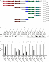
The UBX domain is necessary and sufficient to bind to p97. (A) Schematic representation of the D. discoideum UBXD9 domain organization and of the analyzed UBXD9 truncation constructs. N, N-terminus; C, C-terminus; LCR, Low complexity region, UBL1, Ubiquitin-like domain 1; LHU, Low Homology UBX domain; CC, Coiled coil domain; UBX, Ubiquitin regulatory X domain. Numbers indicate amino acid positions of UBXD9. (B) Pull-down experiments with recombinant p97 and full-length or truncated GST-UBXD9 coupled to glutathione beads. GST coupled to glutathione beads was used as negative control. Representative Western blots with anti p97 antibodies of supernatants (SN) and pellets (P) are shown. The position of p97 is indicated. (C) Quantification of p97 in the SN and P fractions. The sum of the values of SN and P for each experiment was taken as 100%. The bar graphs represent mean values and standard deviations (SD) of at least three independent experiments. For statistical analysis, the Dunnett’s multiple comparison test, implemented in GraphPad Prism as post hoc analysis, was performed. ***p ≤ 0.001; *p ≤ 0.05. ns, not significant.
FIGURE 4
FIGURE 4
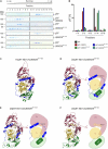
The helical lariat structure of UBXD9 is pivotal for its disassembly activity. (A) Sucrose density gradient sedimentation with p97 alone (top panel) or with equimolar amounts of p97 and full-length UBXD9 (2nd panel from top) or p97 and UBXD9261–573 (3rd and 4th panels from top), or p97 and UBXD9331–573 (5th and 6th panels from top), or p97 and UBXD9384–573 (lower two panels). Before loading the samples on the sucrose density gradient (0.3–1.1 M sucrose), they were incubated with 1 mM ATP for 30 min. Fractions were removed consecutively after centrifugation and analyzed by SDS-PAGE and Coomassie Brilliant Blue staining. p97 monomers or dimers are present in fractions 5–8 and p97 hexamers in fractions 11–14. Fractions are numbered, starting with #1 at 0.3 M sucrose. The positions of p97, full-length UBXD9 and UBXD9 truncation constructs are indicated. (B) Bar graphs depicting the quantification of p97 in fractions 1–4, 5–8, 9–10, 11–14, and 15–18 of the different experiments. The bar graphs represent mean values of at least three independent experiments. The determined total amount of p97 in all 18 fractions was set to 100%. (C) Structural model of the Hs p97-ND1:Hs UBXD9317–496 (VCP:ASPL; PDB: 5ifs) complex. UBXD9317–496 is shown in green and ochre and the ND1 domains of p97 (VCP) are colored yellow and dark pink, respectively. The region N-terminal of the UBX domain forms a loop, which encompasses two α-helices (α1 and α0; blue), an extended linker (blue) and a β-strand (β0, blue). The α-helices and the linker embrace the N domain of p97 and the β0-strand appears to be crucial for the closure of the lariat structure. The helical lariat probably interferes with the D1:D1’ inter-monomeric interaction of the p97 hexamer, causing its disassembly (Arumughan et al., 2016; Banchenko et al., 2019). (D–F) Homology models and schematic representation of the D. discoideum p97-ND1:UBXD9320–522 (D), p97-ND1:UBXD9331–522 (E), and p97-ND1:UBXD9384–522 (F) complexes representing our truncation constructs UBXD9261–573, UBXD9331–573, and UBXD9384–573, respectively. Since our modeling was based on the Hs-p97-ND1:Hs-UBXD9317–496 (VCP:ASPL; PDB: 5ifs) complex, the structural models could only be generated from amino acids 320–522 of D. discoideum UBXD9.
FIGURE 5
FIGURE 5
Blue Native PAGE analysis of p97, full-length UBXD9 as well as UBXD9 truncation constructs. Representative Blue Native PAGE gels of purified recombinant non-tagged D. discoideum proteins. Specific bands are marked by asterisks. (A) p97, UBXD9, UBXD91–336 and UBXD9261–573. (B) UBXD9331–573 and UBXD9384–573.
FIGURE 6
FIGURE 6
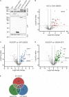
Interaction proteomics. (A) Verification of the BirA-UBXD9, GFP, GFP-UBXD9, and UBXD9-GFP expressing strains by western blotting using the polyclonal UBXD9 (top panel), the polyclonal BirA (2nd panel from top), and the monoclonal GFP (3rd panel from top) antibodies. Detection of actin (bottom panel) with the monoclonal actin antibody was used as loading control. (B–D) Volcano plots depicting p-value versus fold change (FC) for all proteins identified by mass spectrometry in AX2 versus AX2/BirA-UBXD9 expressing cells (B), AX2/GFP versus AX2/GFP-UBXD9 expressing cells (C) and AX2/GFP versus AX2/UBXD9-GFP expressing cells (D). Putative UBXD9 interacting proteins with a fold change ≥ 1.4 and a p ≤ 0.05 are depicted as red, blue or green dots, respectively. Dots representing UBXD9, p97, and glutamine synthetase type III (GSIII) are indicated. The plots were done using GraphPad prism. (E) Venn diagram of significantly enriched proteins in the pull-down experiments of AX2/BirA-UBXD9 (red), AX2/GFP-UBXD9 (blue), and AX2/UBXD9-GFP (green) cells. The intersections of the circles provide the number of proteins that were identified in two or all three approaches. Only those proteins with a fold change ≥ 1.4 and a p ≤ 0.05 were used as input.
FIGURE 7
FIGURE 7
Gene ontology (GO) term enrichment analysis of proteins identified as putative interaction partners of UBXD9 in at least two experimental setups. The proteins, which were identified in at least two experimental approaches (listed in Table 2), were used as input for GO analysis. The analysis was performed with PANTHER version 15.0. For the enriched categories –Log10 p-value and fold enrichment (FE) are given.
FIGURE 8
FIGURE 8
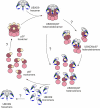
Structure-based model for the disassembly of the p97 hexamer by UBXD9. We propose, that a D. discoideum UBXD9 hexamer binds to a p97 hexamer and forms in the first step a hetero-dodecamer. The course of the dissociation of this complex is still unclear and possibly proceeds via intermediate steps. This could result in hetero-tetramers, which then dissociate to heterodimers and finally UBXD9 and p97 monomers are released. These are then free for another round of oligomerization. The p97 D1 and D2 domains are depicted in dark and light pink, respectively, and the N domain in yellow. UBXD9 is shown schematically with the UBX domain in green. The blue cylinders represent the α0 and α1 helices and the blue arrow the β0 strand.

Similar articles

Cited by

References

    1. Abraham M. J., Murtola T., Schulz R., Páll S., Smith J. C., Hess B., et al. (2015). GROMACS: high performance molecular simulations through multi-level parallelism from laptops to supercomputers. SoftwareX 1-2 19–25. 10.1016/j.softx.2015.06.001 - DOI
    1. Alexandru G., Graumann J., Smith G. T., Kolawa N. J., Fang R., Deshaies R. J. (2008). UBXD7 binds multiple ubiquitin ligases and implicates p97 in HIF1alpha turnover. Cell 134 804–816. - PMC - PubMed
    1. Arhzaouy K., Strucksberg K. H., Tung S. M., Tangavelou K., Stumpf M., Faix J., et al. (2012). Heteromeric p97/p97R155C complexes induce dominant negative changes in wild-type and autophagy 9-deficient Dictyostelium strains. PLoS One 7:e46879. 10.1371/journal.pone.0046879 - DOI - PMC - PubMed
    1. Arumughan A., Roske Y., Barth C., Forero L. L., Bravo-Rodriguez K., Redel A., et al. (2016). Quantitative interaction mapping reveals an extended UBX domain in ASPL that disrupts functional p97 hexamers. Nat. Commun. 7:13047. - PMC - PubMed
    1. Banchenko S., Arumughan A., Petrovic S., Schwefel D., Wanker E. E., Roske Y., et al. (2019). Common mode of remodeling AAA ATPases p97/CDC48 by their disassembling cofactors ASPL/PUX1. Structure 27 1830–1841.e3. - PubMed