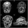
Case Report: Complex Congenital Brain Anomaly in a BBxHF Calf-Clinical Signs, Magnetic Resonance Imaging, and Pathological Findings
- PMID: 34631846
- PMCID: PMC8492953
- DOI: 10.3389/fvets.2021.700527
Case Report: Complex Congenital Brain Anomaly in a BBxHF Calf-Clinical Signs, Magnetic Resonance Imaging, and Pathological Findings
Abstract
This case report describes the clinical signs, magnetic resonance imaging (MRI) findings and associated (histo)pathological findings in a crossbred Belgian Blue calf with congenital complex brain anomaly. The calf was presented with non-progressive signs (including cerebellar ataxia) since it was born, suggestive of a multifocal intracranial lesion. A congenital anomaly was suspected and after hematology, biochemistry, serology, and cerebrospinal fluid analysis, a magnetic resonance imaging study was performed. The following suspected abnormalities were the principal changes identified: severe hydrocephalus, porencephaly, suspected partial corpus callosum agenesis (CCA), and increased fluid signal between the folia of the cerebellum. Post-mortem examination predominately reflected the MRI findings. The origin for these malformations could not be identified and there was no evidence of a causative infectious agent. Corpus callosum abnormalities have been reported in bovids before and have been linked to bovine viral diarrhea virus (BVDV) infections, as have several other central nervous system anomalies in this species. In this case, BVDV was deemed an unlikely causative agent based on serology test results and lack of typical histopathological signs. The etiology of the congenital anomaly present in this bovine calf remains unknown.
Keywords: bovid; cerebellar ataxia; corpus callosum agenesis; hydrocephalus; porencephaly.
Copyright © 2021 Veenema, Santifort, Kuijpers, Seijger and Hut.
Conflict of interest statement
The authors declare that the research was conducted in the absence of any commercial or financial relationships that could be construed as a potential conflict of interest.
Figures


Similar articles
-
Clinical, MRI, and histopathological findings of congenital focal diplomyelia at the level of L4 in a female crossbred calf.BMC Vet Res. 2020 Oct 21;16(1):398. doi: 10.1186/s12917-020-02580-4. BMC Vet Res. 2020. PMID: 33087102 Free PMC article.
-
Perosomus elumbis in a Holstein calf infected with bovine viral diarrhea virus.Tierarztl Prax Ausg G Grosstiere Nutztiere. 2013;41(6):387-91. Tierarztl Prax Ausg G Grosstiere Nutztiere. 2013. PMID: 24326794
-
Accuracy of prenatal ultrasound in the diagnosis of corpus callosum anomalies.J Matern Fetal Neonatal Med. 2021 Feb;34(3):439-444. doi: 10.1080/14767058.2019.1609931. Epub 2019 Apr 29. J Matern Fetal Neonatal Med. 2021. PMID: 31035852
-
Role of magnetic resonance imaging in fetuses with mild or moderate ventriculomegaly in the era of fetal neurosonography: systematic review and meta-analysis.Ultrasound Obstet Gynecol. 2019 Aug;54(2):164-171. doi: 10.1002/uog.20197. Epub 2019 Jul 11. Ultrasound Obstet Gynecol. 2019. PMID: 30549340
-
Magnetic Resonance Imaging Findings in Fetal Corpus Callosal Developmental Abnormalities: A Pictorial Essay.J Pediatr Neurosci. 2020 Oct-Dec;15(4):352-357. doi: 10.4103/jpn.JPN_174_19. Epub 2021 Jan 19. J Pediatr Neurosci. 2020. PMID: 33936297 Free PMC article. Review.
Cited by
-
3T Magnetic resonance imaging and computed tomography of the bovine carpus.BMC Vet Res. 2022 Jun 22;18(1):236. doi: 10.1186/s12917-022-03346-w. BMC Vet Res. 2022. PMID: 35733155 Free PMC article.
References
-
- Coppock RW, Dziwenka MM. Chapter 84: Teratogeneses in livestock. In: Gupta RC. editor. Reproduction and Developmental Toxicology. London: Academic Press; (2011). p. 1127–37. 10.1016/B978-0-12-382032-7.10084-0 - DOI
-
- Cho DY, Leipold HW. Agenesis of corpus callosum in calves. Cornell Vet. (1978) 68:99–107. - PubMed
Publication types
LinkOut - more resources
Full Text Sources

