A Pyramid Architecture-Based Deep Learning Framework for Breast Cancer Detection
- PMID: 34631877
- PMCID: PMC8500767
- DOI: 10.1155/2021/2567202
A Pyramid Architecture-Based Deep Learning Framework for Breast Cancer Detection
Abstract
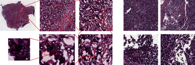
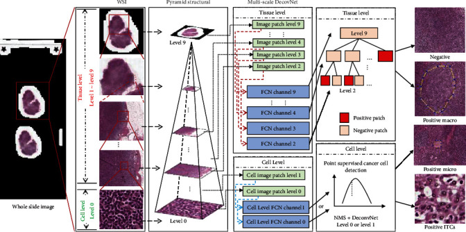
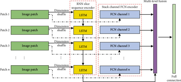
Breast cancer diagnosis is a critical step in clinical decision making, and this is achieved by making a pathological slide and gives a decision by the doctors, which is the method of final decision making for cancer diagnosis. Traditionally, the doctors usually check the pathological images by visual inspection under the microscope. Whole-slide images (WSIs) have supported the state-of-the-art diagnosis results and have been admitted as the gold standard clinically. However, this task is time-consuming and labour-intensive, and all of these limitations make low efficiency in decision making. Medical image processing protocols have been used for this task during the last decades and have obtained satisfactory results under some conditions; especially in the deep learning era, it has exhibited the advantages than those in the shallow learning period. In this paper, we proposed a novel breast cancer region mining framework based on deep pyramid architecture from multilevel and multiscale breast pathological WSIs. We incorporate the tissue- and cell-level information together and integrate these into a LSTM model for the final sequence modelling, which successfully keeps the WSIs' integration and is not mentioned by the prevalence frameworks. The experiment results demonstrated that our proposed framework greatly improved the detection accuracy than that only using tissue-level information.
Copyright © 2021 Dong Sui et al.
Conflict of interest statement
The authors declare that they have no conflicts of interest.
Figures


Similar articles
-
RegWSI: Whole slide image registration using combined deep feature- and intensity-based methods: Winner of the ACROBAT 2023 challenge.Comput Methods Programs Biomed. 2024 Jun;250:108187. doi: 10.1016/j.cmpb.2024.108187. Epub 2024 Apr 22. Comput Methods Programs Biomed. 2024. PMID: 38657383
-
Deep learning approaches for breast cancer detection in histopathology images: A review.Cancer Biomark. 2024;40(1):1-25. doi: 10.3233/CBM-230251. Cancer Biomark. 2024. PMID: 38517775 Free PMC article. Review.
-
A deep learning based multiscale approach to segment the areas of interest in whole slide images.Comput Med Imaging Graph. 2021 Jun;90:101923. doi: 10.1016/j.compmedimag.2021.101923. Epub 2021 Apr 15. Comput Med Imaging Graph. 2021. PMID: 33894669
-
Recognizing basal cell carcinoma on smartphone-captured digital histopathology images with a deep neural network.Br J Dermatol. 2020 Mar;182(3):754-762. doi: 10.1111/bjd.18026. Epub 2019 Aug 22. Br J Dermatol. 2020. PMID: 31017653
-
Deep learning in mammography and breast histology, an overview and future trends.Med Image Anal. 2018 Jul;47:45-67. doi: 10.1016/j.media.2018.03.006. Epub 2018 Mar 26. Med Image Anal. 2018. PMID: 29679847 Review.
Cited by
-
Artificial Intelligence in Oncology: Current Landscape, Challenges, and Future Directions.Cancer Discov. 2024 May 1;14(5):711-726. doi: 10.1158/2159-8290.CD-23-1199. Cancer Discov. 2024. PMID: 38597966 Free PMC article. Review.
-
Deep Learning Application for Effective Classification of Different Types of Psoriasis.J Healthc Eng. 2022 Jan 15;2022:7541583. doi: 10.1155/2022/7541583. eCollection 2022. J Healthc Eng. 2022. PMID: 35075392 Free PMC article.
-
Magnifying Networks for Histopathological Images with Billions of Pixels.Diagnostics (Basel). 2024 Mar 1;14(5):524. doi: 10.3390/diagnostics14050524. Diagnostics (Basel). 2024. PMID: 38472996 Free PMC article.
-
Artificial intelligence in histopathology: enhancing cancer research and clinical oncology.Nat Cancer. 2022 Sep;3(9):1026-1038. doi: 10.1038/s43018-022-00436-4. Epub 2022 Sep 22. Nat Cancer. 2022. PMID: 36138135 Review.
-
The Role of Artificial Intelligence in Early Cancer Diagnosis.Cancers (Basel). 2022 Mar 16;14(6):1524. doi: 10.3390/cancers14061524. Cancers (Basel). 2022. PMID: 35326674 Free PMC article. Review.
References
-
- Gomes D. S., Porto S. S., Balabram D., Gobbi H. Inter-observer variability between general pathologists and a specialist in breast pathology in the diagnosis of lobular neoplasia, columnar cell lesions, atypical ductal hyperplasia and ductal carcinoma in situ of the breast. Diagnostic Pathology . 2014;9(1):121–121. doi: 10.1186/1746-1596-9-121. - DOI - PMC - PubMed
MeSH terms
LinkOut - more resources
Full Text Sources
Other Literature Sources
Medical