

Mechanisms of instantaneous inactivation of SARS-CoV-2 by silicon nitride bioceramic
- PMID: 34632359
- PMCID: PMC8485720
- DOI: 10.1016/j.mtbio.2021.100144
Mechanisms of instantaneous inactivation of SARS-CoV-2 by silicon nitride bioceramic
Abstract
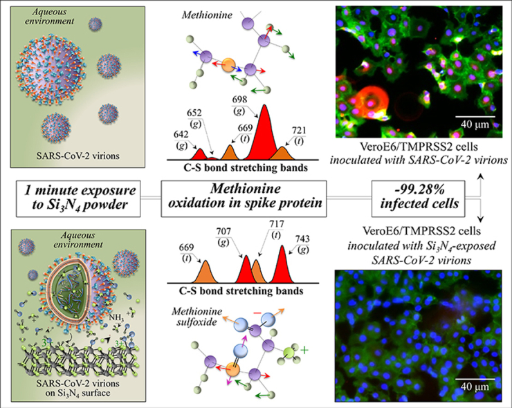
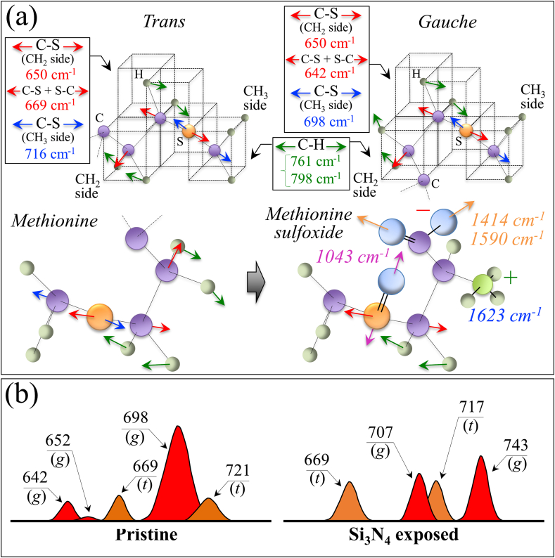
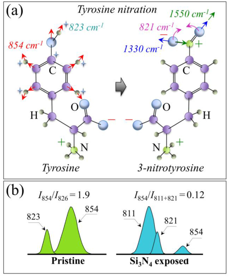
The hydrolytic processes occurring at the surface of silicon nitride (Si3N4) bioceramic have been indicated as a powerful pathway to instantaneous inactivation of SARS-CoV-2 virus. However, the virus inactivation mechanisms promoted by Si3N4 remain yet to be elucidated. In this study, we provide evidence of the instantaneous damage incurred on the SARS-CoV-2 virus upon contact with Si3N4. We also emphasize the safety characteristics of Si3N4 for mammalian cells. Contact between the virions and micrometric Si3N4 particles immediately targeted a variety of viral molecules by inducing post-translational oxidative modifications of S-containing amino acids, nitration of the tyrosine residue in the spike receptor binding domain, and oxidation of RNA purines to form formamidopyrimidine. This structural damage in turn led to a reshuffling of the protein secondary structure. These clear fingerprints of viral structure modifications were linked to inhibition of viral functionality and infectivity. This study validates the notion that Si3N4 bioceramic is a safe and effective antiviral compound; and a primary antiviral candidate to replace the toxic and allergenic compounds presently used in contact with the human body and in long-term environmental sanitation.
Keywords: Ammonia; Hydrolysis; SARS-CoV-2; Silicon nitride; Surface chemistry; Virus.
© 2021 The Author(s).
Conflict of interest statement
The authors declare that they have no known competing financial interests or personal relationships that could have appeared to influence the work reported in this paper.
Figures
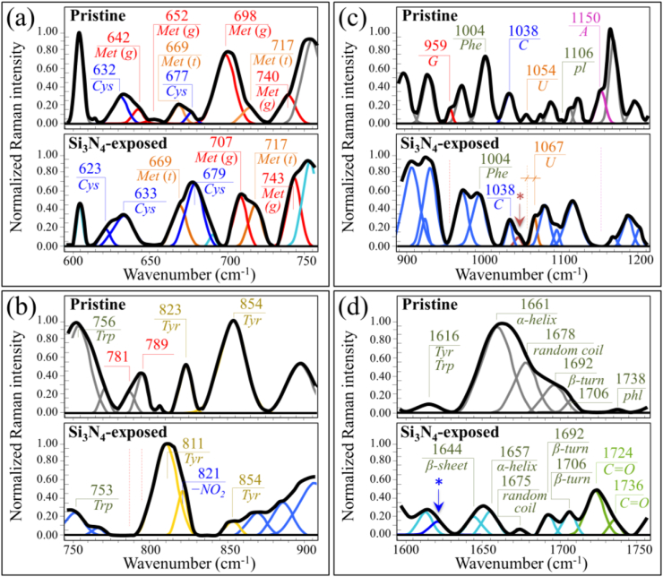
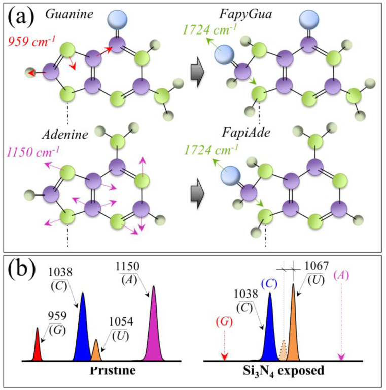
 O stretching mode appearing at 1724 cm−1. In (b), a comparison is shown of Voigtian components representing fingerprint signals (and respective frequencies) of RNA purine and pyrimidines before and after exposure of the virions to Si3N4 powder.
Similar articles
-
Instantaneous Inactivation of Herpes Simplex Virus by Silicon Nitride Bioceramics.Int J Mol Sci. 2023 Aug 10;24(16):12657. doi: 10.3390/ijms241612657. Int J Mol Sci. 2023. PMID: 37628838 Free PMC article.
-
Raman Fingerprints of the SARS-CoV-2 Delta Variant and Mechanisms of Its Instantaneous Inactivation by Silicon Nitride Bioceramics.ACS Infect Dis. 2022 Aug 12;8(8):1563-1581. doi: 10.1021/acsinfecdis.2c00200. Epub 2022 Jul 12. ACS Infect Dis. 2022. PMID: 35819780
-
Raman signatures of type A and B influenza viruses: molecular origin of the "catch and kill" inactivation mechanism mediated by micrometric silicon nitride powder.RSC Chem Biol. 2025 Jan 22;6(2):182-208. doi: 10.1039/d4cb00237g. eCollection 2025 Feb 5. RSC Chem Biol. 2025. PMID: 39850321 Free PMC article.
-
Therapeutic potential of green tea catechin, (-)-epigallocatechin-3-O-gallate (EGCG) in SARS-CoV-2 infection: Major interactions with host/virus proteases.Phytomed Plus. 2023 Feb;3(1):100402. doi: 10.1016/j.phyplu.2022.100402. Epub 2022 Dec 30. Phytomed Plus. 2023. PMID: 36597465 Free PMC article. Review.
-
Protein post-translational modification in SARS-CoV-2 and host interaction.Front Immunol. 2023 Jan 13;13:1068449. doi: 10.3389/fimmu.2022.1068449. eCollection 2022. Front Immunol. 2023. PMID: 36713387 Free PMC article. Review.
Cited by
-
In Situ Raman Analysis of Biofilm Exopolysaccharides Formed in Streptococcus mutans and Streptococcus sanguinis Commensal Cultures.Int J Mol Sci. 2023 Apr 3;24(7):6694. doi: 10.3390/ijms24076694. Int J Mol Sci. 2023. PMID: 37047667 Free PMC article.
-
Photochemical synthesis of pink silver and its use for monitoring peptide nitration via surface enhanced Raman spectroscopy (SERS).Amino Acids. 2022 Sep;54(9):1261-1274. doi: 10.1007/s00726-022-03178-w. Epub 2022 Jun 22. Amino Acids. 2022. PMID: 35731286
-
Viral Transmission in Sea Food Systems: Strategies for Control and Emerging Challenges.Foods. 2025 Mar 20;14(6):1071. doi: 10.3390/foods14061071. Foods. 2025. PMID: 40232102 Free PMC article. Review.
-
Instantaneous Inactivation of Herpes Simplex Virus by Silicon Nitride Bioceramics.Int J Mol Sci. 2023 Aug 10;24(16):12657. doi: 10.3390/ijms241612657. Int J Mol Sci. 2023. PMID: 37628838 Free PMC article.
-
Targeting the YXXΦ Motifs of the SARS Coronaviruses 1 and 2 ORF3a Peptides by In Silico Analysis to Predict Novel Virus-Host Interactions.Biomolecules. 2022 Jul 29;12(8):1052. doi: 10.3390/biom12081052. Biomolecules. 2022. PMID: 36008946 Free PMC article.
References
-
- Pezzotti G. Silicon nitride: a bioceramic with a gift. ACS Appl. Mater. Interfaces. 2019;11:26619–26636. - PubMed
LinkOut - more resources
Full Text Sources
Miscellaneous
