. 2021 Sep;2(3):031402.
doi: 10.1063/5.0057071. Epub 2021 Sep 30.

High-throughput assays show the timescale for phagocytic success depends on the target toughness

High-throughput assays show the timescale for phagocytic success depends on the target toughness

Layla A Bakhtiari et al. Biophys Rev (Melville). 2021 Sep.

Abstract

Phagocytic immune cells can clear pathogens from the body by engulfing them. Bacterial biofilms are communities of bacteria that are bound together in a matrix that gives biofilms viscoelastic mechanical properties that do not exist for free-swimming bacteria. Since a neutrophil is too small to engulf an entire biofilm, it must be able to detach and engulf a few bacteria at a time if it is to use phagocytosis to clear the infection. We recently found a negative correlation between the target elasticity and phagocytic success. That earlier work used time-consuming, manual analysis of micrographs of neutrophils and fluorescent beads. Here, we introduce and validate flow cytometry as a fast and high-throughput technique that increases the number of neutrophils analyzed per experiment by two orders of magnitude, while also reducing the time required to do so from hours to minutes. We also introduce the use of polyacrylamide gels in our assay for engulfment success. The tunability of polyacrylamide gels expands the mechanical parameter space we can study, and we find that high toughness and yield strain, even with low elasticity, also impact the phagocytic success as well as the timescale thereof. For stiff gels with low-yield strain, and consequent low toughness, phagocytic success is nearly four times greater when neutrophils are incubated with gels for 6 h than after only 1 h of incubation. In contrast, for soft gels with high-yield strain and consequent high toughness, successful engulfment is much less time-sensitive, increasing by less than a factor of two from 1 to 6 h incubation.


Figures

FIG. 1.
FIG. 1.
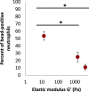
High-throughput measurements of phagocytic success using flow cytometry preserves previously published trends. The elastic modulus of alginate hydrogels negatively correlates with the neutrophil association with the beads that were initially embedded in gels, as expected from our previous study. ANOVA significance testing yields a p-value for the group of p = 0.000 19. Horizontal bars and accompanying asterisks indicate p-values from Student two-tailed T-test between connected pairs of data: *p < 0.05 (N = 8 experiments per data point, with 20 000 neutrophils per sample in each experiment).
FIG. 2.
FIG. 2.
Representative (a) frequency and (b) strain sweeps from bulk oscillatory rheology measurements on polyacrylamide gels. Elastic moduli for each sample are shown with a solid marker, while viscous moduli are shown with hollow markers. Gels made with 8% acrylamide have an average elastic modulus of 5320 Pa, while those made with 3% acrylamide are two orders of magnitude softer with an average elastic modulus of 43 Pa. Elastic and viscous moduli are values corresponding to the measurement at 1% strain within the plateau region of each gel. In both gel types, the viscous modulus is one order of magnitude lower than the elastic modulus, indicating that the elastic properties dominate, and gels behave primarily as viscoelastic solids. We note that the differences in mechanical properties for polyacrylamide gels are attributed to not just the acrylamide concentration displayed in the legend, but also the crosslinker concentration (see methods for details).
FIG. 3.
FIG. 3.
Comparison of gel mechanics across various polyacrylamide and alginate hydrogels. Each gel is characterized by oscillatory rheology to obtain the material's elastic modulus (a), yield strain (b), yield stress (c), and toughness (d). Gels containing 3% acrylamide are significantly tougher than both those containing 8% acrylamide and those containing alginate ( T=258Jm3 as compared to =62Jm3). The high toughness arises from a very high-yield strain. The differences in mechanical properties for polyacrylamide gels are attributed to not just the acrylamide concentration, but also the crosslinker concentration (see methods for details). Gels containing 3% acrylamide did not yield during testing, and thus this analysis provides a lower bound estimate of the gel's mechanical properties. Horizontal bars and accompanying asterisks indicate p-values from the Student two-tailed T-test between the groups they connect: * p < 0.05, ** p < 0.005 (N = 3).
FIG. 4.
FIG. 4.
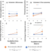
Phagocytic success and fold change for microscopy-based assay [(a) and (c)] and high-throughput assay [(b) and (d)] with cells from volunteer 1. Data for individual experiments are shown with a dashed line, and the average from each gel group is shown with a solid line. Initial high-throughput measurements demonstrate similar trends to the microscopy-based assay after 1 and 3 h incubation. Cells incubated on gels with 8% acrylamide experienced a larger increase in phagocytic success after extended incubation times than cells incubated on gels with 3% acrylamide. A significant fold change at the 3 h time marker was, however, not observed for cells from volunteer 1 with either acrylamide gel using the high-throughput method. This highlights the need for more extensive studies involving neutrophils isolated from different healthy donors. ANOVA significance testing yields a p-value for the group of all gels of (A) p = 0.01. Horizontal bars with accompanying asterisk indicate p-values from using the Student two-tailed T-test to compare the results from the same type of gel at 1 and 3 h; gel type is indicated with asterisk color. Asterisks without horizontal bars indicate that data for one gel type at one timepoint are being tested with a null hypothesis that the fold change is unity. *p < 0.05. N = 3 for all measurements.
FIG. 5.
FIG. 5.
Phagocytic success (top row) and fold change (bottom row) for microscopy-based assay [(a) and (e)] and high-throughput assay [(b) and (f)] with cells from volunteer 2, and additional high-throughput measurements with cells from volunteers 1 [(c) and (g)] and 3 [(d) and (h)]. Data for individual experiments are shown with a dashed line, and the average from each gel group is shown in bold. Using the microscopy-based assay, we see some successful engulfment from 8% acrylamide gels (orange markers) after extending the time the neutrophils are incubated on gel samples for up to 6 h. Such a strong change in phagocytic success after extending incubation time to 6 h is not seen for neutrophils incubated on 3% acrylamide gels (blue markers). ANOVA significance testing yields a p-value for the group of all gels of (a) p = 0.02, (e) p = 0.01, and (h) p = 0.05. Horizontal bars and accompanying asterisk indicate p-values from Student two-tailed T-test for gel samples shown with the corresponding color. Horizontal bars indicate two datasets from the same gel type and different timepoints are being compared. Asterisks without horizontal bars indicate that data from one gel type at one timepoint are being compared with the null hypothesis of unity. Black asterisks (with a vertical line) indicate that data from both gel types, at the same timepoint, are being compared with each other. *p < 0.05, **p < 0.005, ***p< 0.000 5. For volunteer 1: for microscopy-based data, N = 4 for data displayed in blue and N = 5 for data displayed in orange; for high-throughput data, N = 6 for all measurements. For volunteers 2 and 3, N = 3 for all measurements.

References

    1. Amulic B., Cazalet C., Hayes G. L., Metzler K. D., and Zychlinsky A., Annu. Rev. Immunol. 30, 459–489 (2012).10.1146/annurev-immunol-020711-074942 - DOI - PubMed
    1. Borregaard N., Immunity 33(5), 657–670 (2010).10.1016/j.immuni.2010.11.011 - DOI - PubMed
    1. Kolaczkowska E. and Kubes P., Nat. Rev. Immunol. 13(3), 159–175 (2013).10.1038/nri3399 - DOI - PubMed
    1. Silva M. and Correia-Neves M., Front. Immunol. 3, 174 (2012).10.3389/fimmu.2012.00174 - DOI - PMC - PubMed
    1. Richards D. M. and Endres R. G., Biophys. J. 107(7), 1542–1553 (2014).10.1016/j.bpj.2014.07.070 - DOI - PMC - PubMed

LinkOut - more resources