. 2021 Nov 1;224(21):jeb242639.
doi: 10.1242/jeb.242639. Epub 2021 Nov 5.

Swimming kinematics and performance of spinal transected lampreys with different levels of axon regeneration

Affiliations

Swimming kinematics and performance of spinal transected lampreys with different levels of axon regeneration

Jacob Fies et al. J Exp Biol. .

Abstract

Axon regeneration is critical for restoring neural function after spinal cord injury. This has prompted a series of studies on the neural and functional recovery of lampreys after spinal cord transection. Despite this, there are still many basic questions remaining about how much functional recovery depends on axon regeneration. Our goal was to examine how swimming performance is related to degree of axon regeneration in lampreys recovering from spinal cord transection by quantifying the relationship between swimming performance and percent axon regeneration of transected lampreys after 11 weeks of recovery. We found that while swimming speeds varied, they did not relate to percent axon regeneration. In fact, swimming speeds were highly variable within individuals, meaning that most individuals could swim at both moderate and slow speeds, regardless of percent axon regeneration. However, none of the transected individuals were able to swim as fast as the control lampreys. To swim fast, control lampreys generated high amplitude body waves with long wavelengths. Transected lampreys generated body waves with lower amplitude and shorter wavelengths than controls, and to compensate, transected lampreys increased their wave frequencies to swim faster. As a result, transected lampreys had significantly higher frequencies than control lampreys at comparable swimming velocities. These data suggest that the control lampreys swam more efficiently than transected lampreys. In conclusion, there appears to be a minimal recovery threshold in terms of percent axon regeneration required for lampreys to be capable of swimming; however, there also seems to be a limit to how much they can behaviorally recover.

Keywords: Petromyzon marinus; Anguilliform; Neuromuscular.


Conflict of interest statement

Competing interests The authors declare no competing or financial interests.

Figures

Fig. 1.
Fig. 1.
Reticulospinal (RS) axon regeneration in the lamprey spinal cord. (A) Schematic of a larval lamprey with the site of spinal cord transection indicated with a red dashed line for a 5th gill or mid-body transection. (B) A montage of confocal z-projections stitched together of a control, sham uninjured spinal cord with axons labeled by a 10 kDa Alexa Fluor 488 dextran, showing fairly uniform labeling along the length of the spinal cord. (C,D) Labeling of axons ∼10.5 weeks post injury in a 5th gill transected and a mid-body transected animal shows sparser axon labeling in the region caudal to the lesion site in comparison to the rostral region, indicating the amount of axon regeneration. Note that the amount of axon regeneration is comparable between the 5th gill and mid-body transected spinal cords. Scale bar in D applies to B–D. Rostral (R) is to the left and caudal (C) is to the right.
Fig. 2.
Fig. 2.
Comparison of swimming performance and body kinematic parameters of lampreys. (A) Swimming speed of lampreys versus the degree of axon regeneration (%) after recovering for 10.5 weeks from complete spinal transection (regression analysis, d.f.=1, P>0.05). (B) Comparison of mean swimming speeds among treatments (ANOVA, d.f.=2, P>0.05). (C–E) Comparison of (C) wave frequency, (D) wavelength and (E) wave amplitude versus the degree of spinal cord regeneration (%; regression including 5th gill and mid-body, d.f.=1, P>0.05). Asterisks indicate that the control group kinematics were significantly different than the transected groups (Holm–Šidák post hoc comparison, P<0.05).
Fig. 3.
Fig. 3.
Comparison of body and swimming kinematics of lampreys. (A) Sequential images of different lamprey showing the progression of a body wave (indicated by white arrow) moving from head to tail. Notice the control lamprey has only one large wave traveling along the body at a time, whereas all the transected lampreys, regardless of swimming velocity (see D), have multiple smaller waves moving along the body. (B) Tracking of the movement of the head of the lamprey for 1 s. Notice the distance traveled and the evenness versus unevenness of the lateral motion of the heads through time. (C) Distance the different lampreys traveled over 1 s. (D) Velocity of the different lampreys over 1 s. Notice the regular swim cycles of the control lampreys (blue) versus the more erratic motion of the transected lampreys (orange).
Fig. 4.
Fig. 4.
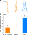
Change in body wave amplitude as the wave travels from head to tail. (A) Change in midline of representative lampreys over time. (B) Mean change in amplitude [as ratio of amplitude at the tail (caudal) and the head (rostral)] among treatments. Lowercase letters indicate significantly different treatment groups (Holm–Šidák post hoc comparison, P<0.05).
Fig. 5.
Fig. 5.
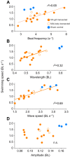
Body kinematic variables versus swimming speeds of 5th gill transected (filled orange circles), mid-body transected (open circles) and control lampreys (filled blue circles). (A) Tail-beat frequency, (B) wavelength and (C) wave speed were all positively related to swimming speed for the transected lampreys (regression analysis, P<0.01). (D) Wave amplitude of the traveling body waves was not significantly related to swimming speed (regression analysis, P>0.1).
Fig. 6.
Fig. 6.
Effects of percent regeneration on Strouhal number (St) and stride length (body lengths traveled per wave) for transected and control lampreys. (A) St of lampreys with different levels of regenerated spinal cord. Dotted blue lines highlight the region where studies have shown animals and flapping foils to have the highest propulsive efficiency. The 5th gill lampreys fell outside the optimal range of St, whereas the control and the one mid-body lamprey fall within the optimal range. (B) The 5th gill transected lampreys had significantly higher St than the mid-body transected and control lampreys (Holm–Šidák post hoc comparison, P<0.05). (C) Stride length of lampreys with different levels of regenerated spinal cord. (D) Comparison of the stride lengths among treatments; different lowercase letters designate significantly different groups (Holm–Šidák post hoc comparison, P<0.05).

Similar articles

Cited by

References

    1. Ajmani, N., Yasmin, T., Docker, M. F. and Good, S. V. (2021). Transcriptomic analysis of gonadal development in parasitic and non-parasitic lampreys (Ichthyomyzon spp.), with a comparison of genomic resources in these non-model species. G3 11, jkab030. 10.1093/g3journal/jkab030 - DOI - PMC - PubMed
    1. Armstrong, J., Zhang, L. and McClellan, A. D. (2003). Axonal regeneration of descending and ascending spinal projection neurons in spinal cord-transected larval lamprey. Exp. Neurol. 180, 156-166. 10.1016/S0014-4886(02)00057-2 - DOI - PubMed
    1. Becker, M. and Parker, D. (2019). Time course of functional changes in locomotor and sensory systems after spinal cord lesions in lamprey. J. Neurophysiol. 121, 2323-2335. 10.1152/jn.00120.2019 - DOI - PubMed
    1. Blight, A. R. (1977). The muscular control of vertebrate swimming movements. Biol. Rev. 52, 181-218. 10.1111/j.1469-185X.1977.tb01349.x - DOI
    1. Cohen, A. H., Mackler, S. A. and Selzer, M. E. (1986). Functional regeneration following spinal transection demonstrated in the isolated spinal cord of the larval sea lamprey. Proc. Natl. Acad. Sci. USA 83, 2763-2766. 10.1073/pnas.83.8.2763 - DOI - PMC - PubMed

Publication types