. 2021 Oct 10;69(1):27.
doi: 10.1007/s00005-021-00632-7.

Cyclosporine A, in Contrast to Rapamycin, Affects the Ability of Dendritic Cells to Induce Immune Tolerance Mechanisms

Affiliations

Cyclosporine A, in Contrast to Rapamycin, Affects the Ability of Dendritic Cells to Induce Immune Tolerance Mechanisms

Maja Machcińska et al. Arch Immunol Ther Exp (Warsz). .

Abstract

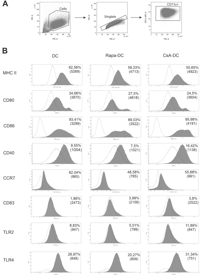
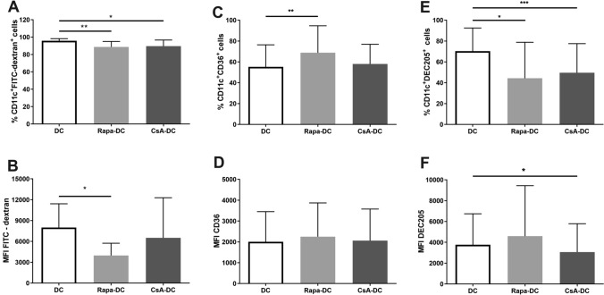
Following organ transplantation, it is essential that immune tolerance is induced in the graft recipient to reduce the risk of rejection and avoid complications associated with the long-term use of immunosuppressive drugs. Immature dendritic cells (DCs) are considered to promote transplant tolerance and may minimize the risk of graft rejection. The aim of the study was to evaluate the effects of immunosuppressive agents: rapamycin (Rapa) and cyclosporine A (CsA) on generation of human tolerogenic DCs (tolDCs) and also to evaluate the ability of these cells to induce mechanisms of immune tolerance. tolDCs were generated in the environment of Rapa or CsA. Next, we evaluated the effects of these agents on surface phenotypes (CD11c, MHC II, CD40, CD80, CD83, CD86, CCR7, TLR2, TLR4), cytokine production (IL-4, IL-6, IL-10, IL-12p70, TGF-β), phagocytic capacity and resistant to lipopolysaccharide activation of these DCs. Moreover, we assessed ability of such tolDCs to induce T cell activation and apoptosis, Treg differentiation and production of Th1- and Th2-characteristic cytokine profile. Data obtained in this study demonstrate that rapamycin is effective at generating maturation-resistant tolDCs, however, does not change the ability of these cells to induce mechanisms of immune tolerance. In contrast, CsA affects the ability of these cells to induce mechanisms of immune tolerance, but is not efficient at generating maturation-resistant tolDCs.

Keywords: Cyclosporine A; Dendritic cells; Immune tolerance; Immunosuppressive agents; Rapamycin; T cells.


Conflict of interest statement

The authors declare no conflict of interest.

Figures

Fig. 1
Fig. 1
Effects of Rapa and CsA on the expression of DCs surface markers. Immature DCs were differentiated in the environment of immunosuppressive agents: Rapa (Rapa-DC) or CsA (CsA-DC) and without drugs (DC). Gating strategy for flow cytometric analysis of DCs: a time gate was initially applied to exclude any electronic noise and artifact (not shown here). Next, based on size and granularity, DCs were gated in a forward scatter area (FSC-A) versus side scatter area (SSC-A) plot. Then, doublet cells were excluded using FSC-A/FSC-height (FSC-H) parameters. Within the singlet cell population CD11c+ DCs were gated (A), followed by expression of individual markers shown on representative histograms for DC, Rapa-DC and CsA-DC (B). The averages of the percentage of positively labelled CD11c+ DCs and mean fluorescence intensity in bracket 7–14 different donors are reported on each histogram. The white shade indicates FMO control. Representative dot plots are presented
Fig. 2
Fig. 2
Effects of Rapa and CsA on the cytokine production by immature DCs. Immature DCs were differentiated in the environment of immunosuppressive agents: Rapa (Rapa-DC) or CsA (CsA-DC) and without drugs (DC). Cytokine expression was measured by intracellular staining (IL-4; A) or by ELISA of culture supernatants (IL-10, TGF-β and IL-6; BD, respectively). Results are the averages ± SD of the percentage of positively labelled CD11c+ DCs (A; n = 5) or cytokine secretion (BD; n = 19). **p ≤ 0.01; p values were calculated by Wilcoxon matched pair test
Fig. 3
Fig. 3
Effects of Rapa and CsA on phagocytic capacity of DCs. Immature DCs were differentiated in the environment of immunosuppressive agents: Rapa (Rapa-DC) or CsA (CsA-DC) and without drugs (DC). Cellular FITC-dextran uptake (A, B; n = 7) and the expression of phagocytosis receptors: CD36 (C, D; n = 10) and DEC205 (F, G; n = 11) were determined by flow cytometry. Results are the averages ± SD of the percentage of positively labelled CD11c+ DCs and mean fluorescence intensity (MFI). *p ≤ 0.05, **p ≤ 0.01, ***p ≤ 0.001; p values were calculated by Wilcoxon matched pair test
Fig. 4
Fig. 4
Effects of Rapa and CsA on the cytokine production by LPS-activated DCs. Immature DCs generated in the environment of: Rapa (Rapa-DC + LPS), CsA (CsA-DC + LPS) and without drugs (DC + LPS) were activated with LPS. Additionally, DCs, which differentiated without drugs, were activated with LPS simultaneously with Rapa (DC + LPS + Rapa) or CsA (DC + LPS + CsA). Cytokine expression was measured by intracellular staining (IL-4; A) or by ELISA of culture supernatants (IL-10, TGF-β and IL-6; BD, respectively). Results are the averages ± SD of the percentage of positively labelled CD11c+ DCs (A) or cytokine secretion (BD) from at least five different donors. *p ≤ 0.05, **p ≤ 0.01, ***p ≤ 0.001; p values were calculated by Wilcoxon matched pair test
Fig. 5
Fig. 5
Effects of Rapa and CsA on PD-L1 expression on DCs. Immature DCs were differentiated in the environment of immunosuppressive agents: Rapa (Rapa-DC) or CsA (CsA-DC) and without drugs (DC). Expression of PD-L1 was determined by flow cytometry. Results are the averages ± SD of the percentage of CD11c+PD-L1+ DCs (A) and PD-L1 mean fluorescence intensity (MFI; B) from nine different donors. *p ≤ 0.05; p values were calculated by Wilcoxon matched pair test
Fig. 6
Fig. 6
Effects of Rapa-DC and CsA-DC on the expression of CD95 on CD4+ and CD8+ T cells. Immature DCs, Rapa-DC and CsA-DC were cocultured with T cells. Expression of CD95 was determined by flow cytometry. Results are the averages ± SD of the percentage of positively labelled CD4+ (A) or CD8+ (C) T cells and mean fluorescence intensity (MFI) for CD4+ (B) or CD8+ (D) T cells, from ten different donors. *p ≤ 0.05, **p ≤ 0.01; p values were calculated by Wilcoxon matched pair test
Fig. 7
Fig. 7
Effects of Rapa-DC and CsA-DC on T regulatory cells populations. Immature DCs, Rapa-DC and CsA-DC were cocultured with T cells. The percentage of CD4+CD25highFoxp3+ (A), CD8+CD25+CD28+ (B) and CD8+CD25CD28 (C) T cells were determined by flow cytometry. Results are the averages ± SD from ten different donors. *p ≤ 0.05; p values were calculated by Wilcoxon matched pair test
Fig. 8
Fig. 8
Effects of Rapa-DC and CsA-DC on the production of Th1- and Th2-characteristic cytokines. Immature DCs, Rapa-DC and CsA-DC were cocultured with T cells. Cytokine expression was measured by ELISA of culture supernatants for the presence of Th1 cytokines: IL-2 (A), IFN-γ (B) and Th2 cytokines: IL-4 (C), IL-5 (D), IL-10 (E), IL-13 (F). Results are the averages ± SD of cytokine secretion from 19 different donors. *p ≤ 0.05, **p ≤ 0.01; p values were calculated by Wilcoxon matched pair test

Similar articles

Cited by

References

    1. Abdul M, Charron D, Haziot A. Selective effects of cyclosporine A on Th2-skewed dendritic cells matured with viral-like stimulus by means of Toll-like receptors. Transplantation. 2008;86:880–884. doi: 10.1097/TP.0b013e3181861f1d. - DOI - PubMed
    1. Adnan E, Matsumoto T, Ishizaki J, et al. Human tolerogenic dendritic cells generated with protein kinase C Inhibitor are optimal for functional regulatory T cell induction – a comparative study. Clin Immunol. 2016;173:96–108. doi: 10.1016/j.clim.2016.09.007. - DOI - PubMed
    1. Bhatia S, Edidin M, Almo SC, et al. B7–1 and B7–2: similar costimulatory ligands with different biochemical, oligomeric and signaling propert. Immunol Lett. 2006;104:70–75. doi: 10.1016/j.imlet.2005.11.019. - DOI - PubMed
    1. Boks MA, Kager-Groenland JR, Haasjes MSP, et al. IL-10-generated tolerogenic dendritic cells are optimal for functional regulatory T cell induction - a comparative study of human clinical-applicable DC. Clin Immunol. 2012;142:332–342. doi: 10.1016/j.clim.2011.11.011. - DOI - PubMed
    1. Cangemi M, Montico B, Faè DA, et al. Dissecting the multiplicity of immune effects of immunosuppressive drugs to better predict the risk of de novo malignancies in solid organ transplant patients. Front Oncol. 2019;9:160. doi: 10.3389/fonc.2019.00160. - DOI - PMC - PubMed