

Cannabidiol-induced activation of the metallothionein pathway impedes anticancer effects of disulfiram and its metabolite CuET
- PMID: 34632694
- PMCID: PMC8978514
- DOI: 10.1002/1878-0261.13114
Cannabidiol-induced activation of the metallothionein pathway impedes anticancer effects of disulfiram and its metabolite CuET
Abstract
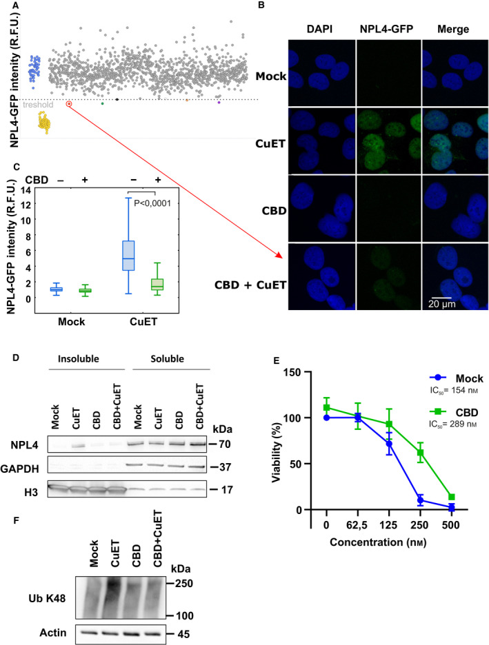
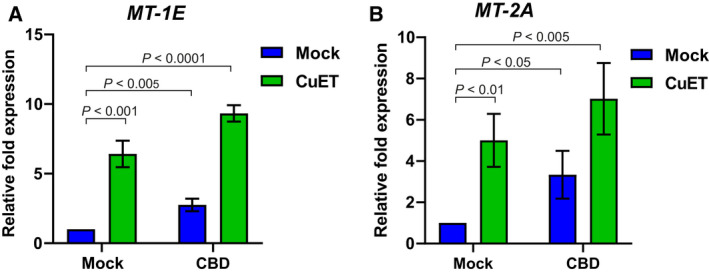
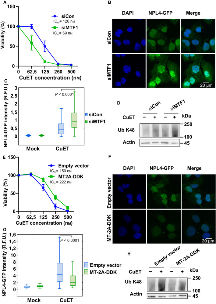
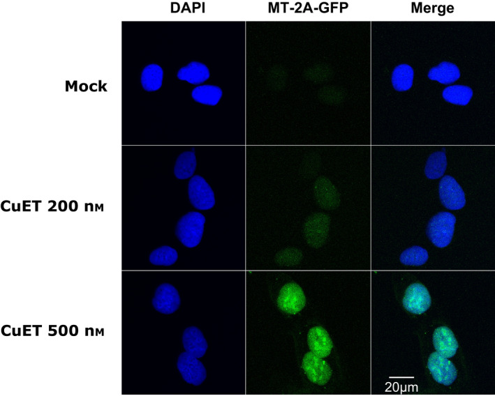
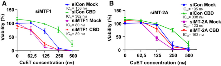
Disulfiram (DSF), an established alcohol-aversion drug, is a candidate for repurposing in cancer treatment. DSF's antitumor activity is supported by preclinical studies, case reports, and small clinical trials; however, ongoing clinical trials of advanced-stage cancer patients encounter variable results. Here, we show that one reason for the inconsistent clinical effects of DSF may reflect interference by other drugs. Using a high-throughput screening and automated microscopy, we identify cannabidiol, an abundant component of the marijuana plant used by cancer patients to mitigate side effects of chemotherapy, as a likely cause of resistance to DSF. Mechanistically, in cancer cells, cannabidiol triggers the expression of metallothioneins providing protective effects by binding heavy metal-based substances including the bis-diethyldithiocarbamate-copper complex (CuET). CuET is the documented anticancer metabolite of DSF, and we show here that the CuET's anticancer toxicity is effectively neutralized by metallothioneins. Overall, this work highlights an example of undesirable interference between cancer therapy and the concomitant usage of marijuana products. In contrast, we report that insufficiency of metallothioneins sensitizes cancer cells toward CuET, suggesting a potential predictive biomarker for DSF repurposing in oncology.
Keywords: CuET; cancer; cannabidiol; disulfiram; metallothionein.
© 2021 The Authors. Molecular Oncology published by John Wiley & Sons Ltd on behalf of Federation of European Biochemical Societies.
Conflict of interest statement
MM, JB, ZS, and MH are co‐inventors on patent EP 17193240.3 and patent application EP 18199181.1, both utilizing CuET formulation into nanoparticles as an anticancer agent. Other authors declare no competing interests.
Figures
Similar articles
-
Diethyldithiocarbamate-copper nanocomplex reinforces disulfiram chemotherapeutic efficacy through light-triggered nuclear targeting.Theranostics. 2020 May 16;10(14):6384-6398. doi: 10.7150/thno.45558. eCollection 2020. Theranostics. 2020. PMID: 32483459 Free PMC article.
-
Nanoscale Copper(II)-Diethyldithiocarbamate Coordination Polymer as a Drug Self-Delivery System for Highly Robust and Specific Cancer Therapy.Mol Pharm. 2020 Aug 3;17(8):2864-2873. doi: 10.1021/acs.molpharmaceut.0c00284. Epub 2020 Jul 1. Mol Pharm. 2020. PMID: 32551674
-
Targeting genotoxic and proteotoxic stress-response pathways in human prostate cancer by clinically available PARP inhibitors, vorinostat and disulfiram.Prostate. 2019 Mar;79(4):352-362. doi: 10.1002/pros.23741. Epub 2018 Nov 29. Prostate. 2019. PMID: 30499118
-
Advancing Cancer Therapy with Copper/Disulfiram Nanomedicines and Drug Delivery Systems.Pharmaceutics. 2023 May 23;15(6):1567. doi: 10.3390/pharmaceutics15061567. Pharmaceutics. 2023. PMID: 37376016 Free PMC article. Review.
-
Recent Advances in Repurposing Disulfiram and Disulfiram Derivatives as Copper-Dependent Anticancer Agents.Front Mol Biosci. 2021 Sep 17;8:741316. doi: 10.3389/fmolb.2021.741316. eCollection 2021. Front Mol Biosci. 2021. PMID: 34604310 Free PMC article. Review.
Cited by
-
Actionable cancer vulnerability due to translational arrest, p53 aggregation and ribosome biogenesis stress evoked by the disulfiram metabolite CuET.Cell Death Differ. 2023 Jul;30(7):1666-1678. doi: 10.1038/s41418-023-01167-4. Epub 2023 May 4. Cell Death Differ. 2023. PMID: 37142656 Free PMC article.
-
Drug-Drug Interactions of Cannabidiol with Standard-of-Care Chemotherapeutics.Int J Mol Sci. 2023 Feb 2;24(3):2885. doi: 10.3390/ijms24032885. Int J Mol Sci. 2023. PMID: 36769206 Free PMC article. Review.
-
Role of Cannabidiol for Improvement of the Quality of Life in Cancer Patients: Potential and Challenges.Int J Mol Sci. 2022 Oct 26;23(21):12956. doi: 10.3390/ijms232112956. Int J Mol Sci. 2022. PMID: 36361743 Free PMC article. Review.
-
Effect of Disulfiram and Copper Plus Chemotherapy vs Chemotherapy Alone on Survival in Patients With Recurrent Glioblastoma: A Randomized Clinical Trial.JAMA Netw Open. 2023 Mar 1;6(3):e234149. doi: 10.1001/jamanetworkopen.2023.4149. JAMA Netw Open. 2023. PMID: 37000452 Free PMC article. Clinical Trial.
-
Disulfiram bolsters T-cell anti-tumor immunity through direct activation of LCK-mediated TCR signaling.EMBO J. 2022 Aug 16;41(16):e110636. doi: 10.15252/embj.2022110636. Epub 2022 May 31. EMBO J. 2022. PMID: 35638332 Free PMC article.
References
-
- Brar SS, Grigg C, Wilson KS, Holder WD, Dreau D, Austin C, Foster M, Ghio AJ, Whorton AR et al. (2004) Disulfiram inhibits activating transcription factor/cyclic AMP‐responsive element binding protein and human melanoma growth in a metal‐dependent manner in vitro, in mice and in a patient with metastatic disease. Mol Cancer Ther 3, 1049–1060. - PubMed
-
- Chen D, Cui QC, Yang H & Dou QP (2006) Disulfiram, a clinically used anti‐alcoholism drug and copper‐binding agent, induces apoptotic cell death in breast cancer cultures and xenografts via inhibition of the proteasome activity. Cancer Res 66, 10425–10433. - PubMed
-
- Dufour P, Lang JM, Giron C, Duclos B, Haehnel P, Jaeck D, Jung JM & Oberling F (1993) Sodium dithiocarb as adjuvant immunotherapy for high risk breast cancer: a randomized study. Biotherapy 6, 9–12. - PubMed
-
- Karamanakos PN, Trafalis DT, Papachristou DJ, Panteli ES, Papavasilopoulou M, Karatzas A, Kardamakis D, Nasioulas G & Marselos M (2017) Evidence for the efficacy of disulfiram and copper combination in glioblastoma multiforme ‐ A propos of a case. J BUON 22, 1227–1232. - PubMed
-
- Lewison EF (1976) Spontaneous regression of breast cancer. Natl Cancer Inst Monogr 44, 23–26. - PubMed
Publication types
MeSH terms
Substances
LinkOut - more resources
Full Text Sources
Miscellaneous
