. 2022 Mar;16(5):1132-1152.
doi: 10.1002/1878-0261.13115. Epub 2021 Nov 11.

Targeting mitochondrial respiration and the BCL2 family in high-grade MYC-associated B-cell lymphoma

Affiliations

Targeting mitochondrial respiration and the BCL2 family in high-grade MYC-associated B-cell lymphoma

Giulio Donati et al. Mol Oncol. 2022 Mar.

Abstract

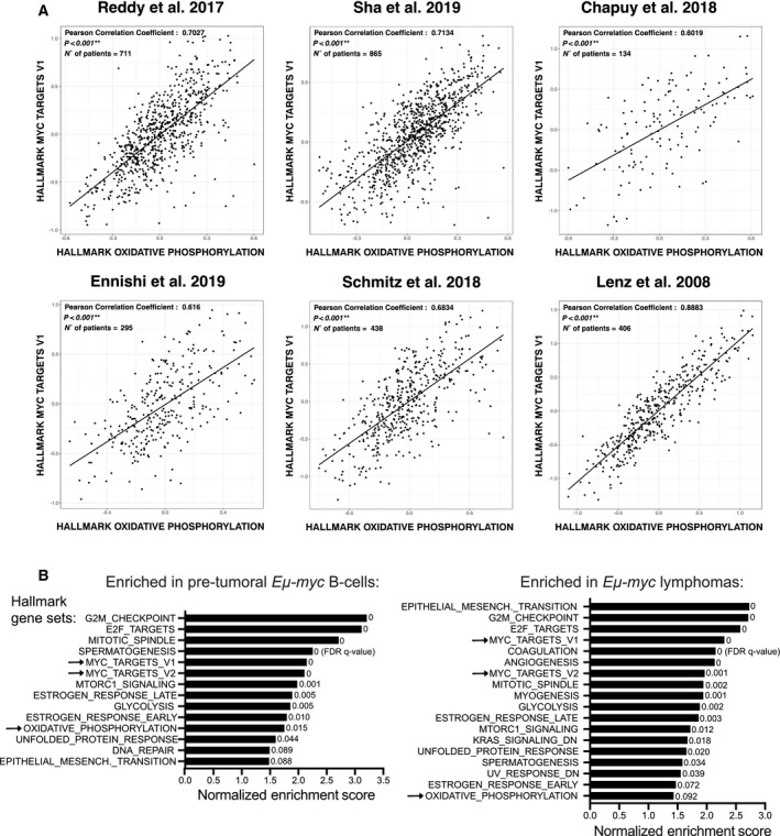
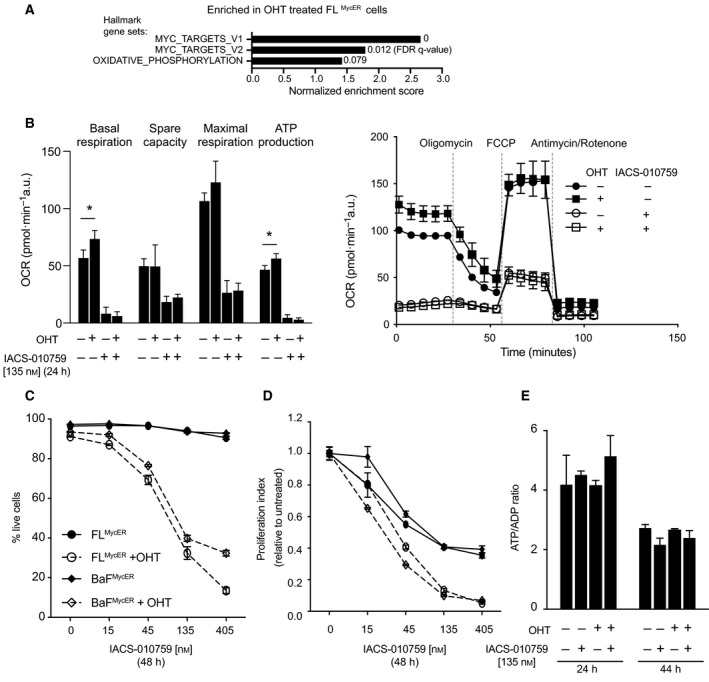
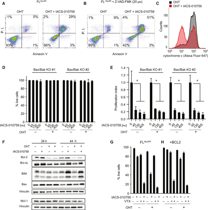
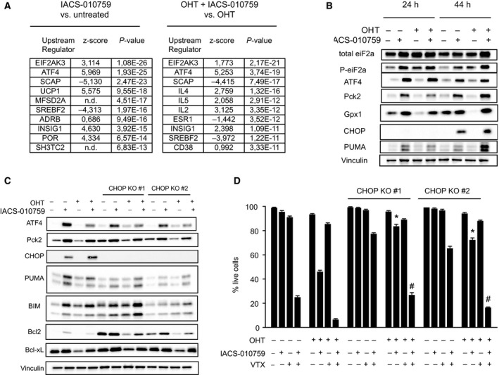
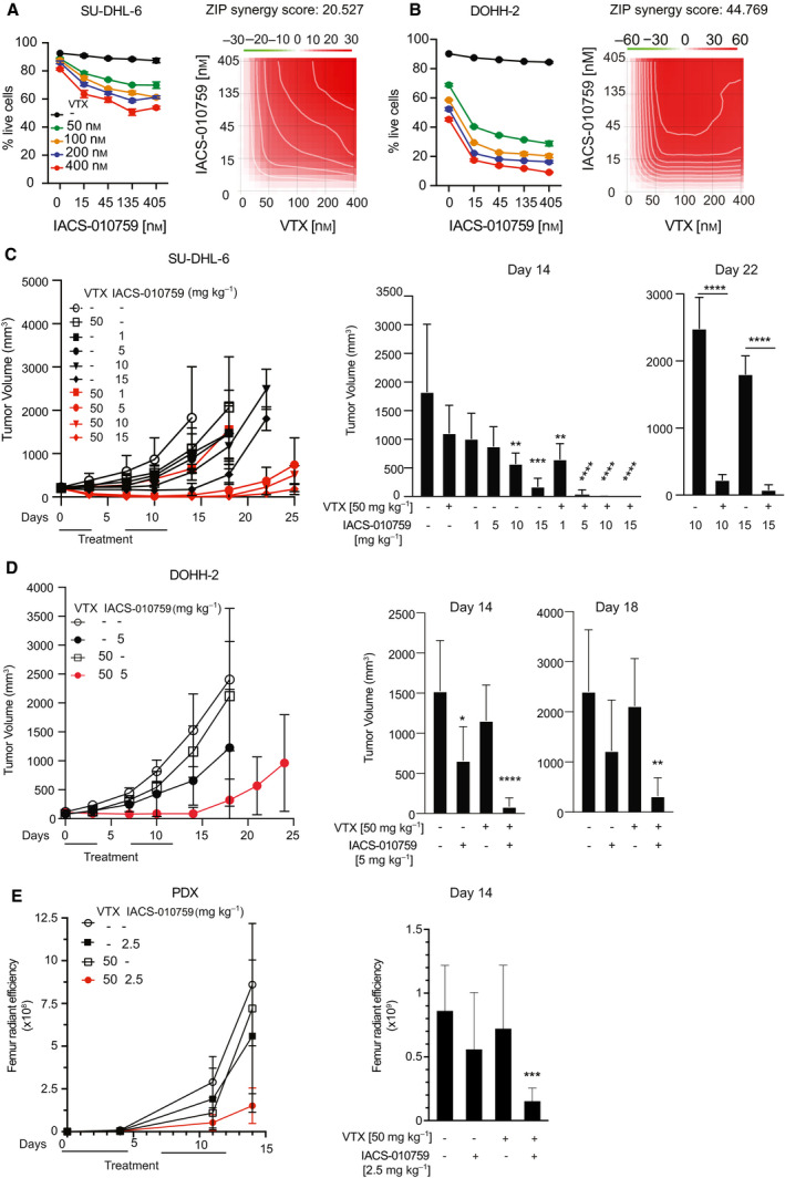
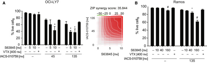
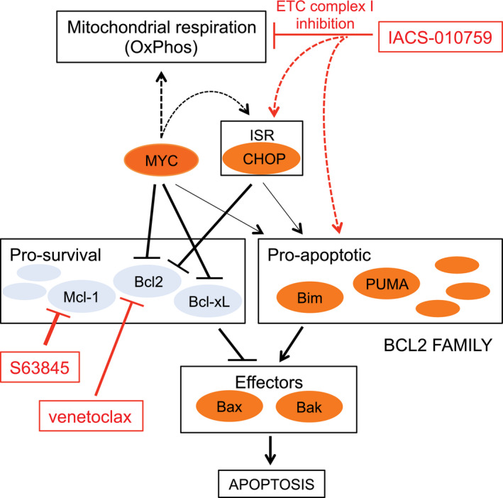
Multiple molecular features, such as activation of specific oncogenes (e.g., MYC, BCL2) or a variety of gene expression signatures, have been associated with disease course in diffuse large B-cell lymphoma (DLBCL), although their relationships and implications for targeted therapy remain to be fully unraveled. We report that MYC activity is closely correlated with-and most likely a driver of-gene signatures related to oxidative phosphorylation (OxPhos) in DLBCL, pointing to OxPhos enzymes, in particular mitochondrial electron transport chain (ETC) complexes, as possible therapeutic targets in high-grade MYC-associated lymphomas. In our experiments, indeed, MYC sensitized B cells to the ETC complex I inhibitor IACS-010759. Mechanistically, IACS-010759 triggered the integrated stress response (ISR) pathway, driven by the transcription factors ATF4 and CHOP, which engaged the intrinsic apoptosis pathway and lowered the apoptotic threshold in MYC-overexpressing cells. In line with these findings, the BCL2-inhibitory compound venetoclax synergized with IACS-010759 against double-hit lymphoma (DHL), a high-grade malignancy with concurrent activation of MYC and BCL2. In BCL2-negative lymphoma cells, instead, killing by IACS-010759 was potentiated by the Mcl-1 inhibitor S63845. Thus, combining an OxPhos inhibitor with select BH3-mimetic drugs provides a novel therapeutic principle against aggressive, MYC-associated DLBCL variants.

Keywords: BCL2; DLBCL; Integrated Stress Response; MYC; OxPhos; chemotherapy.


Conflict of interest statement

The authors declare no conflict of interest.

Figures

Fig. 1
Fig. 1
MYC activity positively correlates with the expression of OxPhos‐associated genes. (A) Correlation between the Hallmark‐OxPhos and MYC‐V1 gene sets across gene expression profiles from six independent human DLBCL patient cohorts [3, 4, 5, 22, 23, 24]. The X and Y axes report the mean expression of all genes in the indicated signature, in each patient sample (dots). Black lines represent linear regression fits to the data points. (B) Biological processes enriched during Eµ‐myc driven lymphomagenesis. Our previous RNA‐seq data [45] were used to address the enrichment of gene sets from the Hallmark collection and the CCC model [2] in pretumoral Eµ‐myc B cells (left) and lymphomas (right), relative to control nontransgenic B cells. The plot shows the enriched genes signatures (FDR q‐value < 0.1), ranked according to their normalized enrichment score. Only a subset of the Hallmark‐associated biological pathways, but none of the CCC signatures, reached this threshold. The arrows indicate the MYC target and OxPhos gene signatures.
Fig. 2
Fig. 2
Elevated MYC activity promotes OxPhos and sensitizes B cells to IACS‐010759. (A) Enrichment of MYC target and OxPhos gene sets among upregulated DEGs in FLMycER treated with OHT (100 nm, 72 h). (B‐E) FLMycER of BaFMycER cells were cell pretreated or not with OHT (100 nm, 48 h), followed by IACS‐010759 treatment at the indicated times and concentrations. (B) Left: basal respiration, spare capacity, maximal respiration, and respiration‐coupled ATP production, averaged from three independent mitochondrial stress test profiles in FLMycER cells, treated as indicated (Error bars: SEM; *P < 0.05 from t‐test between OHT‐treated cells and their respective controls); a representative profile is shown on the right (Error bars: SD; n = 10). (C) Percentage of live cells and (D) Proliferation index in FLMycER and BaFMycER cells. In both panels, OHT‐primed samples treated with ≥ 45 nm IACS‐010759 showed significant differences relative to their controls (P < 0.0001); note that in (D) each IACS‐010759‐treated sample was normalized to its untreated control (either with, or without OHT). Error bars: SD (n = 3). Cell count and viability were determined by propidium iodide staining. (E) ATP/ADP ratios in FLMycER cells, treated as indicated. Error bars: SD (n = 3).
Fig. 3
Fig. 3
IACS‐010759 activates intrinsic apoptosis in Myc‐overexpressing cells. FLMycER cells were sequentially treated with OHT (100 nm, 48 h), followed by IACS‐010759 (135 nm, unless otherwise indicated; all 48 h). (A) Apoptotic cell death was assayed by FACS analysis of propidium iodide (P.I.) and Annexin V staining (both shown as arbitrary fluorescence units). One representative experiment is shown, from two performed in FLMycER and BaFMycER cells. (B) As in (A) with the addition of 20 µm Z‐VAD‐FMK together with IACS‐010759. (C) Mitochondrial retention of cytochrome c, evaluated by anti‐cyt. c staining and FACS analysis of digitonin‐permeabilized FLMycER cells. (D) Percentage of live cells and (E) proliferation index (as defined in Fig. 2) in two independent Bax/Bak double knockout FLMycER clones pretreated or not with OHT, then treated with IACS‐010759 for 48 h. Error bars: SD (n = 3). *P < 0.001 vs. IACS‐010759‐untreated control (one‐way ANOVA). (F) Immunoblot analysis of the indicated BCL2‐family members in FLMycER cells pretreated or not with OHT, followed by IACS‐010759 at the indicated times. Vinculin was used as loading control. Note that Mcl‐1 and its own vinculin control (bottom) are from a different experiment, generated during the revision of our manuscript. (G, H) Percentage of live cells in parental FLMycER (G) and FLMycER cells overexpressing BCL2 (H). The cells were pretreated or not with OHT (100 nm, 48 h), then treated for 48 h with IACS‐010759 (135 nm) and/or venetoclax (VTX; 100 nm), as indicated. Error bars: SD (n = 3).
Fig. 4
Fig. 4
The Integrated Stress Response mediates IACS‐010759‐induced cell death. FLMycER cells were pretreated or not with OHT (100 nm, 48 h), followed by IACS‐010759 (135 nm, 24 h). (A) Gene expression was profiled by RNA‐seq, as defined in the Materials and methods section: the table shows the 10 most significantly enriched Upstream Regulators identified through Ingenuity Pathway Analysis of IACS‐010759‐responsive genes, either with (right) or without OHT (left). n.d.: not determined. P values were calculated according to the Fisher’s exact test. (B) Immunoblot analysis of ISR components in FLMycER cells after treatment with IACS‐010759 for 24 or 44 h as indicated. Vinculin was used as loading control. (C) Immunoblot analysis of parental and CHOP‐knockout FLMycER cells after 44 h of IACS‐010759 treatment. (D) Percentage of live cells, confronting parental FLMycER cells with two CHOP‐knockout clones, treated with OHT, followed by IACS‐010759 and/or venetoclax (VTX; 100 nm), as indicated. Error bars: SD (n = 3). *P < 0.0001 from t‐test between each CHOP KO clone and the parental cells, both treated with OHT and IACS‐101759; # P < 0.0001 from t‐test for each CHOP KO clone between cells treated with OHT, IACS‐101759 and VTX, relative to those treated with OHT and IACS‐101759.
Fig. 5
Fig. 5
Combinatorial action of IACS‐010759 and BH3‐mimetic compounds against MYC‐associated lymphomas. (A) Left: percentage of live SU‐DHL‐6 cells after 24 h treatment with the indicated concentrations of IACS‐010759 and venetoclax (VTX). Error bars: SD (n = 3). Right: drug interaction landscape and synergy score for the two drugs calculated according to the ZIP model. Note that a positive ZIP score (> 10) signifies a synergistic interaction. The landscape identifies the doses at which the drugs either synergize (red) or antagonize each‐other (green)—the latter not observed here. (B) As in (A), for DOHH‐2 cells. (C) Tumor progression in CD1 nude mice bearing subcutaneous SU‐DHL‐6 tumors treated by oral gavage with the indicated daily doses of IACS‐010759 and/or venetoclax. Tumor volumes (mm3) were monitored at the indicated time‐points. Error bars: SD; n = 5 animals per group. The mean values, standard deviations and statistical significance for individual groups are visualized on the right: *P < 0.05; **P < 0.01; ***P < 0.001; ****P < 0.0001 (one‐way ANOVA relative to the untreated control at day 14, to the VTX‐only group at day 22). (D) As in (C) for subcutaneous DOHH‐2 tumors (all relative to untreated control). (E) Tumor progression in NSG mice injected with the luciferase‐positive PDX line DFBL‐20954‐V3‐mCLP. Tumor development in individual mice was monitored by in vivo imaging through quantification of bilateral femur radiant efficiency. After randomization, the animals were subjected to treatment with 2.5 mg·kg−1 IACS‐010759 and/or 50 mg·kg−1 venetoclax (VTX). Error bars: SD; n = 4/5 animals per group. Right: mean values and SD at day 14; ***P < 0.001 (one‐way ANOVA).
Fig. 6
Fig. 6
Combinatorial action of IACS‐010759 and the Mcl‐1 inhibitor S63845 in DLBCL and BL cell lines. (A) OCI‐LY7 and (B) Ramos cells were treated for 24 h with IACS‐010759 and either venetoclax or S63845, at the indicated concentrations. The graphs show the percentage of live cells after treatment. Error bars: SD (n = 3). *P < 0.01; # P < 0.0001 vs. untreated control (one‐way ANOVA). The heatmap in (A) shows the drug interaction landscape and synergy score for IACS‐010759 and S63845 (as in Fig. 5A).
Fig. 7
Fig. 7
Combinatorial targeting of OxPhos and BCL2‐family proteins in MYC‐associated lymphoma. Schematic summary of the pharmaco‐genetic interactions described in this work. Dashed arrows represent indirect effects; the connections between MYC, the integrated stress response (ISR), and the pro‐apoptotic BCL2 arm (thin arrows) were described in other studies (see text) and may further reinforce the sensitivity of MYC‐overexpressing cells to ETC inhibitors. Among a number of other pathways, MYC supports mitochondrial respiration (Oxphos): inhibition of this process, and in particular of ETC complex I by IACS‐010759 is synthetic‐lethal with MYC, pointing to Oxphos as an important effector in MYC‐induced tumorigenesis. Mechanistically, IACS‐010759 induces apoptosis through activation of the ISR and in particular its pro‐apoptotic effector CHOP and may independently impact BCL2‐family proteins. MYC sensitizes to apoptosis by modulating the expression of BCL2‐family members (and, not shown here, activation of the ARF/p53 pathway). Hence, the ISR and MYC activity converge on the BCL2 family to lower the apoptotic threshold upon IACS‐010759 treatment. This model provides a coherent rationale for the effects reported in this work, including (a) MYC‐induced sensitization of B cells to killing by IACS‐010759 and (b) the cooperative cytotoxic action of IACS‐010759 and BH3‐mimetic compounds, in particular venetoclax in MYC/BCL2 DHL cells, and S63845 in Mcl‐1‐expressing DLBCL and Burkitt’s lymphoma cells.

Similar articles

Cited by

References

    1. Liu Y & Barta SK (2019) Diffuse large B‐cell lymphoma: 2019 update on diagnosis, risk stratification, and treatment. Am J Hematol 94, 604–616. - PubMed
    1. Monti S, Savage KJ, Kutok JL, Feuerhake F, Kurtin P, Mihm M, Wu B, Pasqualucci L, Neuberg D, Aguiar RC et al. (2005) Molecular profiling of diffuse large B‐cell lymphoma identifies robust subtypes including one characterized by host inflammatory response. Blood 105, 1851–1861. - PubMed
    1. Reddy A, Zhang J, Davis NS, Moffitt AB, Love CL, Waldrop A, Leppa S, Pasanen A, Meriranta L, Karjalainen‐Lindsberg ML et al. (2017) Genetic and functional drivers of diffuse large B cell lymphoma. Cell 171, 481–494 e15. - PMC - PubMed
    1. Chapuy B, Stewart C, Dunford AJ, Kim J, Kamburov A, Redd RA, Lawrence MS, Roemer MGM, Li AJ, Ziepert M et al. (2018) Molecular subtypes of diffuse large B cell lymphoma are associated with distinct pathogenic mechanisms and outcomes. Nat Med 24, 679–690. - PMC - PubMed
    1. Schmitz R, Wright GW, Huang DW, Johnson CA, Phelan JD, Wang JQ, Roulland S, Kasbekar M, Young RM, Shaffer AL et al. (2018) Genetics and pathogenesis of diffuse large B‐cell lymphoma. N Engl J Med 378, 1396–1407. - PMC - PubMed

Publication types

MeSH terms

Substances