. 2021 Oct;9(5):e00857.
doi: 10.1002/prp2.857.

Recombinant botulinum neurotoxin serotype A1 in vivo characterization

Affiliations

Recombinant botulinum neurotoxin serotype A1 in vivo characterization

Cindy Périer et al. Pharmacol Res Perspect. 2021 Oct.

Abstract

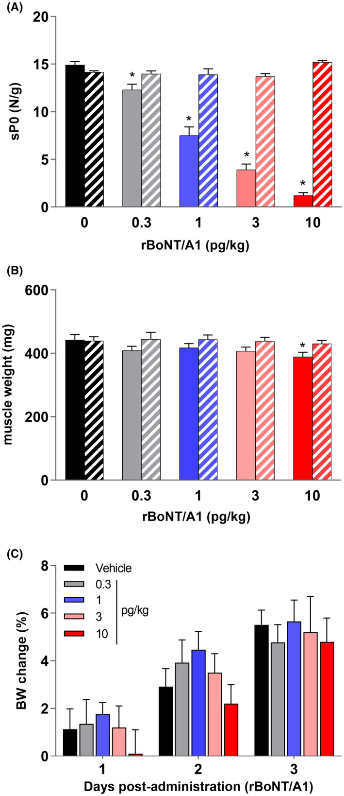
Clinically used botulinum neurotoxins (BoNTs) are natural products of Clostridium botulinum. A novel, recombinant BoNT type A1 (rBoNT/A1; IPN10260) has been synthesized using the native amino acid sequence expressed in Escherichia coli and has previously been characterized in vitro and ex vivo. Here, we aimed to characterize rBoNT/A1 in vivo and evaluate its effects on skeletal muscle. The properties of rBoNT/A1 following single, intramuscular administration were evaluated in the mouse and rat digit abduction score (DAS) assays and compared with those of natural BoNT/A1 (nBoNT/A1). rBoNT/A1-injected tibialis anterior was assessed in the in situ muscle force test in rats. rBoNT/A1-injected gastrocnemius lateralis (GL) muscle was assessed in the compound muscle action potential (CMAP) test in rats. The rBoNT/A1-injected GL muscle was evaluated for muscle weight, volume, myofiber composition and immunohistochemical detection of cleaved SNAP25 (c-SNAP25). Results showed that rBoNT/A1 and nBoNT/A1 were equipotent and had similar onset and duration of action in both mouse and rat DAS assays. rBoNT/A1 caused a dose-dependent inhibition of muscle force and a rapid long-lasting reduction in CMAP amplitude that lasted for at least 30 days. Dose-dependent reductions in GL weight and volume and increases in myofiber atrophy were accompanied by immunohistochemical detection of c-SNAP25. Overall, rBoNT/A1 and nBoNT/A1 exhibited similar properties following intramuscular administration. rBoNT/A1 inhibited motoneurons neurotransmitter release, which was robust, long-lasting, and accompanied by cleavage of SNAP25. rBoNT/A1 is a useful tool molecule for comparison with current natural and future modified recombinant neurotoxins products.

Keywords: SNAP25; botulinum; muscle; neurotoxin; recombinant; rodent.


Conflict of interest statement

Cindy Périer, Vincent Martin, Sylvie Cornet, Christine Favre‐Guilmard, Marie‐Noelle Rocher, Alban Vignaud, Matthew Beard, Stephane Lezmi, and Mikhail Kalinichev are current or former employees of Ipsen. The research was funded by Ipsen.

Figures

FIGURE 1
FIGURE 1
(A) Mean peak digit abduction score (DAS) curves of rBoNT/A1 or nBoNT/A1 (0.89–10 pg/mouse) 2–3 days post‐administration into the gastrocnemius–soleus muscle complex of male CD‐1 mice. (B) Time course of DAS following i.m. injection of rBoNT/A1 (left panel) and nBoNT/A1 (right panel) or corresponding vehicle (GPB). (C) Percentage change in body weight (vs. Day 0) after i.m. administration of rBoNT/A1 (left panel) or nBoNT/A1 (right panel) in the same experiments. Doses closest to the ED50 and DAS 4 values are in green and red, respectively. All values are mean ± SEM from two independent experiments (n = 6/dose/experiment). *p < 0.05 compared with the vehicle‐treated group (black)
FIGURE 2
FIGURE 2
(A) rBoNT/A1 and nBoNT/A1 dose–response (0.5–10 pg/rat) in the digit abduction score (DAS) assay following a single i.m. injection into the peroneal muscle complex in rats. The curves correspond to mean peak DAS responses observed 2–3 days post‐administration. (B) Time course of DAS following i.m. injection of either rBoNT/A1 or nBoNT/A1 (0.5–10 pg/rat) or vehicle (GPB) into the peronei muscle in rats. (C) Percentage change in body weight (vs. day 0) after i.m. administration of rBoNT/A1 (left panel) and nBoNT/A1 (right panel) in the same experiments. Doses closest to the ED50 and DAS 4 values are in green and red, respectively. All values are mean ± SEM from two independent experiments (n = 6/dose/experiment)
FIGURE 3
FIGURE 3
(A) The maximal muscle force (N/g) generated by the tibialis anterior (TA) following electrical stimulation of the sciatic nerve, performed 3 days after i.m. injection of rBoNT/A1 (0.3–10 pg/kg) or vehicle (GPB). (B) Weight of the TA muscle (mg) at the end of the experiment. Solid color bars represent the injected (ipsilateral) muscle, whereas striped bars represent the non‐injected (contralateral) muscle. (C) Percent change in body weight (vs. day 0) on days 1–3 post‐injection of rBoNT/A1 and vehicle (GPB). All values are mean ± SEM (n = 6/group). *p < 0.05 compared with contralateral leg (A and B)
FIGURE 4
FIGURE 4
(A) The compound muscle action potential (CMAP) amplitude (mV) measured from the lateral gastrocnemius of female Sprague‐Dawley rats injected with rBoNT/A1 (0.5 pg/kg) or vehicle (GPB) across days 1–30 postadministration (n = 8–12/group). (B) Representative CMAP recordings at baseline (Day 7) and 24 h (Day 1) after vehicle or rBoNT/A1 0.5 pg/kg administration
FIGURE 5
FIGURE 5
Female Sprague‐Dawley rats were injected with either rBoNT/A1 (1–200 pg/kg) or vehicle (GPB) into the lateral gastrocnemius, and both ipsilateral (black) and contralateral (gray) muscles were harvested 30 days later for determination of weight (mg; A) and volume (mL; B). *p < 0.05 compared with vehicle‐injected animals
FIGURE 6
FIGURE 6
The muscle fiber size distribution in the right gastrocnemius lateralis in female Sprague‐Dawley rats 30 days after they were injected with rBoNT/A1 (1–200 pg/kg) or vehicle (GPB; n = 6/group; A). The myofiber size distribution was analyzed from a cross‐section of the harvested muscle. The area frequency distribution was calculated in intervals of 0.1 × 103 µm2 ± SEM. (B) The representative images of the muscle cross‐section with reticulin staining from female Sprague‐Dawley rats injected with vehicle (GPB) or rBoNT/A1 (50 and 200 pg/kg) 30 days earlier (B, scale bar: 60 µm)
FIGURE 7
FIGURE 7
(A) Representative images of SNAP25 protein, BoNT/A1 cleavage site and recognition sites for antibodies SYSY 111 011 (N‐terminal part) and EF14007 (BoNT/A1 cleavage site). (B) Immunofluorescence colocalization experiment with SYSY 111 011 (green) and EF14007 (red) in injected muscle of vehicle or 200 pg/kg BoNT/A1‐treated animals. DAPI (nuclear counterstain) is in light gray. Scale bars: 50 µm. (C) Representative images of c‐SNAP25 corresponding to the immunohistochemical scoring system at NMJs, terminal nerve endings, and intramuscular nerves (Nerves) in the gastrocnemius lateralis of female Sprague‐Dawley rats following a single i.m. injection of vehicle (GPB) or rBoNT/A1. Illustrations were taken on post‐injection days 3–6 (scale bars: 10 µm)
FIGURE 8
FIGURE 8
Quantification of immunohistochemical labeling of c‐SNAP25 in the lateral gastrocnemius muscle taken from female Sprague‐Dawley rats 30 days after a single, intramuscular injection of rBoNT/A1 (1–200 pg/kg) or vehicle (GPB; n = 6/group). *p < 0.05 compared with vehicle‐treated animals

References

    1. Aoki KR. Preclinical update on BOTOX® (Botulinum toxin type a)‐purified neurotoxin complex relative to other botulinum neurotoxin preparations. Eur J Neurol. 1999;6:S3‐S10. doi:10.1111/j.1468-1331.1999.tb00032.x - DOI
    1. Aoki KR. A comparison of the safety margins of botulinum neurotoxin serotypes A, B, and F in mice. Toxicon. 2001;39(12):1815‐1820. doi:10.1016/S0041-0101(01)00101-5 - DOI - PubMed
    1. Apland JP, Adler M, Oyler GA. Inhibition of neurotransmitter release by peptides that mimic the N‐terminal domain of SNAP‐25. J Protein Chem. 2003;22(2):147‐153. doi:10.1023/A:1023423013741 - DOI - PubMed
    1. Broide RS, Rubino J, Nicholson GS, et al. The rat Digit Abduction Score (DAS) assay: a physiological model for assessing botulinum neurotoxin‐induced skeletal muscle paralysis. Toxicon. 2013;71:18‐24. doi:10.1016/j.toxicon.2013.05.004 - DOI - PubMed
    1. Cornet S, Périer C, Kalinichev M. (2020). Optimization of the rat digit abduction score (DAS) assay: evaluation of botulinum neurotoxin activity in the gastrocnemius lateralis, peronei, and extensor digitorum longus. Toxicon: X, 6, 100029. doi:10.1016/j.toxcx.2020.100029 - DOI - PMC - PubMed

Publication types

MeSH terms

Substances

LinkOut - more resources