

Renal cell markers: lighthouses for managing renal diseases
- PMID: 34632812
- PMCID: PMC8714975
- DOI: 10.1152/ajprenal.00182.2021
Renal cell markers: lighthouses for managing renal diseases
Abstract
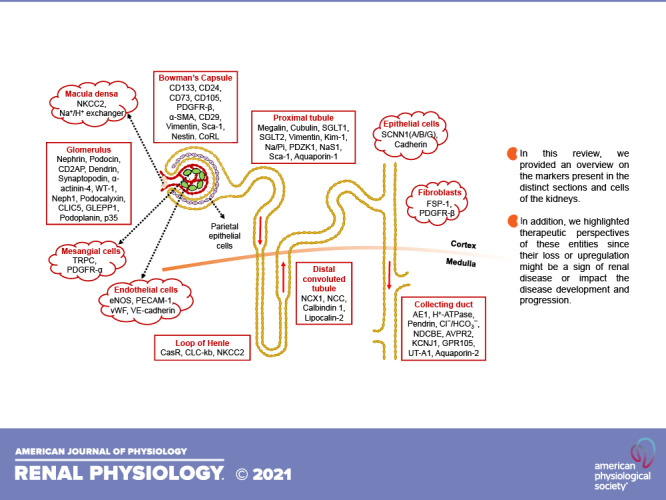
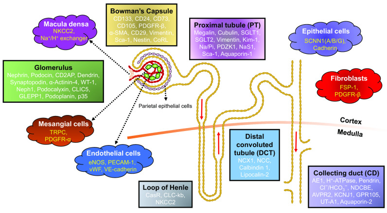
Kidneys, one of the vital organs in our body, are responsible for maintaining whole body homeostasis. The complexity of renal function (e.g., filtration, reabsorption, fluid and electrolyte regulation, and urine production) demands diversity not only at the level of cell types but also in their overall distribution and structural framework within the kidney. To gain an in depth molecular-level understanding of the renal system, it is imperative to discern the components of kidney and the types of cells residing in each of the subregions. Recent developments in labeling, tracing, and imaging techniques have enabled us to mark, monitor, and identify these cells in vivo with high efficiency in a minimally invasive manner. In this review, we summarize different cell types, specific markers that are uniquely associated with those cell types, and their distribution in the kidney, which altogether make kidneys so special and different. Cellular sorting based on the presence of certain proteins on the cell surface allowed for the assignment of multiple markers for each cell type. However, different studies using different techniques have found contradictions in cell type-specific markers. Thus, the term "cell marker" might be imprecise and suboptimal, leading to uncertainty when interpreting the data. Therefore, we strongly believe that there is an unmet need to define the best cell markers for a cell type. Although the compendium of renal-selective marker proteins presented in this review is a resource that may be useful to researchers, we acknowledge that the list may not be necessarily exhaustive.
Keywords: Bowman’s capsule; glomerulus; kidney; nephron; podocytes; proximal tubules.
Conflict of interest statement
J.R. has patents on novel strategies for kidney therapeutics and stands to gain royalties from their commercialization. He is the co-founder of Walden Biosciences (Cambridge, MA), a biotechnology company in which he has financial interest, including stock. Other authors have nothing to disclose and there are no competing interests.
Figures
Similar articles
-
Proteinuria and progression of glomerular diseases.Pediatr Nephrol. 2013 Jul;28(7):1049-58. doi: 10.1007/s00467-012-2335-1. Epub 2012 Nov 3. Pediatr Nephrol. 2013. PMID: 23124512 Review.
-
Diseased renal glomeruli are getting soft. Focus on "Biophysical properties of normal and diseased renal glomeruli".Am J Physiol Cell Physiol. 2011 Mar;300(3):C394-6. doi: 10.1152/ajpcell.00511.2010. Epub 2010 Dec 22. Am J Physiol Cell Physiol. 2011. PMID: 21178112 Free PMC article. No abstract available.
-
Proteinuria: detection and role in native renal disease progression.Transplant Rev (Orlando). 2012 Jan;26(1):3-13. doi: 10.1016/j.trre.2011.10.002. Transplant Rev (Orlando). 2012. PMID: 22137726 Review.
-
Tubulo-interstitial damage in glomerular diseases: its role in the progression of the renal damage.Nephrol Dial Transplant. 1998;13 Suppl 1:80-5. doi: 10.1093/ndt/13.suppl_1.80. Nephrol Dial Transplant. 1998. PMID: 9507504 Review. No abstract available.
-
Renal regulation of sodium excretion. Function in health and in edema-forming states.Arch Intern Med. 1973 Jun;131(6):780-91. Arch Intern Med. 1973. PMID: 4576264 Review. No abstract available.
Cited by
-
Consensus tissue domain detection in spatial omics data using multiplex image labeling with regional morphology (MILWRM).Commun Biol. 2024 Oct 30;7(1):1295. doi: 10.1038/s42003-024-06281-8. Commun Biol. 2024. PMID: 39478141 Free PMC article.
-
Shared hub genes in membranous nephropathy and kidney renal clear cell carcinoma: investigating molecular overlap and tumor progression.Discov Oncol. 2025 Jun 9;16(1):1035. doi: 10.1007/s12672-025-02701-1. Discov Oncol. 2025. PMID: 40489038 Free PMC article.
-
Advancing the clinical assessment of glomerular podocyte pathology in kidney biopsies via super-resolution microscopy and angiopoietin-like 4 staining.Theranostics. 2025 Jan 1;15(3):784-803. doi: 10.7150/thno.101498. eCollection 2025. Theranostics. 2025. PMID: 39776814 Free PMC article.
-
Research progress of natural active compounds on improving podocyte function to reduce proteinuria in diabetic kidney disease.Ren Fail. 2023;45(2):2290930. doi: 10.1080/0886022X.2023.2290930. Epub 2023 Dec 11. Ren Fail. 2023. PMID: 38073545 Free PMC article. Review.
-
Enhanced Levels of Glycosphingolipid GM3 Delay the Progression of Diabetic Nephropathy.Int J Mol Sci. 2023 Jul 12;24(14):11355. doi: 10.3390/ijms241411355. Int J Mol Sci. 2023. PMID: 37511118 Free PMC article.
References
-
- Hebert SC. Nephron heterogeneity. In: Comprehensive Physiology, edited by Terjung R, 2011. doi:10.1002/cphy.cp080120. - DOI
MeSH terms
Substances
LinkOut - more resources
Full Text Sources
Medical