. 2022 Sep 28;128(6):1050-1063.
doi: 10.1017/S0007114521004104. Epub 2021 Oct 11.

The administration of a pre-digested fat-enriched formula prevents necrotising enterocolitis-induced lung injury in mice

Affiliations

The administration of a pre-digested fat-enriched formula prevents necrotising enterocolitis-induced lung injury in mice

Chhinder P Sodhi et al. Br J Nutr. .

Abstract

Necrotising enterocolitis (NEC) is a devastating gastrointestinal disease of prematurity that typically develops after the administration of infant formula, suggesting a link between nutritional components and disease development. One of the most significant complications that develops in patients with NEC is severe lung injury. We have previously shown that the administration of a nutritional formula that is enriched in pre-digested Triacylglyceride that do not require lipase action can significantly reduce the severity of NEC in a mouse model. We now hypothesise that this 'pre-digested fat (PDF) system' may reduce NEC-associated lung injury. In support of this hypothesis, we now show that rearing newborn mice on a nutritional formula based on the 'PDF system' promotes lung development, as evidenced by increased tight junctions and surfactant protein expression. Mice that were administered this 'PDF system' were significantly less vulnerable to the development of NEC-induced lung inflammation, and the administration of the 'PDF system' conferred lung protection. In seeking to define the mechanisms involved, the administration of the 'PDF system' significantly enhanced lung maturation and reduced the production of reactive oxygen species (ROS). These findings suggest that the PDF system protects the development of NEC-induced lung injury through effects on lung maturation and reduced ROS in the lung and also increases lung maturation in non-NEC mice.

Keywords: Infant nutrition; Lung injury; Necrotising enterocolitis; Reactive oxygen species.


Conflict of interest statement

Conflicts of Interest: None of the authors has any conflicts of interest to declare.

Figures

Fig 1.
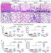
Fig 1.. Administration of formula containing predigested fat prevents NEC-induced lung injury in neonatal mice.
(A-N) representative photomicrographs of Hematoxylin-Eosin (H&E) stained small showing histology and gene expression of pro-inflammatory cytokines, in non-NEC control and NEC mice. (A-E) histology of the small intestine (ileum). (F-J) histology of the lungs. (K-L) qRT-PCR expression of pro-inflammatory cytokines in the small intestine (ileum), (K) Tnf-α and (L) Lcn-2. (M-N) qRT-PCR expression of pro-inflammatory cytokines in the lungs, (M) Tnf-α and (N) Lcn-2. Each dot in scatter-dot plots represents data from an individual mouse, (n≥7 mice/group). Statistical significance was determined by one-way ANOVA, followed by Tukey’s multiple comparisons tests using GraphPad prism 9 software. Scale bars: 100μm.
Fig 2.
Fig 2.. Administration of formula containing predigested fat prevents apoptosis of lung epithelial cells in necrotizing enterocolitis.
(A-E)i-ii representative immunofluorescence images of TUNEL staining (TUNEL green, DAPI nuclei blue), showing apoptosis (white arrows), in lungs of non-NEC control (A-C)i-ii and NEC mice (D-E)i-ii. (F) quantification of TUNEL positive cells, using ImageJ software (2 or more separate areas from each mouse lung section were imaged and quantified, total mice n≥4 mice/group). (G) qRT-PCR of Apoptosis gene Puma (P53 Up-Regulated Modulator Of Apoptosis), each dot in scatter-dot plot represents data from an individual mouse (n≥7 mice/group). Statistical significance was determined by one-way ANOVA, followed by Tukey’s multiple comparisons tests using GraphPad prism 9 software. Scale bars: 25μm.
Fig 3.
Fig 3.. Administration of formula containing predigested fat reduces polymorphonuclear neutrophil (PMNs) accumulation into the neonatal lung.
(A-E)i-ii representatative photomicrographs of Myeloperoxidase (MPO) stained images with DAB staining showing accumulation of PMNs (DAB stained brown cells, black arrows) in lungs of non-NEC controls (A-C)i-ii and NEC (D-E)i-ii mice. (F-G) qRT-PCR expression of of PMNs marker genes, (G) Mpo and (H) neutrophil expressed neutrophil (Elane) (each dot in scatter-dot plots represents data from an individual mouse, n≥7 mice/group). Statistical significance was determined by one-way ANOVA, followed by Tukey’s multiple comparisons tests using GraphPad prism 9 software. Scale bars: 20μm.
Fig 4.
Fig 4.. Administration of formula containing predigested fat prevents loss of lung surfactant proteins in the lungs in neonatal mice with NEC.
(A-J) representative confocal images of surfactant proteins in lungs of non-nec control (A-C, F-H) and NEC (D-E, I-J) mice. (A-E) immunostaiing of surfactant protein A (SP-A, green staining, white arrowheads). (F-J) immunostaining of surfactant protein D (SP-D, red staining, white arrowheads). (K-L) quantification of immunofluorescence intestity of SP-A (K) and SP-D (L) using ImageJ software (each dot in scatter-dot plots represents data from an individual area, 2 or more areas from each mouse lung section IHC were imaged and quantified, n≥4 mice/group). Statistical significance was determined by one-way ANOVA, followed by Tukey’s multiple comparisons tests using GraphPad prism 9 software. Scale bars: 20μm.
Fig 5.
Fig 5.. Administration of formula containing predigested fat prevents the loss of tight junctions in the neonatal lungs of mice with NEC.
(A-E)i-ii representataive confocal images of tight junction protein Zona-Occludin (ZO-1, red staining, white arrowheads) in lungs of non-nec control (A-C)i-ii and NEC (D-E)i-ii mice. (F) quantification of Zo-1 immunofluorescence intestity measured using ImageJ software (each dot represents a diffenent area under focus, 1–2 areas/section/mice, n≥7 mice/group). (G) qRT-PCR expression ZO-1 (each dot represents data from an individual mouse, n≥7 mice/group). Statistical significance was determined by one-way ANOVA, followed by Tukey’s multiple comparisons tests using GraphPad prism 9 software. Scale bars: 25μm.
Fig 6.
Fig 6.. Administration of formula containing predigested fat prevents prevents NEC-induced nitrosylation in the lung.
(A-E)i-ii representative confocal images of 3’-nitrotyrosine (3’-NT, green staining, white arrows) in lungs of non-nec control (A-C)i-ii and NEC (D-E)i-ii mice. (F) quantification of 3’-NT immunofluorescence intestity measured using ImageJ software (each dot represents a diffenent area under focus, 1–2 areas/section/mice, n≥4 mice/group). (G) qRT-PCR of superoxide radical i.e., ROS generation enzyme NADPH oxidase 2 (Nox2) (each dot represents data from an individual mouse, n≥7 mice/group). Statistical significance was determined by one-way ANOVA, followed by Tukey’s multiple comparisons tests using GraphPad prism 9 software. Scale bars: 25um.
Fig 7.
Fig 7.. Intranasal administration of the reactive oxygen species (ROS) scavenger N-acetylcysteine (NAC) prevents NEC and NEC-induced lung injury.
(A-B)i-iii, assessment of NEC histology as shown by representative photomicrographs of H&E stainied histological sections of small intestine (ileum) (Ai-ii), lung (Bi-ii), and qRT-PCR expression of pro-inflammatory cytokine Tnf-a in the small intestine (Aiii) and lung (Biii) in NEC mice either given intranasl saline (NEC) or NAC (NEC+NAC). (Ci-iii) assesement of PMNs infilteration lungs as shown by representative images of myeloperoxidase (MPO) staining (DAB stained brown cells) (Ci-ii) and qRT-PCR expression of neutrophils marker ELANE (Ciii). (Di-Dii) assessments of apoptosis as shown by representative confocal images of TUNEL staining (Di-ii) and TUNEL quatificaion (Diii). (Ei-iii) assesement of oxidative injury as shown by representative confocal images of 3’-NT staining (Ei-ii) and quantification using image J software (Eiii). (F-G)i-ii assesment of surfactant protein levels as shown by representative confocal images of surfactant proteins SP-A (Fi-ii) and SP-D (Gi-ii) along with immunofluorescence intestity quantification of SP-A (Fiii) and SP-D (Giii) using image J software. (Hi-iii) assesement of tight junction proteins as measured by representative confocal images Zona-Occludin (ZO-1) staining (Hi-ii) and qRT-PCR expression of Zo-1 (Hiii). Each bar on the bar graphs represents data from 6–8 mice±SEM. Statistical significance was determined by students;s t-test using GraphPad prism 9 software. Scale bars: Ai-Bi 100μm Ci-Hii, 25μm.
Fig 8.
Fig 8.. Administration of formula containing predigested fat prevents protects against LPS-induced lung injury.
Dot plot charts of qRT-PCR expressions of Pro-inflammtory cytokines Il-6 (A, D), Lcn2 (B, E), and neutrophils enzyme ELANE (C-F), in the lungs of mice reared on the ‘standard fat’ (std fat) and the ‘predigested fat system’(PDF), and treated with LPS via interaperitoneal route (A-C) and intranasal route (D-F). Each dot in scatter-dot plots represents data from an individual mouse (n≥5 mice/group). Statistical significance was determined by one-way ANOVA, followed by Tukey’s multiple comparisons tests using GraphPad prism 9 software.
Fig 9.
Fig 9.. The effects of formula administration on wet/dry lung weights in neonatal mice.
Dot plot charts of total body weight (A), wet lung weight (B), dry lung weight (C), and wet/dry lung weight ratio (D), in mice reared on the ‘standard fat or the ‘predigested fat system’ over a period of 4 days from postnatal 7 to p11. Each dot in scatter-dot plots represents data from an individual mouse (n≥7 mice/group). Statistical significance was determined by one-way ANOVA, followed by Tukey’s multiple comparisons tests using GraphPad prism 9 software.

References

    1. Alganabi M, Lee C, Bindi E et al. (2019) Recent advances in understanding necrotizing enterocolitis. F1000Res 8, 1–8. - PMC - PubMed
    1. Seeman SM, Mehal JM, Haberling DL et al. (2016) Infant and maternal risk factors related to necrotising enterocolitis-associated infant death in the United States. Acta Paediatrica, International Journal of Paediatrics 105, e240–e246. - PubMed
    1. Alsaied A, Islam N, Thalib L (2020) Global incidence of Necrotizing Enterocolitis: a systematic review and Meta-analysis. BMC Pediatrics 20, 1–15. - PMC - PubMed
    1. Patel RM, Denning PW (2015) Intestinal microbiota and its relationship with necrotizing enterocolitis. Pediatr Res 78, 232–238. - PMC - PubMed
    1. Laughon M, O’Shea MT, Allred EN et al. (2009) Chronic lung disease and developmental delay at 2 years of age in children born before 28 weeks’ gestation. Pediatrics 124, 637–648. - PMC - PubMed

Publication types

Substances