. 2021 Oct 29;41(10):BSR20211598.
doi: 10.1042/BSR20211598.

Jian-Pi-Yi-Shen formula enhances perindopril inhibition of chronic kidney disease progression by activation of SIRT3, modulation of mitochondrial dynamics, and antioxidant effects

Affiliations

Jian-Pi-Yi-Shen formula enhances perindopril inhibition of chronic kidney disease progression by activation of SIRT3, modulation of mitochondrial dynamics, and antioxidant effects

Xinhui Liu et al. Biosci Rep. .

Abstract

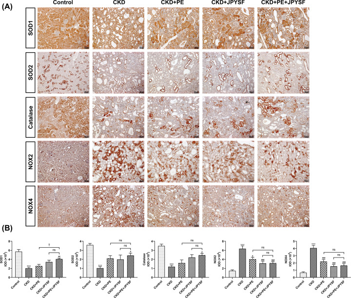
Chronic kidney disease (CKD) is a global public health problem. Renin-angiotensin system (RAS) blockade is the mainstay of CKD therapy with limitations. Jian-Pi-Yi-Shen formula (JPYSF) is a traditional herbal decoction and has been used for treating CKD for decades. The purpose of the present study was to investigate the intervention effects of combined used of perindopril erbumine (PE) and JPYSF on CKD progression and explore their underlying mechanisms. CKD rat model was induced by feeding a diet containing 0.75% w/w adenine for 3 weeks. CKD rats were treated with PE or JPYSF or PE+JPYSF from the induction of CKD and lasted 4 weeks. Renal function was evaluated by serum creatinine (Scr) and blood urea nitrogen (BUN). Pathological lesions were observed by Periodic acid-Schiff (PAS) and Masson's trichrome staining. The protein expression was tested by Western blot and immunohistochemistry analysis. The morphology of mitochondria was observed by transmission electron microscope. The results showed that combined used of PE and JPYSF could better improve renal function and pathological lesions and ameliorate renal fibrosis in CKD rats. Administration of PE and JPYSF enhanced sirtuin 3 (SIRT3) expression, inhibited mitochondrial fission, promoted mitochondrial fusion, and suppressed oxidative stress in the kidney of CKD rats. In conclusion, combined use of PE and JPYSF protected against CKD more effectively than either alone. The underlying mechanism may be associated with activation of SIRT3, modulation of mitochondrial dynamics, and antioxidant effects.

Keywords: Jian-Pi-Yi-Shen formula; chronic kidney disease; mitochondrial dynamics; oxidative stress; perindopril erbumine; sirtuins.


Conflict of interest statement

The authors declare that there are no competing interests associated with the manuscript.

Figures

Figure 1
Figure 1. PE and JPYSF improved renal function in CKD rats
(A) The levels of Scr in different groups. Compared with the control group, Scr level was markedly elevated in CKD rats and declined after PE or JPYSF treatment, especially in the combined treatment group. (B) The levels of BUN in different groups. PE, JPYSF and combined treatment reduced the BUN levels of CKD rats. Data are presented as the means ± SEM, n=4–6 rats per group (***P<0.001 compared with the control group; ##P<0.01, ###P<0.001 compared with the CKD group; P<0.05, compared with the CKD+PE+JPYSF group; ns, no significance).
Figure 2
Figure 2. PE and JPYSF ameliorated renal pathological injury in CKD rats
(A) Representative PAS-stained pictures of each group. (B) Representative Masson’s stained pictures of each group. Obvious renal tubular epithelial cells shedding and tubular dilation in PAS staining and large amounts of collagen fibrils deposition (blue staining) in Masson’s staining were observed in CKD rats. Treatment with PE or JPYSF, especially for combined therapy of PE and JPYSF, significantly ameliorated these pathological lesions. All images are shown at identical magnification, ×200, scale bar = 100 μm.
Figure 3
Figure 3. PE and JPYSF suppressed the expression of fibrotic markers in CKD rats
(A) Representative Western blot images of FN and Col-IV. (B,C) Densitometric analysis of FN and Col-IV protein expression normalized to GAPDH content. The expression of FN and Col-IV were markedly up-regulated in the CKD group. Treatment with PE significantly reduced Col-IV but not FN expression. While treatment with JPYSF or combined use of PE and JPYSF significantly down-regulated both FN and Col-IV expression. Data are presented as the means ± SEM, n=4–6 rats per group (***P<0.001 compared with the control group; #P<0.05, ###P<0.001 compared with the CKD group; ††P<0.01, compared with the CKD+PE+JPYSF group; ns, no significance).
Figure 4
Figure 4. PE and JPYSF enhanced the expression of SIRT3 in CKD rats
(A) Representative Western blot images of SIRT3. (B) Densitometric analysis of SIRT3 protein expression normalized to GAPDH content. (C) Representative IHC images of SIRT3 in different groups. The expression of SIRT3 was significantly reduced in the CKD group. The combination of PE and JPYSF could significantly up-regulate the expression of SIRT3. All images are shown at identical magnification, ×100, scale bar = 100 μm. Data are presented as the means ± SEM, n=4–6 rats per group (***P<0.001 compared with the control group; #P<0.05 compared with the CKD group; ns, no significance).
Figure 5
Figure 5. PE and JPYSF modulated mitochondrial dynamics in CKD rats
(A) Representative Western blot images of Drp-1 and OPA-1. (B,C) Densitometric analysis of Drp-1 and OPA-1 protein expression normalized to GAPDH content. Drp-1 was markedly up-regulated in CKD rats. In contrast, the expression of OPA-1 was significantly down-regulated in CKD rats. Only combined PE and JPYSF treatment could significantly reverse the expression trend of both Drp-1 and OPA-1. (D) Representative TEM images of mitochondria in the kidneys from different groups. Mitochondria fragmented into small, punctuated suborganelles in CKD rats, and this effect was partially reversed in the CKD+PE+JPYSF group. All images are shown at identical magnification, scale bar = 5 μm. Data are presented as the means ± SEM, n=4–6 rats per group (***P<0.001 compared with the control group; #P<0.05, ##P<0.01 compared with the CKD group; ††P<0.01, compared with the CKD+PE+JPYSF group; ns, no significance).
Figure 6
Figure 6. PE and JPYSF suppressed oxidative stress in CKD rats
(A) Representative IHC images of SOD1, SOD2, catalase, NOX2 and NOX4 in different groups. (B) Quantitative analysis of positive staining areas for SOD1, SOD2, catalase, NOX2 and NOX4. Only combination of PE and JPYSF could significantly up-regulate the expression of SOD1, SOD2, and catalase. Treatment with PE or JPYSF or combined treatment could inhibit NOX2 and NOX4 expression in CKD rats. All images are shown at identical magnification, ×100, scale bar = 100 μm. Data are presented as the means ± SEM, n=5 rats per group (***P<0.001 compared with the control group; #P<0.05, ##P<0.01, ###P<0.001 compared with the CKD group; P<0.05, compared with the CKD+PE+JPYSF group; ns, no significance).

Similar articles

Cited by

References

    1. Hill N.R., Fatoba S.T., Oke J.L., Hirst J.A., O’Callaghan C.A., Lasserson D.S.et al. . (2016) Global prevalence of chronic kidney disease - a systematic review and meta-analysis. PLoS ONE 11, e0158765 10.1371/journal.pone.0158765 - DOI - PMC - PubMed
    1. Bello A.K., Alrukhaimi M., Ashuntantang G.E., Bellorin-Font E., Benghanem Gharbi M., Braam B.et al. . (2018) Global overview of health systems oversight and financing for kidney care. Kidney Int. Suppl. 8, 41–51 10.1016/j.kisu.2017.10.008 - DOI - PMC - PubMed
    1. Bello A.K., Levin A., Lunney M., Osman M.A., Ye F., Ashuntantang G.E.et al. . (2019) Status of care for end stage kidney disease in countries and regions worldwide: international cross sectional survey. BMJ 367, l5873 10.1136/bmj.l5873 - DOI - PubMed
    1. Hou F.F., Zhang X., Zhang G.H., Xie D., Chen P.Y., Zhang W.R.et al. . (2006) Efficacy and safety of benazepril for advanced chronic renal insufficiency. N. Engl. J. Med. 354, 131–140 10.1056/NEJMoa053107 - DOI - PubMed
    1. Ruggenenti P., Cravedi P. and Remuzzi G. (2010) The RAAS in the pathogenesis and treatment of diabetic nephropathy. Nat. Rev. Nephrol. 6, 319–330 10.1038/nrneph.2010.58 - DOI - PubMed

Publication types