Review
. 2021 Dec;48(13):4272-4292.
doi: 10.1007/s00259-021-05504-y. Epub 2021 Oct 11.

Targeted optical fluorescence imaging: a meta-narrative review and future perspectives

Affiliations
Review

Targeted optical fluorescence imaging: a meta-narrative review and future perspectives

H M Schouw et al. Eur J Nucl Med Mol Imaging. 2021 Dec.

Abstract

Purpose: The aim of this review is to give an overview of the current status of targeted optical fluorescence imaging in the field of oncology, cardiovascular, infectious and inflammatory diseases to further promote clinical translation.

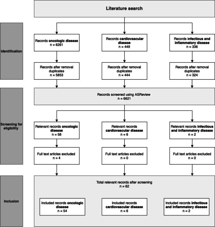
Methods: A meta-narrative approach was taken to systematically describe the relevant literature. Consecutively, each field was assigned a developmental stage regarding the clinical implementation of optical fluorescence imaging.

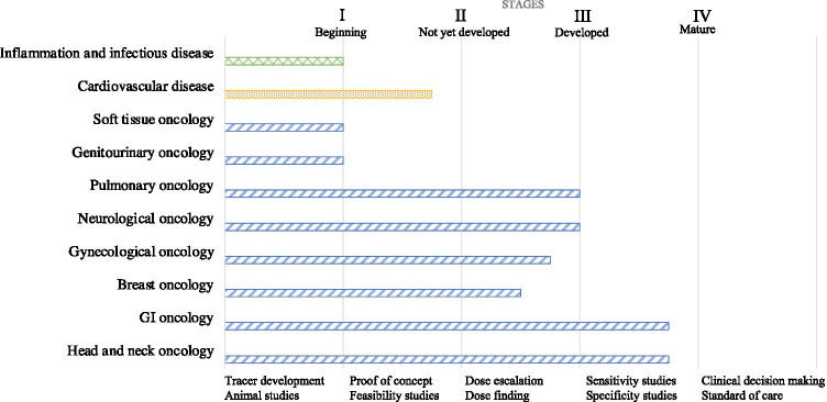
Results: Optical fluorescence imaging is leaning towards clinical implementation in gastrointestinal and head and neck cancers, closely followed by pulmonary, neuro, breast and gynaecological oncology. In cardiovascular and infectious disease, optical imaging is in a less advanced/proof of concept stage.

Conclusion: Targeted optical fluorescence imaging is rapidly evolving and expanding into the clinic, especially in the field of oncology. However, the imaging modality still has to overcome some major challenges before it can be part of the standard of care in the clinic, such as the provision of pivotal trial data. Intensive multidisciplinary (pre-)clinical joined forces are essential to overcome the delivery of such compelling phase III registration trial data and subsequent regulatory approval and reimbursement hurdles to advance clinical implementation of targeted optical fluorescence imaging as part of standard practice.

Keywords: Cardiovascular disease; Fluorescence; Infectious disease; Oncology; Optical fluorescence imaging.


Conflict of interest statement

GMvD is founder, owner and CEO of AxelaRx/TRACER Europe BV, a contract research organisation.

Figures

Fig. 1
Fig. 1
Representation of the concept of fluorescent imaging. Light from a light source, filtered by an excitation filter is deflected by a dichroic mirror before it hits the (tissue) specimen. Consequently, fluorophores in the specimen absorb photons, which results in promotion to an excited state. After losing a part of their energy in the process of vibrational relaxation, the molecule falls back to the ground state by emitting a photon of a lower energy and a longer wavelength. The emitted signal passes through an objective and an emission filter before it hits a detector
Fig. 2
Fig. 2
1a Typical composite camera system using a highly sensitive fluorescence camera (FC) to collect fluorescence images and a colour camera (CC) to collect white-light images through a dichroic mirror (DM) and a common lens (CL). Different light sources may be used for white-light excitation (WL) and fluorescence excitation using a laser source (LS) and common illumination unit (IU). Reprinted by permission from Springer Nature Customer Service Centre GmbH, Springer Nature, Nature Photonics, Tackling standardization in fluorescence molecular imaging, Koch et al. [31] Copyright (2018). 1b Front and back of ICG-NIRF prototype modified action camera with 7.2-mm lens, modified action camera with 7.2-mm lens and bandpass filter and modified LED light with bandpass filter. Reproduced from Yang et al. [32] J. Clin. Med. Copyright 2021 MDPI, Basel, Switzerland. 1c Position of confocal laser endomicroscopy (CLE) scanning probe in an endonasal transsphenoidal approach (arrow points to the CLE probe). Reproduced from Belykh et al. [33] J. Clin. Med. Copyright 2020 MDPI, Basel, Switzerland. 2a Intravenous administration of bevacizumab-800CW 3 days prior to surgery. 2b, c Colour image and corresponding fluorescence image obtained in vivo during surgery to determine potential clinical value. 2d, e Imaging of the fresh surgical specimen, followed by serially slicing. 2f, g Imaging of the fresh tissue slices to determine tumour-to-background ratio based on macro-segmentation, followed by paraffin embedding. 2h, i Imaging of formalin-fixed paraffin-embedded (FFPE) blocks to determine heterogeneity of tracer uptake within a tumour. 2j, k Imaging of 10-μm-thick tissue sections for microsegmentation to reveal microscopic biodistribution and correlation with fluorescence signals from the macroscopic to microscopic level. 2l,m Fluorescence microscopy to determine tracer distribution on a cellular level. Scale bars represent 1 cm, in l, m the scale bar represents 25 μm. Reproduced from Koller et al. [34] Nature Communications Copyright 2018, Springer Nature. 3a Bedside multispectral optoacoustic tomography (MSOT) examination. 3b In the ICG injected limb both lymphatic (green) and blood vessels (red) were detected while in the non-injected limb, only blood vessels (red) could be detected. Reproduced from Giacalone et al. [35] J. Clin. Med. Copyright [2020], MDPI, Basel, Switzerland
Fig. 3
Fig. 3
Flow diagram of literature search
Fig. 4
Fig. 4
The different fields of optical fluorescence imaging and their corresponding stages of clinical implementation

References

    1. Dhawan AP, D’Alessandro B, Fu X. Optical imaging modalities for biomedical applications. IEEE Rev Biomed Eng. 2010;3:69–92. doi: 10.1109/RBME.2010.2081975. - DOI - PubMed
    1. Paulinelli RR, Freitas-Junior R, De Souza Rahal RM, De Pádua Oliveira LF, Vilela MHT, Moreira MAR, Alves KL, Peleja MB, De Resende TCC. A prospective randomized trial comparing patent blue and methylene blue for the detection of the sentinel lymph node in breast cancer patients. Rev Assoc Med Bras. 2017;63:118–123. doi: 10.1590/1806-9282.63.02.118. - DOI - PubMed
    1. Schmid I, Cole SW, Korin YD, Zack JA, Giorgi JV. Detection of cell cycle subcompartments by flow cytometric estimation of DNA-RNA content in combination with dual-color immunofluorescence. Cytometry. 2000;39:108–116. doi: 10.1002/(sici)1097-0320(20000201)39:2<108::aid-cyto3>3.0.co;2-4. - DOI - PubMed
    1. Dumont EA, Reutelingsperger CPM, Smits JFM, Daemen MJAP, Doevendans PAF, Wellens HJJ, Hofstra L. Real-time imaging of apoptotic cell-membrane changes at the single-cell level in the beating murine heart. Nat Med. 2001;7:1352–1355. doi: 10.1038/nm1201-1352. - DOI - PubMed
    1. Joshi BP, Wang TD. Targeted optical imaging agents in cancer: focus on clinical applications. Contrast Media Mol Imaging. 2018 doi: 10.1155/2018/2015237. - DOI - PMC - PubMed

LinkOut - more resources