. 2022 Apr;141(3-4):965-979.
doi: 10.1007/s00439-021-02381-1. Epub 2021 Oct 11.

A pathogenic deletion in Forkhead Box L1 (FOXL1) identifies the first otosclerosis (OTSC) gene

Affiliations

A pathogenic deletion in Forkhead Box L1 (FOXL1) identifies the first otosclerosis (OTSC) gene

Nelly Abdelfatah et al. Hum Genet. 2022 Apr.

Abstract

Otosclerosis is a bone disorder of the otic capsule and common form of late-onset hearing impairment. Considered a complex disease, little is known about its pathogenesis. Over the past 20 years, ten autosomal dominant loci (OTSC1-10) have been mapped but no genes identified. Herein, we map a new OTSC locus to a 9.96 Mb region within the FOX gene cluster on 16q24.1 and identify a 15 bp coding deletion in Forkhead Box L1 co-segregating with otosclerosis in a Caucasian family. Pre-operative phenotype ranges from moderate to severe hearing loss to profound sensorineural loss requiring a cochlear implant. Mutant FOXL1 is both transcribed and translated and correctly locates to the cell nucleus. However, the deletion of 5 residues in the C-terminus of mutant FOXL1 causes a complete loss of transcriptional activity due to loss of secondary (alpha helix) structure. FOXL1 (rs764026385) was identified in a second unrelated case on a shared background. We conclude that FOXL1 (rs764026385) is pathogenic and causes autosomal dominant otosclerosis and propose a key inhibitory role for wildtype Foxl1 in bone remodelling in the otic capsule. New insights into the molecular pathology of otosclerosis from this study provide molecular targets for non-invasive therapeutic interventions.


Conflict of interest statement

The authors declare they have no conflict of interest.

Figures

Fig. 1
Fig. 1
Selected audiograms of otosclerosis cases pre- and post-stapedectomy. a The NL proband (PID II-6) at age 51 showing extensive bilateral air-bone gaps, largely resolved by age 52 after bilateral stapedectomies. PID II-2 at age 63 presents with no measurable hearing after unsuccessful stapedectomy (R), and severe mixed loss (L). PID II-3 at age 38 presents with profound HL and no measurable bone conduction thresholds (bilateral). PID III-2 at age 18 shows normal hearing (L) and residual conductive loss following tympano-mastoidectomy and stapes re-mobilization (R), however, subsequent re-mobilization of right ear ossicles is not sustained and by age 35, conductive HL has progressed bilaterally. b The Ontario case at age 63 shows profound mixed HL (R) and moderate low-frequency sensorineural loss (L). By age 67 and following stapedectomy (R), air-bone gaps are largely resolved, and some bone conduction thresholds are improved (R) and bilateral HL is essentially symmetrical
Fig. 2
Fig. 2
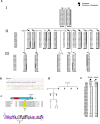
Co-segregation of a 15 bp In-Frame Deletion in Forkhead Box L1 (FOXL1, rs764026385) with Otosclerosis and Alignment of Orthologs. a Partial (core) pedigree from Newfoundland and Labrador (NL) with seven confirmed otosclerosis cases (filled symbols) used to map a new OTSC locus within the FOX gene cluster on chromosome 16q24.1. Asterisk (*) above symbols denote available audiograms. Recombination events to the disease haplotype (black) in siblings PIDs II-2, II-3 and II-5 positioned the causal gene qter of marker D16S518; a recombination in PID III-2 positioned the causal gene pter of D16S413. Taken together, these events refined the disease interval to a 9.96 Mb region on 16q24. Mutation status [Wt = wildtype; Del = FOXL1(rs764026385)] is shown and sex is masked (diamond symbol) to protect privacy. b Electropherogram of subject PID III-2 heterozygous for FOXL1 (rs764026385). c Schematic of mutant Foxl1 (FKH = Fork head domain) with Weblogo display of aa conservation and the five missing residues (yellow) in the C-terminus of Foxl1; species [G. gallus; XP_001231599.2, M. musculus; NP_032050.2, B. taurus;XP_002694802.1, C. lupus; XP_851625.1, H. sapiens; NP_005241.1, P.troglodytes; XP_511154.2]. d Pedigree of Ontario otosclerosis case from the validation cohort identified as heterozygous for FOXL1 (rs764026385). e Comparison of disease haplotype (black) within the critical disease interval (D16S520-D16S413) reveals haplotype sharing between the NL proband and the Ontario case, both are heterozygous for FOXL1 (rs764026385)
Fig. 3
Fig. 3
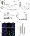
Structural and functional investigation of the 15-bp FOXL1 in-frame deletion. a Computational modeling displaying wildtype Foxl1 (top left) and misfolded Foxl1 (bottom left), the result of disrupting the hydrophobic core (arrow) producing a randomly coiled structure. Secondary structure (right) experimentally probed by circular dichroism (CD) in both wildtype and mutant Foxl1 produced recombinantly in E. coli. Note that wildtype Foxl1 appears to have twice as much helix as the mutant (arrow), the C-terminus of mutant Foxl1 is mostly random coil (48% of the helicity of wildtype). b In osteoblasts (hFOB1.19), both FOXL1 (hFOB-FOXL1-Mut) and the wildtype (hFOB-FOXL1) constructs express RNA to a high level (Non = non-transfection control; FOXL1-Mock = empty vector control). Bar graph represents the mean FOXL1 mRNA relative expression ± SD of three independent experiments. GAPDH was used as an endogenous control. c In osteoblasts (hFOB1.19), cytoplasmic and nuclear extract proteins were prepared and immunoblotted using a FOXL1 antibody (ab83000) using αTubulin (DM1A + DM1B) and P84 (5E10) as cytoplasmic and nuclear loading controls, respectively (left). Protein levels assessed by Western blotting show significantly increased (**p < 0.01) expression (right) of FOXL1 mutant (FOXL1-Mut) compared with FOXL1 wildtype (FOXL1-WT). FOXL1 levels were normalized to P84 and the average band intensity after normalization is presented in the bar graph. Error bars represent the ± SD of three independent experiments. d Expression of wildtype and mutant FOXL1:GFP fusion proteins in HEK293 cells. Transfection of constructs containing wildtype FOXL1;egfp fusion proteins demonstrates that wildtype FOXL1:egfp (panel B) localizes to the nucleus (panel A), as evidenced by their co-localization (panel C). Similarly, expression of mutant FOXL1:egfp fusion proteins (panel E) localizes to the nucleus (panel D) as evidenced by their colocalization (panel F). Expression of empty vector egfp [no FOXLQ ORF, (panel H)] localizes to both the nucleus (panel G) and cytoplasm, as evidenced by egfp detection within and beyond the DAPI stain (panel I). e In HEK293 cells, transfection of constructs containing the wild-type FOXL1 ORF (WT FOXL1) increased transcription from the FOXL1 reporter by 42% over endogenous levels using a luciferase transcription assay. Transfection with the mutant ORF does not induce transcription from the reporter (FOXL1-15bpMUT), indicating a loss of transcriptional activity (p = 7 × 10–9). Bar graph represents pixel intensity ± SD of three independent experiments

References

    1. Abdelfatah N (2014) The genetic aetiology of otosclerosis in the population of Newfoundland and Labrador. Doctoral dissertation. Memorial University of Newfoundland, Canada
    1. Babcock TA, Liu XZ. Otosclerosis: From Genetics to Molecular Biology. Otolaryngol Clin North Am. 2018;51:305–318. doi: 10.1016/j.otc.2017.11.002. - DOI - PubMed
    1. Bamshad MJ, Nickerson DA, Chong JX. Mendelian Gene Discovery: Fast and Furious with No End in Sight. Am J Hum Genet. 2019;105:448–455. doi: 10.1016/j.ajhg.2019.07.011. - DOI - PMC - PubMed
    1. Bel Hadj Ali I, Thys M, Beltaief N, Schrauwen I, Hilgert N, Vanderstraeten K, Dieltjens N, Mnif E, Hachicha S, Besbes G, Ben Arab S, Van Camp G. A new locus for otosclerosis, OTSC8, maps to the pericentromeric region of chromosome 9. Hum Genet. 2008;123:267–272. doi: 10.1007/s00439-008-0470-3. - DOI - PubMed
    1. Brownstein Z, Goldfarb A, Levi H, Frydman M, Avraham KB. Chromosomal mapping and phenotypic characterization of hereditary otosclerosis linked to the OTSC4 locus. Arch Otolaryngol Head Neck Surg. 2006;132:416–424. doi: 10.1001/archotol.132.4.416. - DOI - PubMed

MeSH terms

Substances