. 2021 Oct 11;15(10):e0009873.
doi: 10.1371/journal.pntd.0009873. eCollection 2021 Oct.

Regulation of innate immune responses in macrophages differentiated in the presence of vitamin D and infected with dengue virus 2

Affiliations

Regulation of innate immune responses in macrophages differentiated in the presence of vitamin D and infected with dengue virus 2

Jorge Andrés Castillo et al. PLoS Negl Trop Dis. .

Abstract

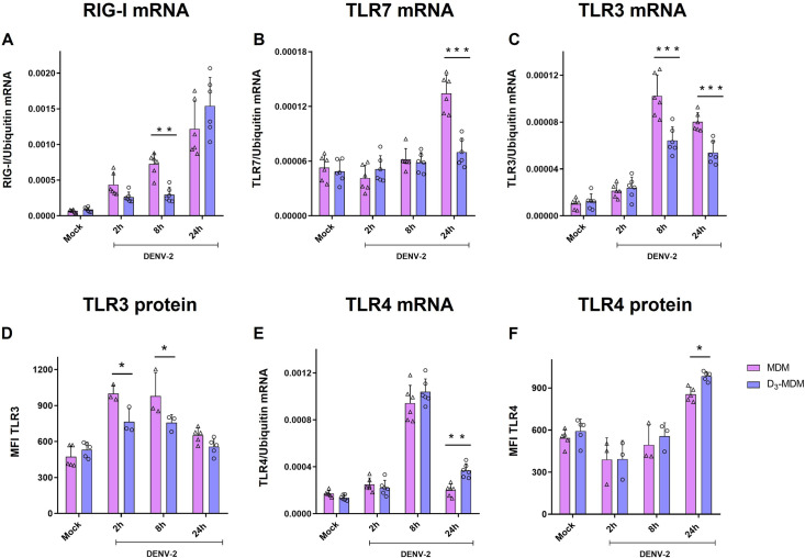
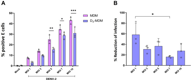
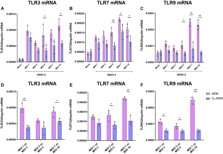
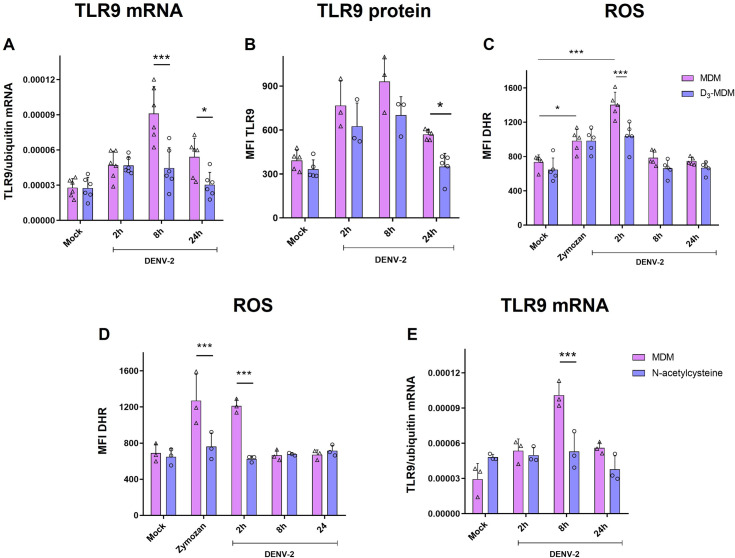
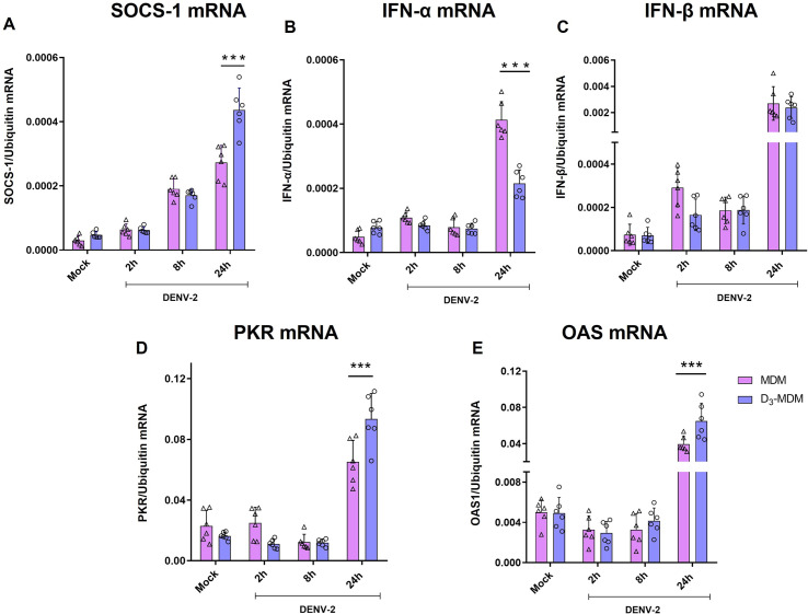
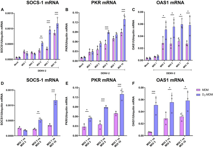
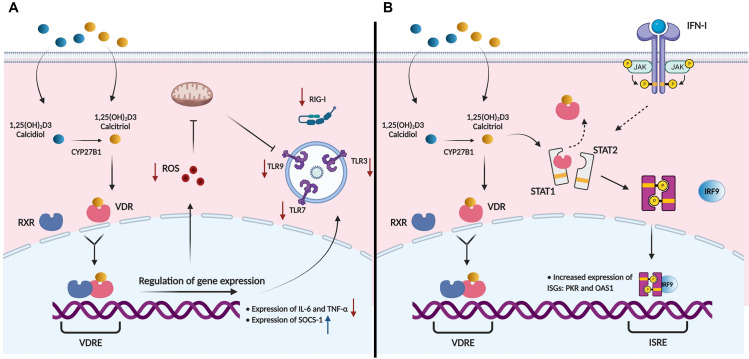
A dysregulated or exacerbated inflammatory response is thought to be the key driver of the pathogenesis of severe disease caused by the mosquito-borne dengue virus (DENV). Compounds that restrict virus replication and modulate the inflammatory response could thus serve as promising therapeutics mitigating the disease pathogenesis. We and others have previously shown that macrophages, which are important cellular targets for DENV replication, differentiated in the presence of bioactive vitamin D (VitD3) are less permissive to viral replication, and produce lower levels of pro-inflammatory cytokines. Therefore, we here evaluated the extent and kinetics of innate immune responses of DENV-2 infected monocytes differentiated into macrophages in the presence (D3-MDMs) or absence of VitD3 (MDMs). We found that D3-MDMs expressed lower levels of RIG I, Toll-like receptor (TLR)3, and TLR7, as well as higher levels of SOCS-1 in response to DENV-2 infection. D3-MDMs produced lower levels of reactive oxygen species, related to a lower expression of TLR9. Moreover, although VitD3 treatment did not modulate either the expression of IFN-α or IFN-β, higher expression of protein kinase R (PKR) and 2'-5'-oligoadenylate synthetase 1 (OAS1) mRNA were found in D3-MDMs. Importantly, the observed effects were independent of reduced infection, highlighting the intrinsic differences between D3-MDMs and MDMs. Taken together, our results suggest that differentiation of MDMs in the presence of VitD3 modulates innate immunity in responses to DENV-2 infection.


Conflict of interest statement

The authors have declared that no competing interests exist.

Figures

Fig 1
Fig 1. VitD3 modulates the expression of TLRs in D3-MDMs.
The MDMs were differentiated in the presence of VitD3 (0.1 nM) for 6 days (D3-MDMs) and then infected with DENV-2 with an MOI of 5 for 2, 8, or 24 h. Expression of RIG I (A), TLR7 (B), TLR3 (C), and TLR4 (E) was measured by qPCR in MDMs and D3-MDMs using the gene encoding ubiquitin as a housekeeper gene. Data are expressed as fold change relative to mock-treated MDMs and D3-MDMs from each time point. Expression of TLR3 (D) and TLR4 (F) protein was measured by flow cytometry and expressed as mean fluorescence intensity. Six different donors were analyzed. Differences were identified using a two-way ANOVA with a Bonferroni posthoc test and a 95% confidence interval (***p < 0.001, **p < 0.01, *p < 0.05).
Fig 2
Fig 2. Reduction of E-positive cells in D3-MDMs infected with different MOI of DENV-2.
The MDMs were differentiated in the presence of VitD3 (0.1 nM) for 6 days (D3-MDMs) and then infected with DENV-2 with different MOI for 24 h. Infection was evaluated through flow cytometry and expressed as % E-positive cells. Three different donors were analyzed. Differences were obtained with a two-way ANOVA with a Bonferroni posthoc test and a 95% confidence interval (***p < 0.001, **p < 0.01, *p < 0.05).
Fig 3
Fig 3. Regulation of TLR expression is independent of the MOI used.
The MDMs were differentiated in the presence of VitD3 (0.1 nM) for 6 days (D3-MDMs) and then infected with DENV-2 with different MOI for 24 h. Expression of TLR3 (A and D), TLR7 (B and E), and TLR9 (C and F) were measured by qPCR in MDMs and D3-MDMs using ubiquitin as housekeeping. Data are expressed as fold change relative to mock-treated MDMs and D3-MDMs. Three different donors were analyzed. Differences were obtained with a two-way ANOVA with a Bonferroni posthoc test and a 95% confidence interval (***p < 0.001, **p < 0.01, *p < 0.05).
Fig 4
Fig 4. VitD3 downregulates TLR9 expression and ROS production in D3-MDMs.
MDMs were differentiated in the presence of VitD3 (0.1 nM) for 6 days (D3-MDMs) and then infected with DENV-2 with an MOI of 5 for 2, 8, or 24 h. Expression of TLR9 (A) was measured by qPCR in MDMs and D3-MDMs using the gene encoding ubiquitin as a housekeeper gene. Data are expressed as fold change relative to mock-treated MDMs and D3-MDMs from each time point. Expression at the protein level of TLR9 (B) was measured by flow cytometry and expressed as mean fluorescence intensity. The production of ROS was measured by flow cytometry through DHR 123 in MDMs and D3-MDMs infected with DENV-2 with an MOI of 5 for 2, 8, and 24 h, or stimulated with 10 μg/mL of zymosan (C). The MDMs were pre-treated with N-acetylcysteine at 10 mM for 1 h and then infected with DENV-2 with an MOI of 5 for 2, 8, and 24 h or treated with 10 μg/mL of zymosan. Production of ROS was measured by flow cytometry through DHR 123) (D) and the TLR9 expression was measured by qPCR (E). Six different donors (A, B, and C) or three different donors (D and E) were analyzed. Differences were obtained with a two-way ANOVA with a Bonferroni posthoc test and a 95% confidence interval (***p < 0.001, **p < 0.01, *p < 0.05).
Fig 5
Fig 5. VitD3 upregulates the expression of SOCS-1 and ISGs in D3-MDMs.
The MDMs were differentiated in the presence of VitD3 (0.1 nM) for 6 days (D3-MDMs) and then infected with DENV-2 for 2, 8, or 24 h. Expression of SOCS-1 (A), IFN α (B), IFN β (C), PKR (D), and OAS1 (E) were measured by qPCR in MDMs and D3-MDMs using the gene encoding ubiquitin as a housekeeper gene. Data are expressed as fold change relative to mock-treated MDMs and D3-MDMs from each time point. Six different donors were analyzed. Differences were obtained with a two-way ANOVA with a Bonferroni post-hoc test and a 95% confidence interval (***p < 0.001, **p < 0.01).
Fig 6
Fig 6. Regulation of SOCS-1 and ISGs expression is independent of the MOI used.
The MDMs were differentiated in the presence of VitD3 (0.1 nM) for 6 days (D3-MDMs) and then infected with DENV-2 with different MOIs for 24 h. Expression of SOCS-1 (A and D), PKR (B and E), and OAS1 (C and F) were measured by qPCR in MDMs and D3-MDMs using ubiquitin as a housekeeper. Data are expressed as fold change relative to mock-treated MDMs and D3-MDMs. Three different donors were analyzed. Differences were obtained with a two-way ANOVA with a Bonferroni posthoc test and a 95% confidence interval (***p < 0.001, **p < 0.01, *p < 0.05).
Fig 7
Fig 7. Hypothetical model of VitD3 effects in D3-MDMs during DENV-2 infection.
(A) VitD3 can be found as inactive form 25(OH)2D2 (calcidiol), as active form 1,25(OH)2D3 (calcitriol), or be converted from inactive to active form within the cell by the action of CYP7B1. VitD3 interacts with the vitamin D receptor (VDR) present in the nuclei and associates then, with the nuclear receptor retinoid X receptor (RXR). Together, they act as transcription factors that will increase the expression of genes that have vitamin D response elements (VDRE) [58,59]. Genomic effects of VitD3 during DENV-2 infection include decreased expression of IL-6, TNF-α, RIG-I, TLR3, TLR7 and decreased production of reactive oxygen species (ROS). Since ROS can induce mitochondrial damage and then increase activation and expression of TLR9 [28], VitD3 reduced TLR9 expression indirectly. (B) As a non-genomic effect, VitD3 can bind to VDR that is initially bound to STAT1 [29], and indirectly enhance the activity of the JAK-STAT signaling pathway and IFN-I activity, thus increasing expression of PKR and OAS1.

Similar articles

Cited by

References

    1. Bhatt S, Gething PW, Brady OJ, Messina JP, Farlow AW, Moyes CL, et al.. The global distribution and burden of dengue. Nature. 2013;496: 504–7. doi: 10.1038/nature12060 - DOI - PMC - PubMed
    1. (WHO) WHO. Dengue. Guidelines for diagnosis, treatment, prevention and control. 2009. - PubMed
    1. (WHO) WHO. Dengue vaccine: WHO position paper–July 2016. Wkly Epidemiol Rec. 2016;91: 349–64. doi: 10.1371/jour - DOI - PubMed
    1. Guzman MG, Harris E. Dengue. Lancet. 2014;6736: 1–13. doi: 10.1016/S0140-6736(14)60572-9 - DOI
    1. Nasirudeen a M a, Wong HH, Thien P, Xu S, Lam K-P, Liu DX. RIG-I, MDA5 and TLR3 synergistically play an important role in restriction of dengue virus infection. PLoS Negl Trop Dis. 2011;5: 1–11. doi: 10.1371/journal.pntd.0000926 - DOI - PMC - PubMed

Publication types

MeSH terms