. 2021 Oct 11;17(10):e1009966.
doi: 10.1371/journal.ppat.1009966. eCollection 2021 Oct.

Differential pathogenesis of closely related 2018 Nigerian outbreak clade III Lassa virus isolates

Affiliations

Differential pathogenesis of closely related 2018 Nigerian outbreak clade III Lassa virus isolates

Derek R Stein et al. PLoS Pathog. .

Abstract

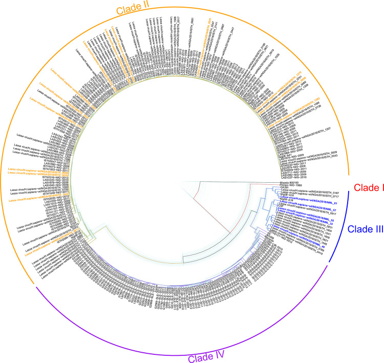
Nigeria continues to experience ever increasing annual outbreaks of Lassa fever (LF). The World Health Organization has recently declared Lassa virus (LASV) as a priority pathogen for accelerated research leading to a renewed international effort to develop relevant animal models of disease and effective countermeasures to reduce LF morbidity and mortality in endemic West African countries. A limiting factor in evaluating medical countermeasures against LF is a lack of well characterized animal models outside of those based on infection with LASV strain Josiah originating form Sierra Leone, circa 1976. Here we genetically characterize five recent LASV isolates collected from the 2018 outbreak in Nigeria. Three isolates were further evaluated in vivo and despite being closely related and from the same spatial / geographic region of Nigeria, only one of the three isolates proved lethal in strain 13 guinea pigs and non-human primates (NHP). Additionally, this isolate exhibited atypical pathogenesis characteristics in the NHP model, most notably respiratory failure, not commonly described in hemorrhagic cases of LF. These results suggest that there is considerable phenotypic heterogeneity in LASV infections in Nigeria, which leads to a multitude of pathogenesis characteristics that could account for differences between subclinical and lethal LF infections. Most importantly, the development of disease models using currently circulating LASV strains in West Africa are critical for the evaluation of potential vaccines and medical countermeasures.


Conflict of interest statement

The authors have declared that no competing interests exist.

Figures

Fig 1
Fig 1. Phylogenetics of five 2018 Nigerian outbreak Lassa virus isolates.
BEAST 2.5.1 was used to produce a time-aware phylogeny of the full S segment of 287 LASV isolates, in addition to the 5 viruses sequenced in this study. Mopeia virus strain AN 21366 was used to root the tree. The light blue trees show 101 posterior draws, displaying the linkage and divergence uncertainty. The main (colored) tree is the median tree, as calculated using TreeAnnotator. The isolates sequenced in this study are highlighted in blue (Clade III), other contemporary isolates (Nigeria, 2018) are highlighted in orange.
Fig 2
Fig 2. Infection of strain 13 guinea pigs with three 2018 Nigerian outbreak Lassa virus isolates.
Groups of 6 guinea pigs (2 female, 4 males) were infected with 1x10⁴ TCID50 of each Nigerian outbreak isolate; NML-33 (orange), NML-46 (black), NML-57 (green) and measured for signs of disease including survival (a), weight loss (b), and temperature (c). Serum and tissues (liver, lung, and spleen) were also collected from moribund animals and assayed for infectious virus (d). Data are presented as mean values with error bars indicating SEM.
Fig 3
Fig 3. Infection of cynomolgus macaques with 2018 Nigerian outbreak Lassa virus isolates.
Groups of 4 female macaques were infected with 1x10⁴ TCID50 of each Nigerian outbreak isolate; NML-33 (orange) and NML-57 (green) along with 2 control animals (Josiah; black) and measured for signs of disease including survival (a), clinical score (b), temperature (c), and weight loss (d). Quantities of viral RNA (copies/ml) was monitored every three days by qRT-PCR (e). Data are presented as mean values with error bars indicated SEM.
Fig 4
Fig 4. Physical appearances of macaques with a terminal 2018 Nigerian Lassa virus clade III isolate (NML-33).
Perimortem observations included severe facial swelling (a), epistaxis (b), and mild petechiae (c) in NHPs infected with the 2018 NML-33 Nigerian Lassa virus clade III isolate.
Fig 5
Fig 5. Infectious virus and gross pathological findings in Nigerian Lassa virus infected macaques.
Infectious virus was assayed in the serum of infected macaques every three days (a) as well as from moribund (open circle) and surviving animals (closed circle) (b); NML-33 (orange), NML-57 (green), Josiah (black). NML-33 animals exhibited severe respiratory symptoms compared to NML-57 with significant pulmonary lesions and hemorrhage (c) and (d). Data are presented as mean values with error bars indicated SEM.
Fig 6
Fig 6. Histopathology of lung, liver and splenic lesions.
Thin sections of formalin fixed tissue specimens collected at the time of severe disease from NHPs infected with Lassa virus Josiah, NML-33 and NML-57 isolates were prepared and stained using standard histopathological methods (Hematoxylin & Eosin, 200X bar = 20μm). Specimens from representative animals demonstrated a. Lung—Josiah—normal b. Lung—NML-33—intestinal pneumonia: alveolar septa expanded by edema, fibrin and inflammatory cells (yellow arrow) (fibrin = black arrow) (asterisk = edema) c. Lung—NML-57normal d. Liver—Josiah—focal minimal fibrin deposits and neutrophilic infiltrates (yellow arrow) e. Liver—NML-33—multifocal fibrin deposits with neutrophilic histiocytic infiltrates (yellow arrows) f. Liver -NML-57—normal g. Spleen—Josiah—normal h. Spleen NML-33—multifocal red pulp necrosis (black arrows) i. Spleen—NML-57—normal.
Fig 7
Fig 7. Biochemical parameters following Nigerian Lassa virus challenge.
EDTA treated blood samples were collected from NHPs immediately prior to (0 dpi) and every three days post-Lassa virus infection and monitored for albumin (a), alkaline phosphatase (b), alanine aminotransferase (c), amylase (d), total bilirubin (e), blood urea nitrogen (f), calcium (g), phosphorus (h), creatinine (i), glucose (j), sodium (k), potassium (l), total protein (m), and globulin (n). Data are represented as individual data points for each animal and time point.
Fig 8
Fig 8. Coagulation profiles following Lassa virus infection of macaques.
Citrate plasma were obtained from NHP blood samples collected every three days post-Lassa virus infection and monitored for Fibrinogen (a), aPTT (b), Thrombin (c), Prothrombin (d), Protein C (e), and Protein S (d).
Fig 9
Fig 9. Hematological profiles in macaques following Lassa virus infection.
EDTA treated blood samples were collected from NHPs every three days post-Lassa virus infection and monitored for white blood cells (a), lymphocytes (b), monocytes (c), neutrophils (d), eosinophils (e), basophils (f), red blood cells (g), hemoglobin (h), hematocrit (i), mean corpuscular volume (j), mean corpuscular hemoglobin (k), mean corpuscular hemoglobin volume (l), red blood cell distribution width (m), platelets (n), plateletcrit (o), mean platelet volume (p), and platelet distribution width (q). Data are represented as individual data points for each animal and time point.
Fig 10
Fig 10. Determination of immune cell, cytokine and chemokine profile during Lassa virus infection of macaques.
Flow cytometric analysis was conducted on each individual NHP at their respective time point (lines represent means for each group). Naïve CD4+ (a) and CD8+ (d) cells, memory CD4+ (b) and CD8+ (e) cells, T-regulatory cells (c), and Natural Killer cells (f) are shown. Serum was collected from LASV-infected NHPs and a Luminex assay detecting 29 analytes was performed on each sample. A heat map showing the fold change in each analyte at the terminal time point for each macaque is shown. Data are represented as individual data points for each animal and time point.
Fig 11
Fig 11. Cytokine and chemokine profiles during Lassa virus infection of macaques.
Cytokine/Chemokine analysis was conducted using a 29-plex Luminex assay on each individual NHP at their respective time point. Shown are IFN-gamma (a), IL-12 (b), IL-15 (c), IL-1b (d), IL-1RA (e), IL-6 (f), ITAC (g), MIG (h), TNF (i).

Similar articles

Cited by

References

    1. Frame JD, Baldwin JM, Gocke DJ, Troup JM. Lassa fever, a new virus disease of man from West Africa. I. Clinical description and pathological findings. Am J Trop Med Hyg. 1970;19: 670–676. doi: 10.4269/ajtmh.1970.19.670 - DOI - PubMed
    1. Ogbu O, Ajuluchukwu E, Uneke CJ. Lassa fever in West African sub-region: An overview. Journal of Vector Borne Diseases. 2007. pp. 1–11. - PubMed
    1. Ilori EA, Furuse Y, Ipadeola OB, Dan-Nwafor CC, Abubakar A, Womi-Eteng OE, et al.. Epidemiologic and clinical features of lassa fever outbreak in Nigeria, january 1-may 6, 2018. Emerg Infect Dis. 2019;25: 1066–1074. doi: 10.3201/eid2506.181035 - DOI - PMC - PubMed
    1. Chika-Igwenyi NM, Harrison RE, Psarra C, Gilcuesta J, Gulamhusein M, Onwe EO, et al.. Early onset of neurological features differentiates two outbreaks of lassa fever in ebonyi state, nigeria during 2017–2018. PLoS Negl Trop Dis. 2021;15. doi: 10.1371/journal.pntd.0009169 - DOI - PMC - PubMed
    1. Babalola SO, Babatunde JA, Remilekun OM, Amaobichukwu AR, Abiodun AM, Jide I, et al.. Lassa virus rna detection from suspected cases in Nigeria, 2011–2017. Pan Afr Med J. 2019;34. doi: 10.11604/pamj.2019.34.76.16425 - DOI - PMC - PubMed