

Measurements of OCT Angiography Complement OCT for Diagnosing Early Primary Open-Angle Glaucoma
- PMID: 34634501
- PMCID: PMC9405474
- DOI: 10.1016/j.ogla.2021.09.012
Measurements of OCT Angiography Complement OCT for Diagnosing Early Primary Open-Angle Glaucoma
Abstract
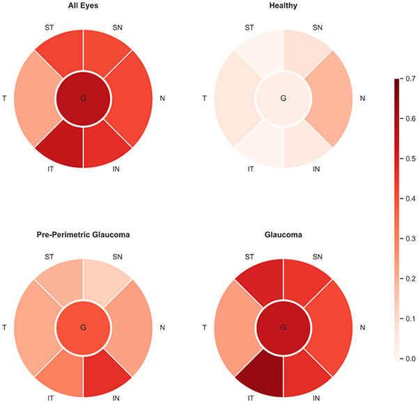
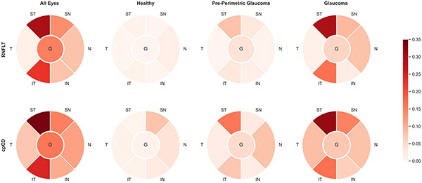
Purpose: To compare measurements of global and regional circumpapillary capillary density (cpCD) with retinal nerve fiber layer (RNFL) thickness and characterize their relationship with visual function in early primary open-angle glaucoma (POAG).
Design: Cross-sectional study.
Participants: Eighty healthy eyes, 64 preperimetric eyes, and 184 mild POAG eyes from the Diagnostic Innovations in Glaucoma Study.
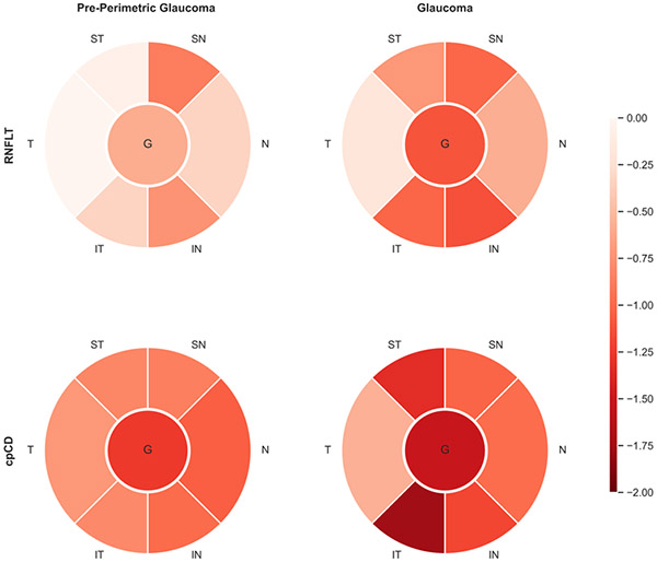
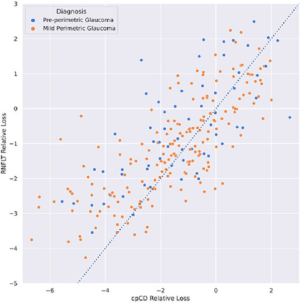
Methods: Global and regional RNFL thickness and cpCD measurements were obtained using OCT and OCT angiography (OCTA). For direct comparison at the individual and diagnostic group level, RNFL thickness and capillary density values were converted to a normalized relative loss scale.
Main outcome measures: Retinal nerve fiber layer thickness and cpCD normalized loss at the individual level and diagnostic group. Global and regional areas under the receiver operating characteristic curve (AUROC) for RNFL thickness and cpCD to detect preperimetric glaucoma and glaucoma, R2 for the strength of associations between RNFL thickness function and capillary density function in diagnostic groups.
Results: Both global and regional RNFL thickness and cpCD decreased progressively with increasing glaucoma severity (P < 0.05, except for temporal RNFL thickness). Global and regional cpCD relative loss values were higher than those of RNFL thickness (P < 0.05) in preperimetric glaucoma (except for the superonasal region) and glaucoma (except for the inferonasal and superonasal regions) eyes. Race, intraocular pressure (IOP), and cpCD were associated with greater cpCD than RNFL thickness loss in early glaucoma at the individual level (P < 0.05). Global measurements of capillary density (whole image capillary density and cpCD) had higher diagnostic accuracies than RNFL thickness in detecting preperimetric glaucoma and glaucoma (P < 0.05; except for cpCD/RNFL thickness comparison in glaucoma [P = 0.059]). Visual function was significantly associated with RNFL thickness and cpCD globally and in all regions (P < 0.05, except for temporal RNFL thickness-function association [P = 0.070]).
Conclusions: Associations between capillary density and visual function were found in the regions known to be at highest risk for damage in preperimetric glaucoma eyes and all regions of mild glaucoma eyes. In early glaucoma, capillary density loss was more pronounced than RNFL thickness loss. Individual characteristics influence the relative magnitudes of capillary density loss compared with RNFL thickness loss. Retinal nerve fiber layer thickness and microvascular assessments are complementary and yield valuable information for the detection of early damages seen in POAG.
Keywords: Early glaucoma; OCT angiography; Retinal nerve fiber layer; Visual field.
Copyright © 2021 American Academy of Ophthalmology. Published by Elsevier Inc. All rights reserved.
Figures
Similar articles
-
Comparison of Peripapillary Capillary Density in Glaucoma Patients of African and European Descent.Ophthalmol Glaucoma. 2021 Jan-Feb;4(1):51-62. doi: 10.1016/j.ogla.2020.07.005. Epub 2020 Jul 18. Ophthalmol Glaucoma. 2021. PMID: 32693049 Free PMC article.
-
Peripapillary and Macular Vessel Density in Patients with Primary Open-Angle Glaucoma and Unilateral Visual Field Loss.Ophthalmology. 2018 Apr;125(4):578-587. doi: 10.1016/j.ophtha.2017.10.029. Epub 2017 Nov 22. Ophthalmology. 2018. PMID: 29174012 Free PMC article.
-
Peripapillary and Macular Vascular Density in Patients With Preperimetric and Early Primary Open Angle Glaucoma.J Glaucoma. 2022 Sep 1;31(9):724-733. doi: 10.1097/IJG.0000000000002069. Epub 2022 Jun 21. J Glaucoma. 2022. PMID: 36044326
-
Optical Coherence Tomography Angiography in Glaucoma.J Glaucoma. 2020 Apr;29(4):312-321. doi: 10.1097/IJG.0000000000001463. J Glaucoma. 2020. PMID: 32053551 Free PMC article. Review.
-
[Optical coherence tomography and optical coherence tomography angiography for detecting glaucoma progression. Part 1. Study methods, measurement variability and the role of age-related changes].Vestn Oftalmol. 2023;139(1):122-128. doi: 10.17116/oftalma2023139011122. Vestn Oftalmol. 2023. PMID: 36924524 Review. Russian.
Cited by
-
Deep Learning Estimation of 10-2 Visual Field Map Based on Macular Optical Coherence Tomography Angiography Measurements.Am J Ophthalmol. 2024 Jan;257:187-200. doi: 10.1016/j.ajo.2023.09.014. Epub 2023 Sep 19. Am J Ophthalmol. 2024. PMID: 37734638 Free PMC article.
-
Comparative evaluation of OCT with OCTA changes at the optic disc and macula in glaucoma suspect and early glaucoma.Indian J Ophthalmol. 2025 Mar 1;73(Suppl 2):S260-S266. doi: 10.4103/IJO.IJO_2575_23. Epub 2024 Oct 25. Indian J Ophthalmol. 2025. PMID: 39446809 Free PMC article.
-
Macular Oxygen Saturation in Glaucoma Using Retinal Oximetry of Visible Light Optical Coherence Tomography.medRxiv [Preprint]. 2023 Dec 22:2023.12.20.23300300. doi: 10.1101/2023.12.20.23300300. medRxiv. 2023. Update in: Transl Vis Sci Technol. 2025 Feb 03;14(2):12. doi: 10.1167/tvst.14.2.12. PMID: 38196641 Free PMC article. Updated. Preprint.
-
Combining Optical Coherence Tomography and Optical Coherence Tomography Angiography Longitudinal Data for the Detection of Visual Field Progression in Glaucoma.Am J Ophthalmol. 2023 Feb;246:141-154. doi: 10.1016/j.ajo.2022.10.016. Epub 2022 Nov 1. Am J Ophthalmol. 2023. PMID: 36328200 Free PMC article.
-
Macular Oxygen Saturation in Glaucoma Using Retinal Oximetry of Visible Light Optical Coherence Tomography: A Pilot Study.Transl Vis Sci Technol. 2025 Feb 3;14(2):12. doi: 10.1167/tvst.14.2.12. Transl Vis Sci Technol. 2025. PMID: 39913123 Free PMC article.
References
-
- Weinreb RN, Leung CKS, Crowston JG, et al. Primary open-angle glaucoma. Nature Reviews Disease Primers 2016;2:16067. - PubMed
-
- Leske MC, Heijl A, Hussein M, et al. Factors for Glaucoma Progression and the Effect of Treatment: The Early Manifest Glaucoma Trial. Archives of Ophthalmology 2003;121:48–56. - PubMed
