. 2021 Oct 26;12(5):e0250921.
doi: 10.1128/mBio.02509-21. Epub 2021 Oct 12.

The Cyclin Cln1 Controls Polyploid Titan Cell Formation following a Stress-Induced G2 Arrest in Cryptococcus

Affiliations

The Cyclin Cln1 Controls Polyploid Titan Cell Formation following a Stress-Induced G2 Arrest in Cryptococcus

Sophie Altamirano et al. mBio. .

Abstract

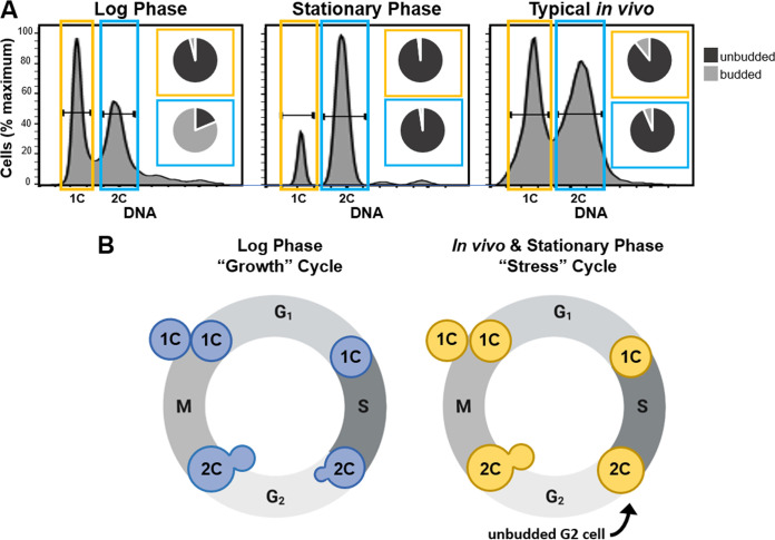
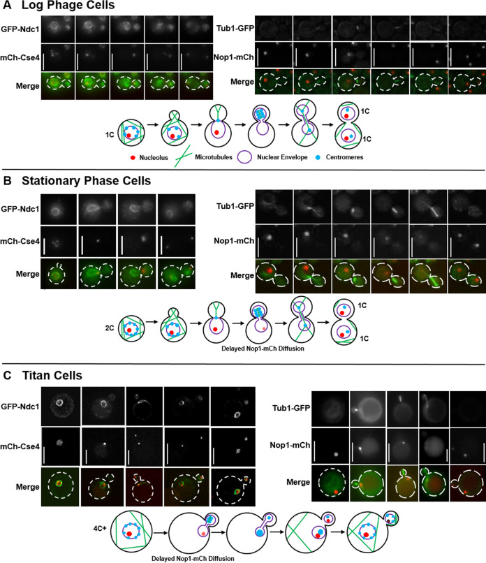
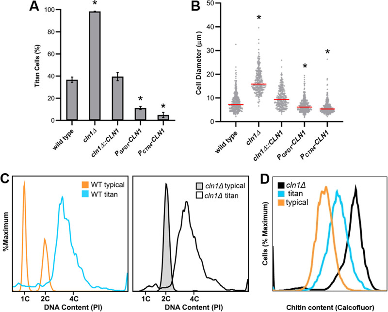
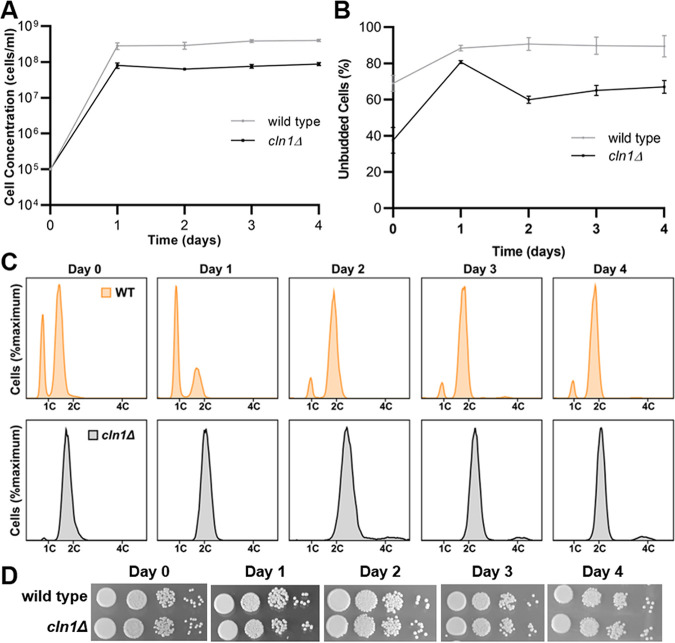
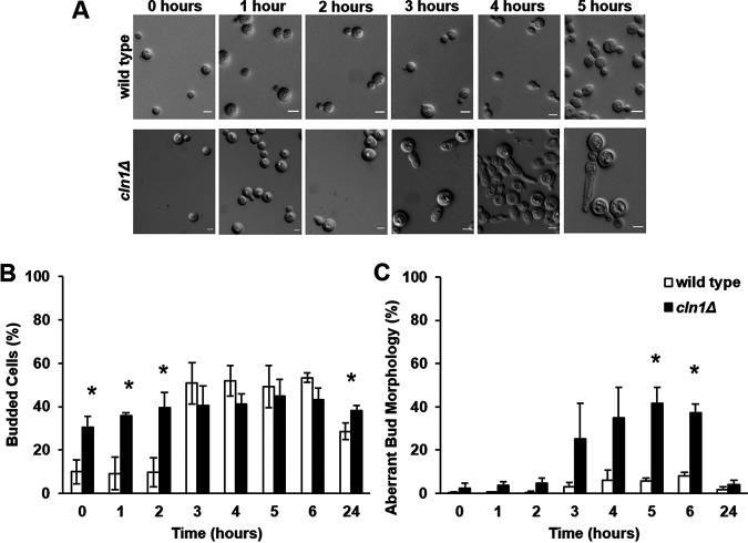
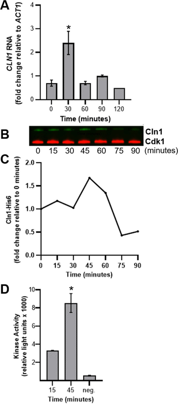
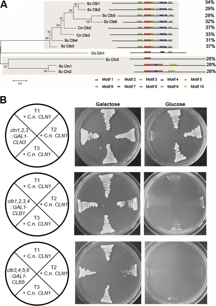
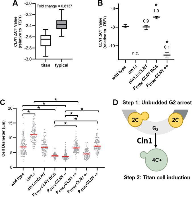
The pathogenic yeast Cryptococcus neoformans produces polyploid titan cells in response to the host lung environment that are critical for host adaptation and subsequent disease. We analyzed the in vivo and in vitro cell cycles to identify key aspects of the C. neoformans cell cycle that are important for the formation of titan cells. We identified unbudded 2C cells, referred to as a G2 arrest, produced both in vivo and in vitro in response to various stresses. Deletion of the nonessential cyclin Cln1 resulted in overproduction of titan cells in vivo and transient morphology defects upon release from stationary phase in vitro. Using a copper-repressible promoter PCTR4-CLN1 strain and a two-step in vitro titan cell formation assay, our in vitro studies revealed Cln1 functions after the G2 arrest. These studies highlight unique cell cycle alterations in C. neoformans that ultimately promote genomic diversity and virulence in this important fungal pathogen. IMPORTANCE Dysregulation of the cell cycle underlies many human genetic diseases and cancers, yet numerous organisms, including microbes, also manipulate the cell cycle to generate both morphologic and genetic diversity as a natural mechanism to enhance their chances for survival. The eukaryotic pathogen Cryptococcus neoformans generates morphologically distinct polyploid titan cells critical for host adaptation and subsequent disease. We analyzed the C. neoformans in vivo and in vitro cell cycles to identify changes required to generate the polyploid titan cells. C. neoformans paused cell cycle progression in response to various environmental stresses after DNA replication and before morphological changes associated with cell division, referred to as a G2 arrest. Release from this G2 arrest was coordinated by the cyclin Cln1. Reduced CLN1 expression after the G2 arrest was associated with polyploid titan cell production. These results demonstrate a mechanism to generate genomic diversity in eukaryotic cells through manipulation of the cell cycle that has broad disease implications.

Keywords: Cryptococcus neoformans; aneuploidy; cell cycle; cryptococcal meningitis; cryptococcosis; cyclins; deneoformans; ploidy; polyploid; polyploidy; titan cell.


Figures

FIG 1
FIG 1
In vivo typical sized cells and in vitro stationary-phase cells are primarily unbudded 2C cells. (A) Analysis of cell morphology with 1C (yellow) and 2C (blue) DNA content in log-phase cells, stationary-phase cells, and typical-size cells isolated from the lungs of mice at 14 days postinfection (referred to as typical in vivo). Cells were grown in the indicated conditions, fixed, and stained with propidium iodide, and then fluorescence-activated cell sorting (FACS) was performed to purify the 1C and 2C cell populations. The resulting purified cells were then analyzed microscopically to determine the proportion of cells containing buds (insets). (B) Schematic representation of the C. neoformans growth cell cycle (blue cells) observed in log-phase cells and the putative stress cell cycle (yellow cells) that is notable for production of unbudded 2C cells that were observed in both stationary-phase cultures in vitro and among typical sized cells in vivo.
FIG 2
FIG 2
Analysis of nuclear dynamics after unbudded G2 arrest shows minimal effect on the ability of stationary and titan cells to complete mitosis and divide. Representative images and corresponding schematic diagrams showing mitotic events in log-phase cells (A), stationary-phase cells (B), and in vivo titan cells (C). Centromere and nuclear envelope dynamics were analyzed in cells expressing the inner kinetochore protein, mCherry-Cse4, and the nuclear envelope protein, GFP-Ndc1. Microtubules and nucleolus dynamics were analyzed in cells expressing the nucleolar protein, Nop1-mCherry, and the tubulin protein, Tub1-GFP. All cells were analyzed by time-lapse microscopy, and titan cells were analyzed using both time-lapse and 2-photon microscopy due to their larger size. Not all representative images of the different cell cycle stages are of the same cell. (A) Consistent with previous studies of log-phase cells (25), the centromeres were not clustered in the mother cell prior to mitosis, microtubules formed the mitotic spindle in the daughter cell, and the centromeres then clustered and moved completely into the daughter. Elongation of the spindle resulted in breakage of the nuclear envelope and loss of the nucleolus staining. After mitosis, half of the centromeres returned to the mother cell, and two separate nucleoli and two separate nuclear envelopes were formed, one in the mother and one in the daughter cell. (B) Stationary-phase cells that had already undergone DNA replication prior to bud emergence underwent mitosis similar to log-phase cells. A minor difference in nucleolus retention time was observed. The nucleolus of the stationary-phase cells faded dramatically but was retained throughout the course of mitotic spindle formation and DNA segregation. (C) In vivo-derived titan cells also underwent a mitosis similar to the log- and stationary-phase cells, including a slightly delayed disappearance of the nucleolus in the mother cell. The titan cells produced typical-sized daughter cells. Scale bars are 5 μm in panels A and B and 10 μm in panel C.
FIG 3
FIG 3
Cln1 negatively regulates in vivo titan cell formation. Mice were infected via inhalation with 5 × 104 cells of the wild-type strain (KN99α), the cln1Δ deletion, the cln1Δ::CLN1 complement, or the overexpression strains PGPD1-CLN1 or PCTR4-CLN1. Titan cell formation in the lungs was analyzed at 3 days postinfection. (A) Percentage of titan cells was determined based on a cell body size threshold of 10 μm, excluding the capsule, for >300 cells per mouse. Error bars indicate standard deviations (SD), n ≥ 3 mice per strain. *, P < 0.05 compared to the wild type by Student's t test with Welch’s correction. (B) Cell body size, excluding the capsule, was also plotted for >300 cells per mouse as a visual representation of the distribution of cell size in the different strains. Median cell size is indicated by the red line. *, P < 0.05 compared to wild type by Kruskal-Wallis with Dunn posttest. (C) cln1Δ cells isolated from the lungs of mice were fixed, stained with propidium iodide, and analyzed by flow cytometry for DNA content to determine cell ploidies within the population. Wild-type in vitro-grown cells (orange), wild-type titan cells (blue), in vitro-grown cln1Δ cells (gray), and a diploid strain (not shown) were used as controls. (D) Wild-type and cln1Δ cells isolated from the lungs of mice were fixed, stained with calcofluor white, and analyzed by flow cytometry to determine cell wall chitin content. The wild-type population was split based on cell size into the typical (orange) and titan (blue) cell subsets for chitin analysis.
FIG 4
FIG 4
cln1Δ cells arrest as 2C cells and maintain viability during nutrient deprivation. Wild-type and cln1Δ cells were grown in YPD liquid medium for 4 days and assessed for entry into stationary phase at various time points based on cell concentration, morphology, viability, and ploidy. Error bars represent SD from three biological replicates. (A) Wild-type (gray) and cln1Δ (black) cells displayed a plateau in cell growth after 1 day of culture. (B) By 1 day, when nutrients became limited, both wild-type and cln1Δ cells were primarily unbudded, while the cln1Δ cells contained more budded cells at the later time points. (C) DNA content analysis showed both strains arrested as 2C cells. The cln1Δ cells were primarily 2C throughout the experiment, while the wild-type cells arrested as 2C cells only after 2 days in YPD liquid media. (D) Spot assays were performed at days 0, 1, 2, 3, and 4 of nutrient deprivation, and no differences in viability were observed.
FIG 5
FIG 5
cln1Δ cells exhibit a transient aberrant budding morphology after release from nutrient deprivation. After 3 days of incubation under nutrient starvation conditions, wild-type and cln1Δ cells were pelleted and resuspended into fresh YPD liquid medium. Error bars represent SD from three biological replicates. (A) The cln1Δ cells exhibited an elongated bud morphological defect and delayed bud formation. Scale bar, 5 μm. (B) While the wild-type cells initiated budding at 3 h posttransfer, no increase in cln1Δ cell budding was observed posttransfer. (C) The cln1Δ cells had a transient aberrant bud morphology that peaked at 5 h posttransfer but was not observed at 24 h. *, P < 0.05 by Student's t test with Welch’s correction.
FIG 6
FIG 6
Cln1 activates Cdk1 to initiate bud formation. (A) CLN1 RNA levels in wild-type cells were analyzed by RT-qPCR at 30-min intervals after release from stationary phase into nutrient-replete media. Expression levels were normalized to the ACT1 housekeeping gene to determine changes in gene expression. Error bars represent SD of the CT values from 3 technical replicates. Data are representative of 3 biological replicates. *, P < 0.05 compared to t = 0 by Student's t test with Welch’s correction. (B and C) Cells were harvested at 15-min intervals, and the amount of Cln1-His6 coprecipitated with Cdk1-Myc was determined at each time point by Western blotting (B) and then quantified based on the 0-min time point (C). (D) Cdk1 kinase activity was determined at 15 and 45 min. The negative control consisted of the 1× kinase buffer only. Error bars represent SD from 3 technical replicates. *, P < 0.05 compared to t = 15 by Student's t test.
FIG 7
FIG 7
Motif analysis of C. neoformans Cln1 shows it contains both S. cerevisiae Cln and Clb motifs but only complements Cln cyclins in S. cerevisiae. (A) Phylogenetic analysis based on protein motifs of the Cdc28 family of cyclins from C. neoformans (Cln1, Clb2, and Clb3) and S. cerevisiae (Cln1, Cln2, Cln3, Clb1, Clb2, Clb3, Clb4, Clb5, and Clb6) showed C. neoformans Cln1 had highest homology to the S. cerevisiae Clb proteins. (B) C. neoformans CLN1 expressed in S. cerevisiae was able to rescue growth of a cln1,2,3 triple but not clb1,2,3,4 or clb3,4,5,6 quadruple mutants. C. neoformans CLN1 was expressed with a constitutive S. cerevisiae promoter, and all cln1,2,3::GAL-CLN3 +C. neoformans CLN1 transformants (T1, T2, or T3 + C.n. CLN1) produced growth on the nonpermissive glucose medium, whereas none of the clb1,2,3,4::GAL1-CLB1 +C. neoformans CLN1 or clb3,4,5,6::GAL-CLB5 +C. neoformans CLN1 transformants had growth on the nonpermissive glucose medium.
FIG 8
FIG 8
Low CLN1 expression after unbudded G2 arrest induces titan cell formation. (A) RT-qPCR showed higher CLN1 levels in typical-size cells than titan cells isolated from the lungs of mice. Expression levels were normalized to both cell number and the TEF1 housekeeping gene to determine change in cycle threshold (ΔCT). Fold decrease in gene expression in titan compared to typical cells was calculated using 2-ΔΔCT. Data presented are the ΔCT values from three biological replicates. P = 0.0004 by Student's t test. (B) RT-qPCR analysis of CLN1 expression in vitro in the wild-type, cln1Δ, and cln1Δ::CLN1 strains and when the copper-repressible promoter strain was grown with 400 μM of the copper chelator BCS (PCTR4-CLN1 BCS) or 25 μM copper (PCTR4-CLN1++). No expression change was detected in the cln1Δ strain (n.c). Fold change compared to the wild type is indicated above each bar. Data presented are the ΔCT values from three technical replicates. *, P ≤ 0.0015 compared to the wild type by one-way ANOVA with a Dunnett correction for multiple comparisons. (C) In vitro titan cell formation with wild-type, cln1Δ, cln1Δ::CLN1, and PCTR4-CLN1 cells showed CLN1 expression only affects titan cell formation after unbudded 2C cells are already formed. In vitro titan cell formation was induced using an initial stationary-phase culture to induce unbudded G2 arrest followed by a second incubation under hypoxic conditions to induce titan cell formation (43). CLN1 expression was manipulated using the PCTR4-CLN1 strain and copper addition during the initial stationary culture (+−), second incubation (−+), both (++), or neither (−−). The cell diameter of at least 100 cells was measured at the end of the protocol to determine in vitro titan cell formation. Median cell size is indicated by the red line. *, P < 0.0001 by Kruskal-Wallis test with Dunn’s posttest correction. (D) Our results show Cln1 is necessary in the stress cell cycle after the unbudded 2C cells are already formed. Loss or reduction of CLN1 expression in cells after unbudded G2 arrest results in polyploid titan cells (green cells) only with concomitant environmental stimuli.

References

    1. Fox DT, Soltis DE, Soltis PS, Ashman TL, Van de Peer Y. 2020. Polyploidy: a biological force from cells to ecosystems. Trends Cell Biol 30:688–694. doi: 10.1016/j.tcb.2020.06.006. - DOI - PMC - PubMed
    1. Øvrebø JI, Edgar BA. 2018. Polyploidy in tissue homeostasis and regeneration. Development 145:dev156034. doi: 10.1242/dev.156034. - DOI - PMC - PubMed
    1. Barlow PW, Sherman MI. 1974. Cytological studies on the organization of DNA in giant trophoblast nuclei of the mouse and the rat. Chromosoma 47:119–131. doi: 10.1007/BF00331800. - DOI - PubMed
    1. Losick VP, Fox DT, Spradling AC. 2013. Polyploidization and cell fusion contribute to wound healing in the adult Drosophila epithelium. Curr Biol 23:2224–2232. doi: 10.1016/j.cub.2013.09.029. - DOI - PMC - PubMed
    1. Gandarillas A, Freije A. 2014. Cycling up the epidermis: reconciling 100 years of debate. Exp Dermatol 23:87–91. doi: 10.1111/exd.12287. - DOI - PubMed

Publication types

LinkOut - more resources