. 2021 Oct 11;28(1):69.
doi: 10.1186/s12929-021-00767-x.

Modular self-assembly system for development of oligomeric, highly internalizing and potent cytotoxic conjugates targeting fibroblast growth factor receptors

Affiliations

Modular self-assembly system for development of oligomeric, highly internalizing and potent cytotoxic conjugates targeting fibroblast growth factor receptors

Marta Poźniak et al. J Biomed Sci. .

Abstract

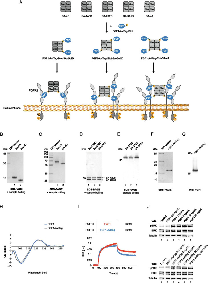
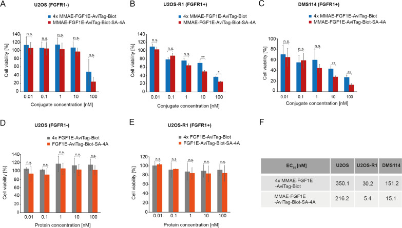
Background: Overexpression of FGFR1 is observed in numerous tumors and therefore this receptor constitutes an attractive molecular target for selective cancer treatment with cytotoxic conjugates. The success of cancer therapy with cytotoxic conjugates largely relies on the precise recognition of a cancer-specific marker by a targeting molecule within the conjugate and its subsequent cellular internalization by receptor mediated endocytosis. We have recently demonstrated that efficiency and mechanism of FGFR1 internalization are governed by spatial distribution of the receptor in the plasma membrane, where clustering of FGFR1 into larger oligomers stimulated fast and highly efficient uptake of the receptor by simultaneous engagement of multiple endocytic routes. Based on these findings we aimed to develop a modular, self-assembly system for generation of oligomeric cytotoxic conjugates, capable of FGFR1 clustering, for targeting FGFR1-overproducing cancer cells.

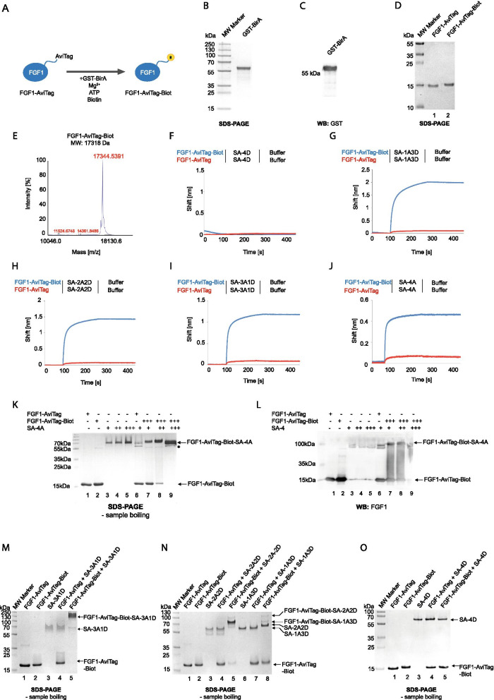
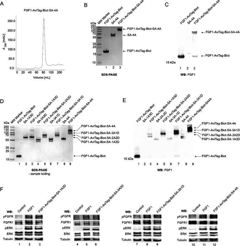
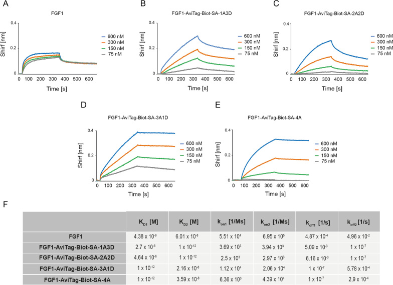
Methods: Engineered FGF1 was used as FGFR1-recognition molecule and tailored for enhanced stability and site-specific attachment of the cytotoxic drug. Modified streptavidin, allowing for controlled oligomerization of FGF1 variant was used for self-assembly of well-defined FGF1 oligomers of different valency and oligomeric cytotoxic conjugate. Protein biochemistry methods were applied to obtain highly pure FGF1 oligomers and the oligomeric cytotoxic conjugate. Diverse biophysical, biochemical and cell biology tests were used to evaluate FGFR1 binding, internalization and the cytotoxicity of obtained oligomers.

Results: Developed multivalent FGF1 complexes are characterized by well-defined architecture, enhanced FGFR1 binding and improved cellular uptake. This successful strategy was applied to construct tetrameric cytotoxic conjugate targeting FGFR1-producing cancer cells. We have shown that enhanced affinity for the receptor and improved internalization result in a superior cytotoxicity of the tetrameric conjugate compared to the monomeric one.

Conclusions: Our data implicate that oligomerization of the targeting molecules constitutes an attractive strategy for improvement of the cytotoxicity of conjugates recognizing cancer-specific biomarkers. Importantly, the presented approach can be easily adapted for other tumor markers.

Keywords: Cancer; Endocytosis; FGF/FGFR; Targeted therapy.


Conflict of interest statement

The authors declare no competing interests.

Figures

Fig. 1
Fig. 1
Basic building blocks for development of SA-FGF1 oligomers. A Strategy for development of FGF1-SA oligomers. In this approach SA tetramers containing from 0 to 4 binding sites for biotinylated ligands are obtained by mixing wild type SA-Alive-HisTag and non-biotin-binding SA-Dead mutant. FGF1-AviTag will be enzymatically biotinylated by GST-BirA and then assembled with various SA scaffolds, leading to formation of FGF1-SA oligomers with distinct potential for clustering of FGFR1. B and C Separately purified SA-Alive-HisTag and non-biotin-binding SA-Dead. The purity and identity of streptavidin variants were confirmed by SDS-PAGE (CBB staining), presence of the HisTag on the SA-Alive version allows for comparison of SA variants composition. To maintain the tetrameric form of the protein, SA samples in C were not subjected to thermal denaturation. D and E Variants of streptavidin tetramers with varying valency. Separately purified SA-Alive-HisTag and SA-Dead were mixed, yielding all possible combinations. Due to the presence of the His-Tag on SA-Alive version (ensuring affinity to metal ions), metal-affinity chromatography was applied to separate various combinations of SA tetramers. The purity and identity of obtained SA variant were confirmed by SDS-PAGE. Upon boiling, samples were separated into monomers (D). The ratio of two SA-bands demonstrates protein tetramers containing from 0 to 4 binding sites for biotinylated ligands. To preserve the tetrameric form of SA, samples in E were not subjected to thermal denaturation. F and G FGF1-AviTag protein was purified by heparin affinity chromatography. Purified protein was analyzed by SDS–PAGE under reducing condition (F) and western blotting (WB) with antibody directed against FGF1 (G). H CD spectra of wild type FGF1 and FGF1-AviTag confirming preservation of 2D structure of FGF1 upon incorporation of AviTag. I BLI comparison of FGF1 and FGF1-AviTag interaction with FGFR1. The extracellular region of FGFR1 was immobilized on BLI sensors and incubated either with FGF1 and FGF1-AviTag. The association and dissociation profiles were measured. J FGF1-AviTag is able to activate FGFR1. Serum-starved NIH3T3 cells were incubated for 15 min with different concentrations of FGF1 (positive control) or FGF1-AviTag in the presence of heparin. Cells were lysed and activation of FGFR1 assessed with western blotting using antibodies recognizing phosphorylated key tyrosine within intracellular FGFR tyrosine kinase domain (pFGFR) and receptor-downstream ERK (detected with antibodies recognizing phosphorylated ERK (pERK). The level of tubulin served as a loading control
Fig. 2
Fig. 2
Assembly of SA-FGF1 oligomers. A BirA-mediated site-specific FGF1-AviTag biotinylation. FGF1 was fused to the AviTag acceptor peptide and this Avi-tagged protein was selectively biotinylated by GST-BirA enzyme, leading to the formation of site-specific mono-biotinylated ligand. Attached biotin is marked in yellow. B and C GST-BirA was purified by glutathione affinity chromatography. Protein purity was analyzed by SDS–PAGE (CBB staining) (B) and western blotting (WB) with anti-GST antibody (C). D Biotinylation of FGF1-AviTag. The efficiency of the biotinylation (seen as upshift in protein migration) and purity of FGF1-AviTag-Biot was analyzed by SDS-PAGE (CBB staining) E Mass spectrometry analysis of purified FGF1-AviTag-Biot. The molecular mass of FGF1-AviTag-biot was assessed by MALDI MS. The theoretical MW of the proteinaceous core of FGF1-AviTag is 17092 Da, and 17,318 Da after biotinylation. F–J BLI comparison of FGF1-AviTag and FGF1-AviTag-Biot interaction with various SA variants. FGF1-AviTag and FGF1-AviTag-Biot were chemically immobilized on BLI sensors and incubated with SA-4D (F), SA-1A3D (G), SA-2A2D (H), SA-3A1D (I), SA-4D (J). The association and dissociation profiles were measured. K and L Assembling of FGF1-AT-Biot-SA-4A oligomer. FGF1E-AviTag and biotinylated variant of this protein were mixed with SA-4A in various ratios and incubated for 5–10 min at RT. Then, proteins mixes were subjected to SDS-PAGE analysis and CBB staining (K) and western blotting (L); *most likely self-assembly form of FGF1-AviTag or partial SA dissociation product upon SDS treatment. M–O Assembling of various FGF1-SA oligomers. Protein components were mixed in 1:1 ratio and incubated for 5–10 min at RT. Then, protein mixes were subjected to SDS-PAGE analysis and CBB staining
Fig. 3
Fig. 3
Purification of functional SA-FGF1 oligomers. A Size exclusion chromatography of FGF1-AviTag-Biot-SA-4A oligomer. The absorbance spectra were monitored at 280 nm. B and C Analysis of purified FGF1-AviTag-Biot-SA-4A complex. To analyze the efficiency of the folding and purity of the complex, SDS-PAGE (B) and western blotting with antibodies recognizing FGF1 (C) were performed. To maintain the tetrameric form of the protein, prepared samples were not subjected to thermal denaturation. D and E Analysis of various purified FGF1-SA oligomers (sequentially from monomer to tetramer). The purified complexes were subjected to SDS-PAGE analysis (D) and western blotting with antibodies recognizing FGF1 (E). Both methods excluded thermal denaturation of samples to maintain the tetrameric form of proteins. F. FGF1-SA oligomers are able to activate FGFR1. Serum-starved NIH3T3 cells were incubated for 15 min with FGF1-AviTag-Biot-SA-1A3D (50 ng/mL), FGF1-AviTag-Biot-SA-2A2D (50 ng/mL), FGF1-AviTag-Biot-SA-3A1D (50 ng/mL) or FGF1-AviTag-Biot-SA-4A (50 ng/mL) and adequately higher concentrations of FGF1, as a control, to provide the cells with equal amounts of FGF1 targeting molecule. Proteins were added in the presence of heparin. Cells were lysed and activation of FGFR1, and receptor-downstream signaling (using antibodies recognizing phosphorylated key tyrosine within intracellular FGFR tyrosine kinase domain (pFGFR) and receptor-downstream ERK (detected with antibodies recognizing phosphorylated ERK (pERK) signaling was assessed with western blotting. The level of tubulin served as a loading control
Fig. 4
Fig. 4
Kinetic parameters of FGF1-SA oligomers interaction with FGFR1. BLI-determined kinetic parameters of the interaction between FGF1 (A), FGF1-AviTag-Biot-SA-1A3D (B), FGF1-AviTag-Biot-SA-2A2D (C), FGF1-AviTag-Biot-SA-3A1D (D) or FGF1-AviTag-Biot-SA-4A (E) and FGFR1, respectively. The extracellular region of FGFR1 was immobilized on BLI sensors and incubated with various concentrations of FGF1-SA oligomers. KD, kon, and koff were calculated using global fitting with the 2:1 “heterogeneous ligand” with ForteBio Data Analysis 11.0 software (F)
Fig. 5
Fig. 5
Development of tetrameric FGF1-SA. A Strategy for generation of the cytotoxic FGF1-SA oligomer. FGF1E-AviTag was conjugated to the cytotoxic compound MMAE via N-terminal cysteine flanked by two lysines. The conjugated protein was then biotinylated and assembled with tetrameric SA-4A to yield a cytotoxic tetrameric conjugate. Conjugated N-terminal cysteine is marked in blue, attached cytotoxic molecules are marked in red and attached biotin is marked in yellow. B and C FGF1E-AviTag protein was purified by heparin affinity chromatography. Using SDS-PAGE (B) and western blotting (C) with antibodies directed against FGF1, the purity and identity of protein were verified. D FGF1E-AviTag-Biot is able to activate FGFR1. Serum-starved NIH3T3 cells were incubated for 15 min with FGF1 (positive control) or with different concentrations of FGF1E-AviTag-Biot with the presence of heparin. Cells were lysed and activation of FGFR1 (pFGFR), and receptor-downstream signaling (pERK) was assessed with western blotting. The signal of non-modified FGFR and ERK served as a loading control. E Conjugation of FGF1E-AviTag with cytotoxic MMAE. The efficiency of the conjugation and biotinylation and purity of obtained MMAE-FGF1E-AviTag-Biot were confirmed by SDS-PAGE and CBB staining. F BLI comparison of MMAE-FGF1E-AviTag and MMAE-FGF1E-AviTag-Biot interaction with SA-4A. Both conjugates were chemically immobilized on BLI sensors and incubated with SA-4A. The association and dissociation profiles were measured. G Assembling of MMAE-FGF1E-AviTag-Biot-SA-4A oligomer. MMAE-FGF1E-AviTag and MMAE-FGF1E-AviTag-Biot were mixed with SA-4A in various ratios and incubated for 5–10 min at RT. Then, proteins mixes were subjected to SDS-PAGE analysis. H Size exclusion chromatography of MMAE-FGF1E-AviTag-Biot-SA-4A oligomer. The absorbance spectra were monitored at 280 nm. I. and J. The efficiency of the folding and purity of MMAE-FGF1E-AviTag-Biot-SA-4A were analyzed with SDS-PAGE (I) and western blotting with antibodies recognizing FGF1 (J). To maintain the tetrameric form of the protein, samples were not subjected to thermal denaturation. K BLI-determined kinetic parameters of the interaction between MMAE-FGF1E-AviTag-Biot-SA-4A and FGFR1. The extracellular region of FGFR1 was immobilized on BLI sensors and incubated with various concentrations of MMAE-FGF1E-AviTag-Biot-SA-4A. KD, kon, and koff were calculated using global fitting with the 2:1 “heterogeneous ligand” with ForteBio Data Analysis 11.0 software
Fig. 6
Fig. 6
Enhanced internalization of tetrameric FGF1-SA conjugates via FGFR1-mediated endocytosis. A Live cell imaging of the kinetics of FGF1E-AviTag-Biot and FGF1E-AviTag-Biot-SA-4A endocytosis. U2OS-R1 cells were incubated on ice for 40 min with Alexa Fluor 488 C5 maleimide-labeled FGF1E-AviTag-Biot in the presence or absence of SA-4A, shifted to 37 °C and imaged live for 60 min using spinning disk confocal microscopy. Images taken at the indicated time points are shown. Scale bar represents 50 μm. B Quantitative analysis of endocytosis of FGF1E-AviTag-Biot and FGF1E-AviTag-Biot-SA-4A. Average values from five independent live cell imaging experiments ± SEM are shown. t-test was used to assess statistical significance; *p < 0.05, **p < 0.01, ***p < 0.0001, n.s.- not significant. C Efficiency of FGF1E-AviTag-Biot and FGF1E-AviTag-Biot-SA-4A internalization analyzed by flow cytometry. Serum-starved U2OS-R1 cells were treated with Alexa Fluor 488 C5 maleimide FGF1E-AviTag-Biot and FGF1E-AviTag-Biot mixed with SA-4A. After 40 min incubation on ice, cells were transferred to 37 °C for 30 min, and then subsequently analyzed by NovoCyte 2060R Flow Cytometer. Average values of three independent experiments ± SD are shown. t-test was used to assess statistical significance (n = 3) * p < 0.05
Fig. 7
Fig. 7
Superior cytotoxicity of the tetrameric FGF1-SA conjugate against FGFR1 producing cells. A–C Cytotoxic potential of MMAE-FGF1E-AviTag-Biot and MMAE-FGF1E-AviTag-Biot-SA-4A was measured in various cell lines: U2OS (A), U2OS-R1 (B) and DMS114 (C). D and E Control cytotoxicity of non-conjugated FGF1E-AviTag-Biot and FGF1E-AviTag-Biot-SA-4A was measured in U2OS (D) and U2OS-R1 (E) cells. All cells were treated with the indicated agents at various concentrations for 96 h and their viability was assessed with the Presto Blue assay (AE). Results are mean values from three independent experiments ± SD. Statistical significance: *p < 0.05, **p < 0.01, ***p < 0.0001, n.s.- not significant. F EC50 values of analyzed proteins were calculated based on the Hill equation using Origin 7 software (Northampton, MA). 4 ×—monomeric non-conjugated FGF1E-AviTag-Biot and monomeric conjugated MMAE-FGF1E-AviTag-Biot were used in fourfold higher concentrations in the experiments to provide cells with equal molar concentrations of drug and targeting molecule

Similar articles

Cited by

References

    1. Zaimy MA, Saffarzadeh N, Mohammadi A, Pourghadamyari H, Izadi P, Sarli A, et al. New methods in the diagnosis of cancer and gene therapy of cancer based on nanoparticles. Cancer Gene Ther. 2017;24:233–243. doi: 10.1038/cgt.2017.16. - DOI - PubMed
    1. Lee YT, Tan YJ, Oon CE. Molecular targeted therapy: treating cancer with specificity. Eur J Pharmacol. 2018;834:188–196. doi: 10.1016/j.ejphar.2018.07.034. - DOI - PubMed
    1. Wang JJ, Lei KF, Han F. Tumor microenvironment: recent advances in various cancer treatments. Eur Rev Med Pharmacol Sci. 2018;22(12):3855–3864. - PubMed
    1. Zeromski J. Significance of tumor-cell receptors in human cancer. Arch Immunol Ther Exp (Warsz) 2002;50(2):105–110. - PubMed
    1. Galluzzi L, Kepp O, Heiden MGV, Kroemer G. Metabolic targets for cancer therapy. Nat Rev Drug Discov. 2013;12(11):829–846. doi: 10.1038/nrd4145. - DOI - PubMed

MeSH terms

Substances