. 2021 Oct 11;11(1):20077.
doi: 10.1038/s41598-021-99568-3.

The mitochondrial signaling peptide MOTS-c improves myocardial performance during exercise training in rats

Affiliations

The mitochondrial signaling peptide MOTS-c improves myocardial performance during exercise training in rats

Jinghan Yuan et al. Sci Rep. .

Abstract

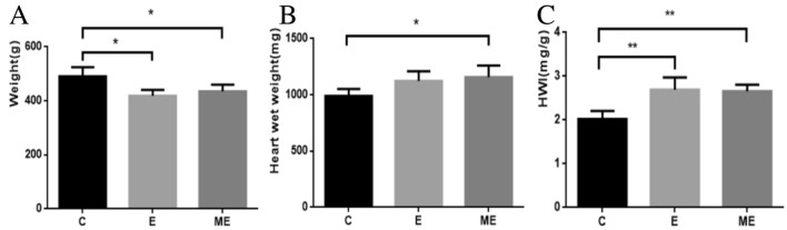
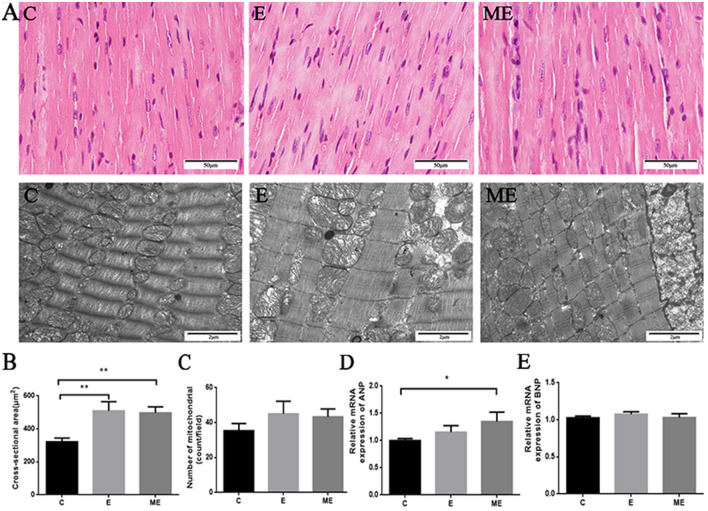
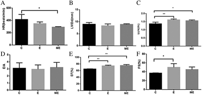
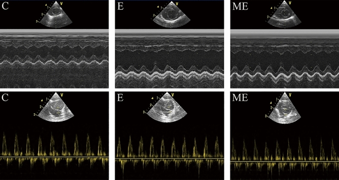
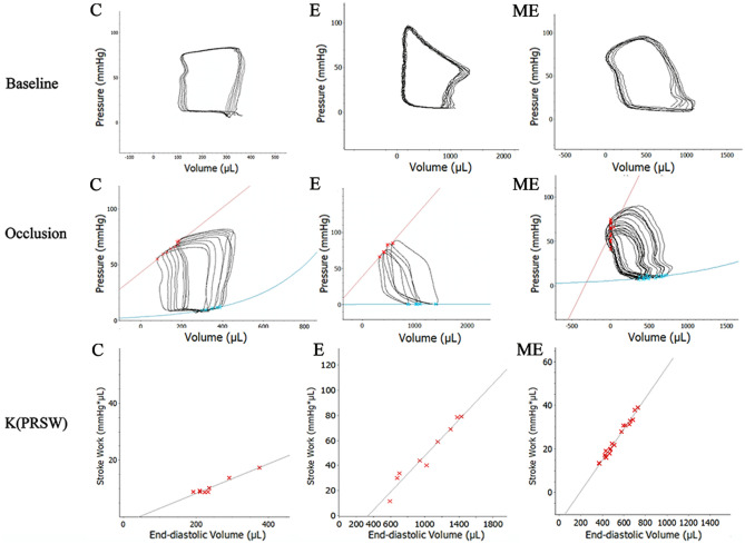
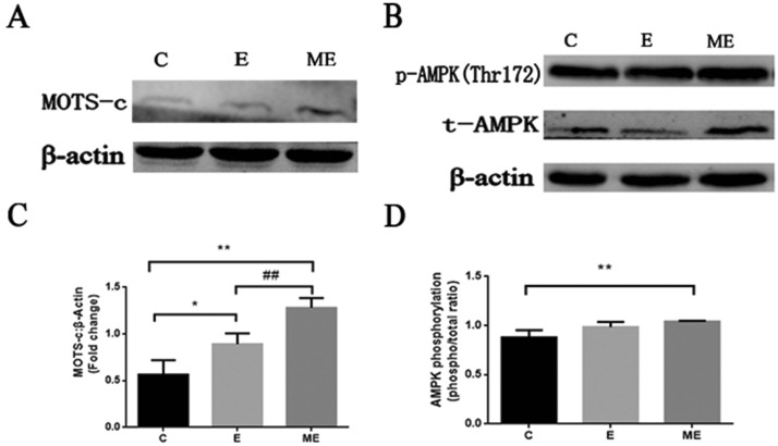
Cardiac remodeling is a physiological adaptation to aerobic exercise and which is characterized by increases in ventricular volume and the number of cardiomyocytes. The mitochondrial derived peptide MOTS-c functions as an important regulator in physical capacity and performance. Exercise elevates levels of endogenous MOTS-c in circulation and in myocardium, while MOTS-c can significantly enhance exercise capacity. However, the effects of aerobic exercise combined with MOTS-c on cardiac structure and function are unclear. We used pressure-volume conductance catheter technique to examine cardiac function in exercised rats with and without treatment with MOTS-c. Surprisingly, MOTS-c improved myocardial mechanical efficiency, enhanced cardiac systolic function, and had a tendency to improve the diastolic function. The findings suggest that using exercise supplements could be used to modulate the cardiovascular benefits of athletic training.


Conflict of interest statement

The authors declare no competing interests.

Figures

Figure 1
Figure 1
Body weight, heart wet weight and heart weight index (HWI). The changes in body weight (A) and heart wet weight (B) of rats in the control group, exercise group and MOTS-c combined exercise group were detected. Calculate the heart wet weight/body weight (HWI) ratio (C) to evaluate the growth response of the heart with or without MOTS-c injection during exercise. *p < 0.05, **p < 0.01, compared to group C.
Figure 2
Figure 2
The effects of MOTS-c and exercise training on rat myocardial structure and hypertrophic genes. (A) is the HE staining (× 400) and TEM (× 20,000) images of the myocardium, which respectively reflect the gross and ultrastructure of the myocardium. The arrow is the lysosome. (B) Quantification of cross-sectional area of cardiomyocytes from H&E-stained sections. (C) Quantitative analysis of the number of myocardial mitochondria in TEM. (D and E) The mRNA expression of ANP and BNP. *p < 0.05, **p < 0.01, compared to group C. C = Control, E = Exercise training, ME = Exercise training combined with MOTS-c treatment.
Figure 3
Figure 3
Changes of cardiac structure and function after MOTS-c. (A) The HR in group ME was lower than in group C. (B) There were no differences in LVIDd among these three groups; (C) The EDV in group E and ME was higher than in group C; (D)There were no differences in E/A among these three groups; (E) The EF in group E and ME was significantly higher than in group C; (F) The FS in group E was higher than in group C. *p < 0.05, **p < 0.01, compared to group C. #p < 0.05, compared to group E.
Figure 4
Figure 4
Changes of M-mode echocardiogram. The blood flow Doppler test records the ratio (E/A) of the peak blood flow velocity E in early diastole and the peak blood flow velocity A in late diastole.
Figure 5
Figure 5
Changes in cardiac hemodynamics after MOTS-c treatment during exercise training. Baseline P–V-loops and after blood flow in the inferior vena cava blood flow was obstructed. Stroke work and the slope of ESPVR (C) [Ees (end-systolic elastance)] in the E and ME were significantly higher than in group C. (C = Control, E = Exercise training, ME = Exercise training combined with MOTS-c treatment).
Figure 6
Figure 6
Changes in the protein expression of MOTS-c, p-AMPK, and t-AMPK in each group of rats. (A and B) are the representative images of MOTS-c, p-AMPK(Thr172), t-AMPK. (C) is the change of MOTS-c compared to β-Actin multiple. (D) is stoichiometric AMPK phosphorylation (phosphor/total ratio). *p < 0.05, **p < 0.01, compared to group C. #p < 0.05, ##p < 0.01, compared to group E. (C = Control, E = Exercise training, ME = Exercise training combined with MOTS-c treatment).

References

    1. Verdoorn KS, Matsuura C, Borges JP. Exercise for cardiac health and regeneration: Killing two birds with one stone. Ann. Transl. Med. 2017;5:S13. doi: 10.21037/atm.2017.03.10. - DOI - PMC - PubMed
    1. Sanchis-Gomar F, Fiuza-Luces C, Lucia A. Exercise as the master polypill of the 21st century for the prevention of cardiovascular disease. Int. J. Cardiol. 2015;181:360–361. doi: 10.1016/j.ijcard.2014.12.048. - DOI - PubMed
    1. Tao L, Bei Y, Zhang H, Xiao J, Li X. Exercise for the heart: Signaling pathways. Oncotarget. 2015;6:20773–20784. doi: 10.18632/oncotarget.4770. - DOI - PMC - PubMed
    1. Bernardo BC, Ooi JYY, Weeks KL, Patterson NL, McMullen JR. Understanding key mechanisms of exercise-induced cardiac protection to mitigate disease: Current knowledge and emerging concepts. Physiol. Rev. 2018;98:419–475. doi: 10.1152/physrev.00043.2016. - DOI - PubMed
    1. Schüttler, D., Clauss, S., Weckbach, L. T. & Brunner, S. Molecular mechanisms of cardiac remodeling and regeneration in physical exercise. Cells8, (2019). - PMC - PubMed

Publication types