. 2022 May;530(7):1099-1111.
doi: 10.1002/cne.25264. Epub 2021 Oct 24.

Ultrastructure of ipsilateral and contralateral tectopulvinar projections in the mouse

Affiliations

Ultrastructure of ipsilateral and contralateral tectopulvinar projections in the mouse

Nazratan Naeem et al. J Comp Neurol. 2022 May.

Abstract

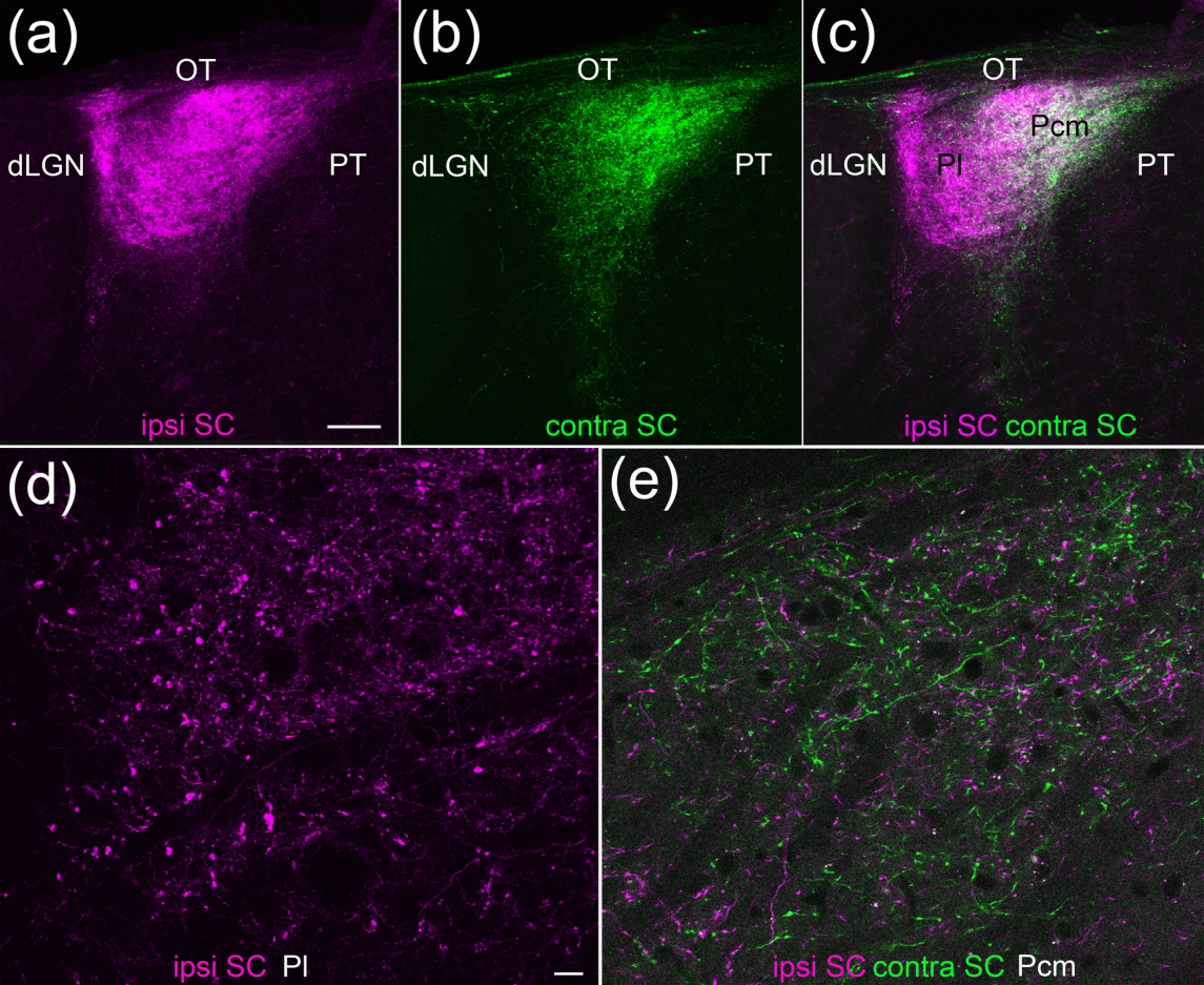
Visual pathways of the brain are organized into parallel channels that code different features of the external environment. In the current study, we investigated the anatomical organization of parallel pathways from the superior colliculus (SC) to the pulvinar nucleus in the mouse. Virus injections placed in the ipsilateral and contralateral SC to induce the expression of different fluorescent proteins define two pulvinar zones. The lateral pulvinar (Pl) receives ipsilateral SC input and the caudal medial pulvinar (Pcm) receives bilateral SC input. To examine the ultrastructure of these projections using transmission electron microscopy, we injected the SC with viruses to induce peroxidase expression within synaptic vesicles or mitochondria. We quantitatively compared the sizes of ipsilateral and contralateral tectopulvinar terminals and their postsynaptic dendrites, as well as the sizes of the overall population of synaptic terminals and their postsynaptic dendrites in the Pl and Pcm. Our ultrastructural analysis revealed that ipsilateral tectopulvinar terminals are significantly larger than contralateral tectopulvinar terminals. In particular, the ipsilateral tectopulvinar projection includes a subset of large terminals (≥ 1 μm2 ) that envelop dendritic protrusions of postsynaptic dendrites. We also found that both ipsilateral and contralateral tectopulvinar terminals are significantly larger than the overall population of synaptic terminals in both the Pl and Pcm. Thus, the ipsilateral tectopulvinar projection is structurally distinct from the bilateral tectopulvinar pathway, but both tectopulvinar channels may be considered the primary or "driving" input to the Pl and Pcm.

Keywords: electron microscopy; pulvinar; superior colliculus; synapse; vesicle; widefield vertical.


Conflict of interest statement

Conflict of interest: The authors declare no competing financial interests.

Figures

Figure 1:
Figure 1:. Identification of virus-labeled and unlabeled synaptic profiles.
Montage images were collected to measure the size of all unlabeled synaptic terminals and postsynaptic dendrites in the Pl and Pcm. a) Low magnification montage of a coronal section through the pulvinar nucleus after collecting 4 high magnification montages in the Pl and 4 high magnification montages in the Pcm. For each high magnification montage, the electron beam caused the tissue to lighten relative to areas that were not montaged at high magnification. b) Pl montage #1. Examples of terminals labeled by a SV-HRP injection in the SC are indicated by black arrows. Examples of mitochondria labeled by a matrix-dAPEX2 virus injection in V1 are indicated with white arrows. c) Example of a labeled mitochondria (white asterisk) in a synaptic terminal resulting from the V1 injection of the matrix-dAPEX2 virus. Below is a synaptic terminal that does not contain mitochondria. The lumens of the vesicles in both terminals are clear. Black arrows indicate synapses. d) Pcm montage #1. Examples of terminals labeled by a SV-HRP injection in the SC are indicated by black arrows. Few labeled mitochondria resulting from a matrix-dAPEX2 virus injection in V1 are present in the Pcm. e) Examples of small unlabeled synaptic terminals that do not contain mitochondria. The lumens of the vesicles in both terminals are clear. Black arrows indicate synapses. f) Example of a large unlabeled terminal that contains unlabeled mitochondria (black asterisks) and vesicles with clear lumens. Black arrow indicates a synapse. g) Example of a synaptic terminal labeled with the SV-HRP virus. Note that the lumens of many of the synaptic vesicles contain a dark reaction product, while the mitochondria are unlabeled (black asterisks). Black arrow indicates a synapse. Scale bar in a = 30 μm. Scale bar in b = 5 μm and also applies to d. Scale bars in c,e,f and g = 400nm. OT, optic tract
Figure 2:
Figure 2:. Ipsilateral and contralateral projections from the superior colliculus (SC) define two zones of the mouse pulvinar nucleus.
Two different virus injections were placed in the left and right SC and confocal images of coronal sections of the pulvinar nucleus were collected. (a-c) The lateral pulvinar (Pl) is primarily innervated by ipsilateral SC terminals (a,c magenta), while the caudal medial pulvinar (Pcm) is innervated by both ipsilateral and contralateral (b,c green) SC terminals (5 μm optical images, 60 μm stack). Scale bar = 100 μm. d,e) High magnification images of virus-labeled tectopulvinar terminals in the Pl (d) and Pcm (e). 1 μm optical images, 6 μm stacks. Scale bar = 10 μm and applies to d and e. dLGN, dorsal lateral geniculate nucleus, OT, optic tract, PT, pretectum.
Figure 3:
Figure 3:. Ultrastructure of ipsilateral tectopulvinar terminals.
(a-i) Terminals in the pulvinar nucleus (Pl and Pcm) labeled by a virus injection in the ipsilateral SC were identified by a dark reaction product in a subset of their synaptic vesicles. Labeled terminals are indicated by magenta overlay, black arrows indicate synapses, and postsynaptic dendrites and dendritic inclusions are indicated by yellow overlay. The labeled tectopulvinar terminals varied in size. The largest labeled terminals contained dendritic protrusions (a-d). e) The asterisk indicates a corticothalamic terminal labeled by a V1 virus injection. Scale bar in a = 400 nm and also applies to f. Scale bar in b = 600nm and also applies to c,d, and g-i.
Figure 4:
Figure 4:. Ultrastructure of contralateral tectopulvinar terminals.
(a-h) Terminals in the pulvinar nucleus (Pcm) labeled by a virus injection in the contralateral SC were identified by a dark reaction product in a subset of their synaptic vesicles. Labeled terminals are indicated by green overlay, black arrows indicate synapses, and postsynaptic dendrites are indicated by blue overlay. Scale bar = 600 nm and applies to all panels.
Figure 5:
Figure 5:. Comparison of the size of ipsilateral and contralateral tectopulvinar terminals and their postsynaptic targets.
Within animal comparison of ipsilateral and contralateral projections from SC to PUL labeled with the SV-HRP virus. (a,b) Quantitative analysis of the size of presynaptic terminal and postsynaptic dendrite sizes. (c,d) Quantitative analysis of the sizes of ipsilateral and contralateral presynaptic terminals, comparing those with and without dendritic protrusions. (e,f) Quantitative analysis of the sizes of ipsilateral and contralateral presynaptic terminals, comparing those with and without mitochondria. Unpaired non-parametric - t test (Mann-Whitney U test) was performed for statistical analysis. ** p ≤ 0.005; *** p ≤ 0.0005; ns = non-significant. Black bars indicate mean and standard deviation.
Figure 6:
Figure 6:. Comparison tectopulvinar synaptic terminals to the overall population of synaptic terminals in the Pl and Pcm.
Within animal comparison of projections from SC to PUL labeled with the SV-HRP virus with surrounding terminals in the PUL not labeled with the virus. (a,b,c) Quantitative analysis of the size of presynaptic terminal sizes, comparing unlabeled terminals with SC to PUL terminals (a), unlabeled terminals in the Pcm with contralateral SC to PUL terminals (b), and unlabeled terminals in the Pl with ipsilateral SC to PUL terminals (c). (d,e,f) Quantitative analysis of the size of postsynaptic dendrite sizes, comparing dendrites contacted by unlabeled terminals in PUL with dendrites contacted by SC to PUL terminals (d), dendrites contacted by unlabeled terminals in the Pcm with dendrites contacted by contralateral SC to PUL terminals (e), and dendrites contacted by unlabeled terminals in the Pl with dendrites contacted by ipsilateral SC to PUL terminals (f). A Mann-Whitney U test was utilized in for the comparison in panels (a,d), and a Kruskal-Wallis test was utilized for comparisons in panels (b,c,e,f). *** p< 0.0001. Black bars indicate mean and standard deviation.

References

    1. Abramson BP, & Chalupa LM (1988). Multiple pathways from the superior colliculus to the extrageniculate visual thalamus of the cat. The Journal of Comparative Neurology, 271(3). 10.1002/cne.902710308 - DOI - PubMed
    1. Baldauf ZB, Chomsung RD, Carden WB, May PJ, & Bickford ME (2005). Ultrastructural analysis of projections to the pulvinar nucleus of the cat. I: Middle suprasylvian gyrus (areas 5 and 7). The Journal of Comparative Neurology, 485(2). 10.1002/cne.20480 - DOI - PMC - PubMed
    1. Baldwin MKL, Balaram P, & Kaas JH (2013). Projections of the superior colliculus to the pulvinar in prosimian galagos ( Otolemur garnettii ) and VGLUT2 staining of the visual pulvinar. Journal of Comparative Neurology, 521(7). 10.1002/cne.23252 - DOI - PMC - PubMed
    1. Baldwin MKL, Wong P, Reed JL, & Kaas JH (2011). Superior colliculus connections with visual thalamus in gray squirrels (Sciurus carolinensis): Evidence for four subdivisions within the pulvinar complex. The Journal of Comparative Neurology, 519(6). 10.1002/cne.22552 - DOI - PMC - PubMed
    1. Basso MA, Bickford ME, & Cang J (2021). Unraveling circuits of visual perception and cognition through the superior colliculus. Neuron, 109(6). 10.1016/j.neuron.2021.01.013 - DOI - PMC - PubMed

Publication types