. 2021 Nov 16;144(20):1629-1645.
doi: 10.1161/CIRCULATIONAHA.121.054182. Epub 2021 Oct 12.

Palmdelphin Regulates Nuclear Resilience to Mechanical Stress in the Endothelium

Affiliations

Palmdelphin Regulates Nuclear Resilience to Mechanical Stress in the Endothelium

Miguel Sáinz-Jaspeado et al. Circulation. .

Abstract

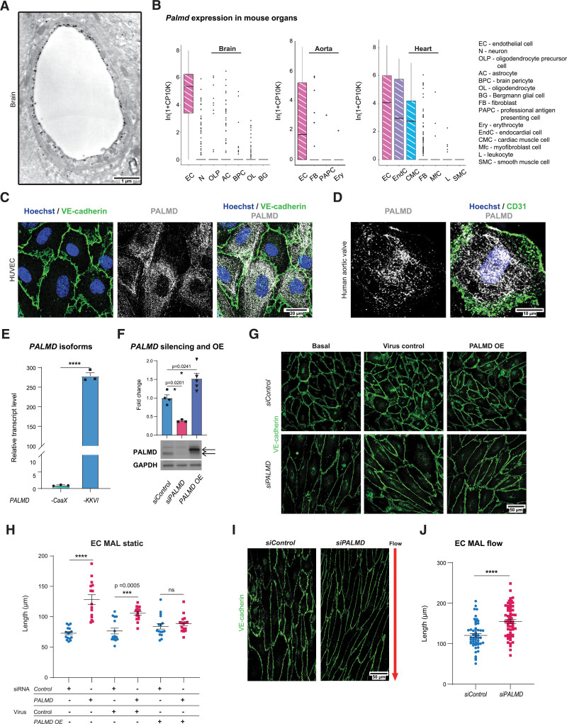
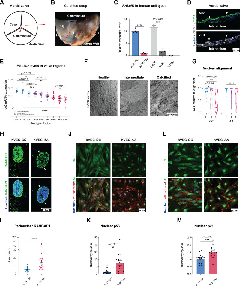
Background: PALMD (palmdelphin) belongs to the family of paralemmin proteins implicated in cytoskeletal regulation. Single nucleotide polymorphisms in the PALMD locus that result in reduced expression are strong risk factors for development of calcific aortic valve stenosis and predict severity of the disease.

Methods: Immunodetection and public database screening showed dominant expression of PALMD in endothelial cells (ECs) in brain and cardiovascular tissues including aortic valves. Mass spectrometry, coimmunoprecipitation, and immunofluorescent staining allowed identification of PALMD partners. The consequence of loss of PALMD expression was assessed in small interferring RNA-treated EC cultures, knockout mice, and human valve samples. RNA sequencing of ECs and transcript arrays on valve samples from an aortic valve study cohort including patients with the single nucleotide polymorphism rs7543130 informed about gene regulatory changes.

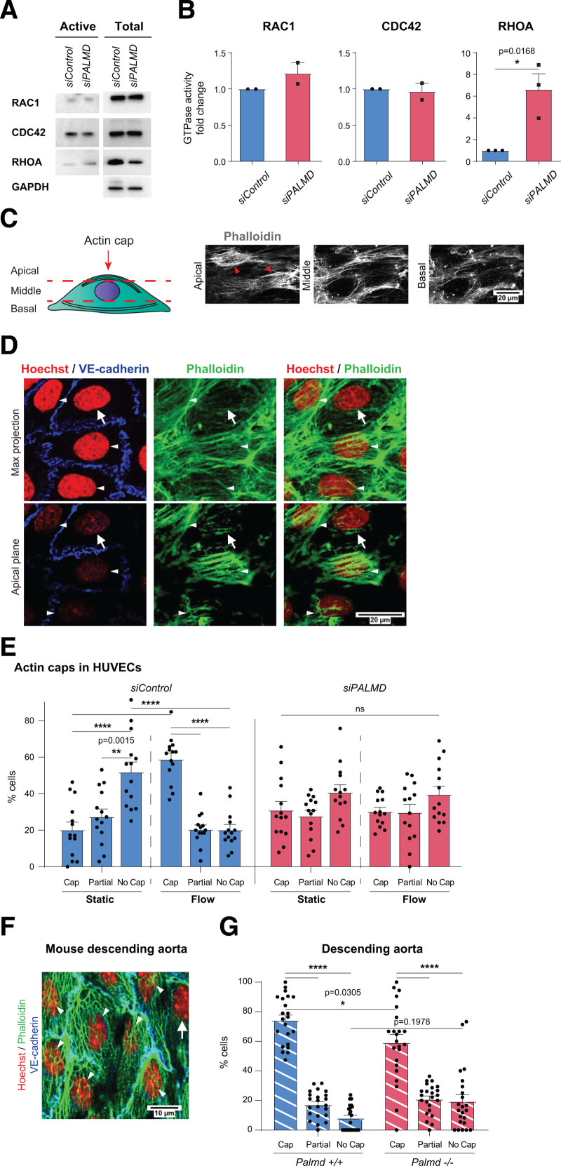
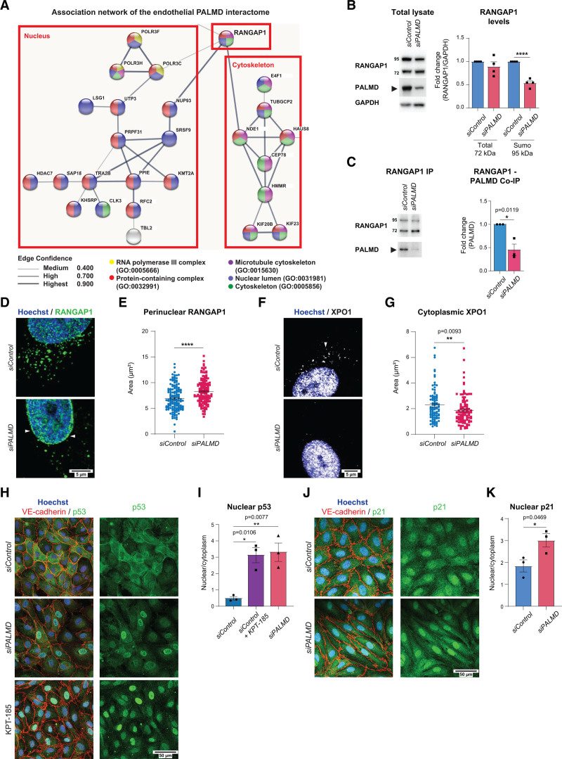
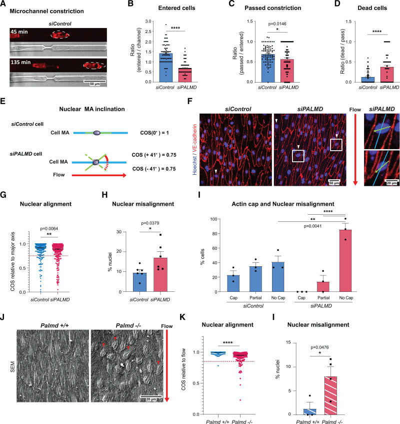
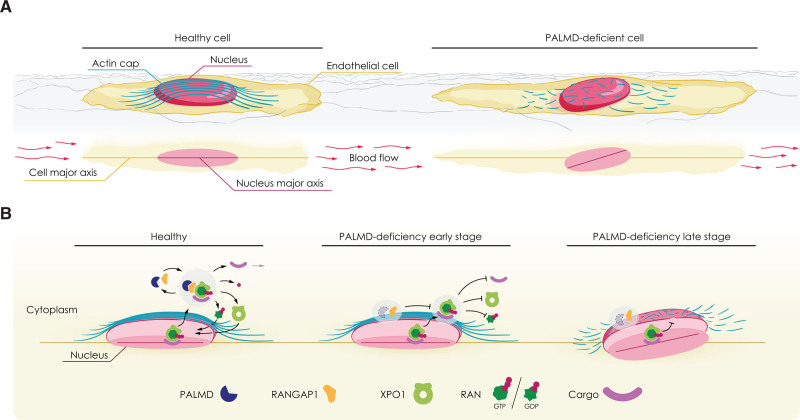
Results: ECs express the cytosolic PALMD-KKVI splice variant, which associated with RANGAP1 (RAN GTP hydrolyase activating protein 1). RANGAP1 regulates the activity of the GTPase RAN and thereby nucleocytoplasmic shuttling via XPO1 (Exportin1). Reduced PALMD expression resulted in subcellular relocalization of RANGAP1 and XPO1, and nuclear arrest of the XPO1 cargoes p53 and p21. This indicates an important role for PALMD in nucleocytoplasmic transport and consequently in gene regulation because of the effect on localization of transcriptional regulators. Changes in EC responsiveness on loss of PALMD expression included failure to form a perinuclear actin cap when exposed to flow, indicating lack of protection against mechanical stress. Loss of the actin cap correlated with misalignment of the nuclear long axis relative to the cell body, observed in PALMD-deficient ECs, Palmd-/- mouse aorta, and human aortic valve samples derived from patients with calcific aortic valve stenosis. In agreement with these changes in EC behavior, gene ontology analysis showed enrichment of nuclear- and cytoskeleton-related terms in PALMD-silenced ECs.

Conclusions: We identify RANGAP1 as a PALMD partner in ECs. Disrupting the PALMD/RANGAP1 complex alters the subcellular localization of RANGAP1 and XPO1, and leads to nuclear arrest of the XPO1 cargoes p53 and p21, accompanied by gene regulatory changes and loss of actin-dependent nuclear resilience. Combined, these consequences of reduced PALMD expression provide a mechanistic underpinning for PALMD's contribution to calcific aortic valve stenosis pathology.

Keywords: aortic valve stenosis; endothelial cells; nucleocytoplasmic transport; palmdelphin.


Figures

Figure 1.
Figure 1.
PALMD is expressed in endothelial cells and affects cell shape. A, Palmd immuno-electron microscopy of a mouse cerebellum capillary. B, Palmd mRNA expression in mouse brain, aorta, and heart from Tabula Muris; values normalized and log-transformed (In[1+CP10K]) as described. Box plot showing 25th to 75th percentiles of expression as boxes and dots representing outliers. Cell type abbreviations and full designations are given. C, HUVECs, immunostaining for VE-cadherin (green) and PALMD (gray). Hoechst 33342 shows nuclei. D, Human aortic valve en face immunostaining for CD31 (green) and PALMD (gray). Hoechst 33342 shows nuclei. E, HUVECs, qPCR of PALMD mRNA splice variants. n=3. ****P<0.0001, unpaired t test. F, PALMD immunoblot shows PALMD silencing and PALMD overexpression (OE) in HUVECs. Quantification of PALMD levels. n=3. *P=0.0201; *P=0.0241, 1-way ANOVA with Tukey correction. Black arrows in the blot indicate Myc-DDK–tagged PALMD protein (upper) and endogenous PALMD (lower), respectively. GAPDH was used for protein normalization. G, HUVECs siControl and siPALMD transduced with control or PALMD lentiviral vectors as indicated. VE-cadherin (green). OE, PALMD overexpression. H, Quantification of major axis length (MAL) in G. n=3, 15 to 16 fields of view (FoV)/condition. ***P=0.0005; ****P<0.0001. Two-way ANOVA with Tukey correction. I, HUVECs exposed to 10 dyn laminar flow, 48 hours. Arrow, flow direction. VE-cadherin (green). J, Quantification of MAL in I. n=3, 55 to 60 FoVs/condition. ****P<0.0001, Mann-Whitney. Mean±SEM shown in all graphs.
Figure 2.
Figure 2.
PALMD regulates perinuclear actin cap formation. A and B, RAC1, CDC42, and RHOA activation assays in siControl, siPALMD HUVECs (left, A). Total lysates with GAPDH for equal loading (right, A). GTPase activities (B). n=3 (RHOA) and 2 (RAC1, CDC42). *P=0.0168, unpaired t test. C, Schematic showing focal points (apical, middle, and basal) defining the actin cap (arrowheads) at the apical point in phalloidin-stained HUVECs. D, Detection of VE-cadherin (blue), phalloidin (green), and Hoechst 33342 (red) in siControl, siPALMD HUVECs with actin caps indicated (white arrowheads) and the lack of actin cap (arrow) by maximum (max) projection (upper) and at the apical plane (lower). E, siControl, siPALMD HUVECs in static and laminar flow. Actin cap status: cap, partial, or no cap. n=3 experiment, 14 fields of view (FoV)/condition. **P=0.0015, ****P<0.0001. Two-way ANOVA with Tukey correction. F, Mouse Palmd+/+ aorta en face, VE-cadherin (blue), Hoechst 33342 (red), and phalloidin (green). Actin cap (arrowheads), without cap (arrow). G, Quantification of actin cap status in Palmd+/+ and Palmd−/− aorta. n=6 aortas and 22 to 23 FoV/genotype. *P=0.0305; ****P<0.0001, 2-way ANOVA with Tukey correction. Mean±SEM shown in all graphs.
Figure 3.
Figure 3.
PALMD interacts with RANGAP1 and affects XPO1-dependent nucleocytoplasmic transport. A, Association network built on the endothelial PALMD interactome. Colors indicate GO terms that include the indicated proteins. Line thickness indicates the confidence score of the known associations. B, RANGAP1 and PALMD immunoblotting on siControl and siPALMD total lysates. Quantification (right) of RANGAP1 (total, 72 kDa and sumoylated, 95 kDa) levels normalized to GAPDH. n=4. ****P<0.0001; unpaired t test. C, RANGAP1 IP and immunoblotting for RANGAP1 and PALMD on from siControl and siPALMD HUVECs shows RANGAP1 (PALMD coimmunoprecipitation; Co-IP). Right, Level of PALMD Co-IP set to 1 in siControl. n=3. *P=0.0119, unpaired t test. D and E, Immunostaining showing perinuclear RANGAP1 (arrowheads), in siControl and siPALMD HUVECs. Quantification in E. n=3, 146 nuclei/condition. ****P<0.0001, Mann-Whitney. F and G, Immunostaining showing cytoplasmic XPO1 (arrowhead) in siControl and siPALMD HUVECs. Quantification in G. n=3 expt, 90 to 100 nuclei/condition. **P=0.0093, Mann-Whitney. H and I, Immunostaining for p53 (green), VE-cadherin (red) in siControl and siPALMD HUVECs treated or not with XPO1 inhibitor KPT-185; Hoechst33342 shows nuclei (H). Quantification of nuclear to cytoplasmic ratio in the presence and absence of PALMD (I). n=3, *P=0.0106, **P=0.0077, 1-way ANOVA with Tukey correction. J and K, Immunostaining for p21 (green) and VE-cadherin (red) in siControl and siPALMD HUVECs; Hoechst33342 shows nuclei (J). Quantification of nuclear to cytoplasmic ratio in the presence and absence of PALMD (K). n=3, *P=0.0469, unpaired t test. Mean±SEM shown in all graphs.
Figure 4.
Figure 4.
PALMD confers resilience to mechanical stress. A, Microchannel restriction point migration. Nuclei, NucRedTM live 647 (outlined) at time points after initiation. B and C, siControl, siPALMD HUVECs entering channel (B), or passing constriction (C). n=4, 74 to 80 channels/100 to 200 cells/expt. ****P<0.0001; *P=0.0146; unpaired t test. D, Dead/passing cells. n=4, 66 to 79 cells/expt. ****P<0.0001; unpaired t test. E, Schematic showing cell body and nuclear major axis (MA) with proper alignment (top). Angle indicating nuclear MA misalignment (below) in cosine (COS). Laminar flow direction indicated by red arrow. F, siControl, siPALMD HUVECs in laminar flow; red arrow indicates flow direction. Arrowheads indicate markedly misaligned nuclei. Boxed regions magnified to the right, showing cell body major axis (blue) and nuclear major axis (green) relative to flow. G, Nuclear alignment shown with COS as outlined in E. Dotted line defines cutoff for nuclear misalignment; COS=0.75. n=3, 416 to 420 nuclei analyzed in 2 fields of view (FoV)/expt. **P=0.0064, Mann-Whitney. H, siControl, siPALMD HUVECs, percent misaligned nuclei with COS≤0.75 as shown in G. n=3, 2 FoV/expt. *P=0.0379; unpaired t test. I, siControl, siPALMD HUVECs with actin cap, partial or no cap in cells with misaligned nuclei; COS≤0.85. n=3. **P=0.0041; ****P<0.0001, 2-way ANOVA with Tukey correction. J, Mouse Palmd+/+ and −/− aorta en face scanning electron microscopy. Red arrow indicates blood flow direction, and arrowheads indicate markedly misaligned nuclei relative to flow. K, Nuclear alignment shown with COS as outlined in E. Dotted line, COS=0.85. n=3 to 4 mice, 240 to 279 nuclei. ****P<0.0001, Mann-Whitney. L, Aorta, percent misaligned nuclei with COS≤0.85 as shown in K. n=3 to 4 mice/genotype. *P=0.0476, unpaired t test. Mean±SEM shown in all graphs.
Figure 5.
Figure 5.
PALMD-deficiency effects on EC in CAVS. A, Schematic of the aortic tricuspid CAVS valve regions: H, healthy; I, intermediate; C, calcified. B, Image of CAVS valve showing macroscopically H, I, and C regions. C, PALMD expression in siControl and siPALMD HUVECs (static condition), human valve endothelial cells (hVECs), human valve interstitial cells (hVICs), and human smooth muscle cells (hSMCs). n=2 (hSMCs), 3 (hVECs, HVICs), 4 (HUVECs). ***P=0.0005; ****P<0.0001; unpaired t test. D, CAVS valve transversal section, CD31 (green), PALMD (gray), Hoechst 33342 (blue), nuclei. PALMD in VECs (arrowheads). E, PALMD expression in 58 patients with CAVS grouped according to genotype; negative for the allele (CC), heterozygotes (CA), and homozygotes for the PALMD SNP rs7543130 (AA), analyzed in H, I, and C regions. n = 9 (CC), 27 (CA), and 22 (AA). *P=0.0435; *P=0.0177; **P=0.0015; **P=0.0014; ***P=0.0004; ****P<0.0001, 2-way ANOVA with Tukey correction. F, CAVS samples of CC and AA (SNP rs7543130), en face scanning electron microscopy. G, Nuclear MA alignment relative to flow direction, with cosine (COS) in CC and AA genotypes. n=50 (H)/170 (I)/30 (C) nuclei. *P=0.0184; **P=0.0038; ****P<0.0001, 1-way ANOVA with Tukey correction. H and I, hVECs of CC and AA genotype showing perinuclear RANGAP1. n=CC (29 nuclei), AA (52 nuclei). ****P<0.0001, Mann-Whitney. J and K, Immunostaining for p53 (green), VE-cadherin (red) in hVECs with CC and AA genotypes. Hoechst33342 (blue) shows nuclei (J). Quantification (K) shows p53 nuclear to cytoplasmic ratio. n=3, 15 fields of view (FoV).**P=0.0013, unpaired t test. L and M, Immunostaining for p21 (green), VE-cadherin (red) in patient-derived VECs with CC and AA genotypes. Hoechst33342 (blue) shows nuclei (L). Quantification (M) shows p21 nuclear to cytoplasmic ratio. n=3, 15 FoV. ***P=0.0010, unpaired t test. Mean±SEM shown in all graphs.
Figure 6.
Figure 6.
PALMD regulates nucleocytoplasmic shuttling via RANGAP1; short- and long-term consequences of PALMD deficiency. Schematics outlining effects of PALMD deficiency. A, Flow-exposed healthy cell (left) with actin cap protecting the nucleus and alignment of cell body and nuclear major axes. In PALMD-deficiency (right), the actin cap fails to form, and the nucleus becomes misaligned. B, In the healthy nucleus, the XPO1-cargo complex binds RAN-GTP for export to the cytoplasm, where PALMD/RANGAP1 catalyzes RAN’s GTPase activity (left). XPO1 releases its cargo followed by recycling of XPO1 and RAN-GDP back to the nucleus. In early-stage PALMD deficiency (middle), RANGAP1 assumes a perinuclear localization, RAN remains GTP-bound, and XPO1 retains its cargo. In late-stage PALMD deficiency (right), disturbed nucleocytoplasmic shuttling results in loss of actin cap, reduced nuclear resilience, and nuclear misalignment.

References

    1. Hultqvist G, Ocampo Daza D, Larhammar D, Kilimann MW. Evolution of the vertebrate paralemmin gene family: ancient origin of gene duplicates suggests distinct functions. PLoS One. 2012; 7:e41850. doi: 10.1371/journal.pone.0041850 - PMC - PubMed
    1. Hu B, Petrasch-Parwez E, Laue MM, Kilimann MW. Molecular characterization and immunohistochemical localization of palmdelphin, a cytosolic isoform of the paralemmin protein family implicated in membrane dynamics. Eur J Cell Biol. 2005; 84:853–866. doi: 10.1016/j.ejcb.2005.07.002 - PubMed
    1. Kalebic N, Gilardi C, Stepien B, Wilsch-Bräuninger M, Long KR, Namba T, Florio M, Langen B, Lombardot B, Shevchenko A, et al. . Neocortical expansion due to increased proliferation of basal progenitors is linked to changes in their morphology. Cell Stem Cell. 2019; 24:535–550.e9. doi: 10.1016/j.stem.2019.02.017 - PubMed
    1. Nie Y, Chen H, Guo C, Yuan Z, Zhou X, Zhang Y, Zhang X, Mo D, Chen Y. Palmdelphin promotes myoblast differentiation and muscle regeneration. Sci Rep. 2017; 7:41608. doi: 10.1038/srep41608 - PMC - PubMed
    1. Dashzeveg N, Taira N, Lu ZG, Kimura J, Yoshida K. Palmdelphin, a novel target of p53 with Ser46 phosphorylation, controls cell death in response to DNA damage. Cell Death Dis. 2014; 5:e1221. doi: 10.1038/cddis.2014.176 - PMC - PubMed

Publication types

MeSH terms