. 2021 Oct 12;19(1):154.
doi: 10.1186/s43141-021-00238-8.

Role of "dual-personality" fragments in HEV adaptation-analysis of Y-domain region

Affiliations

Role of "dual-personality" fragments in HEV adaptation-analysis of Y-domain region

Zoya Shafat et al. J Genet Eng Biotechnol. .

Abstract

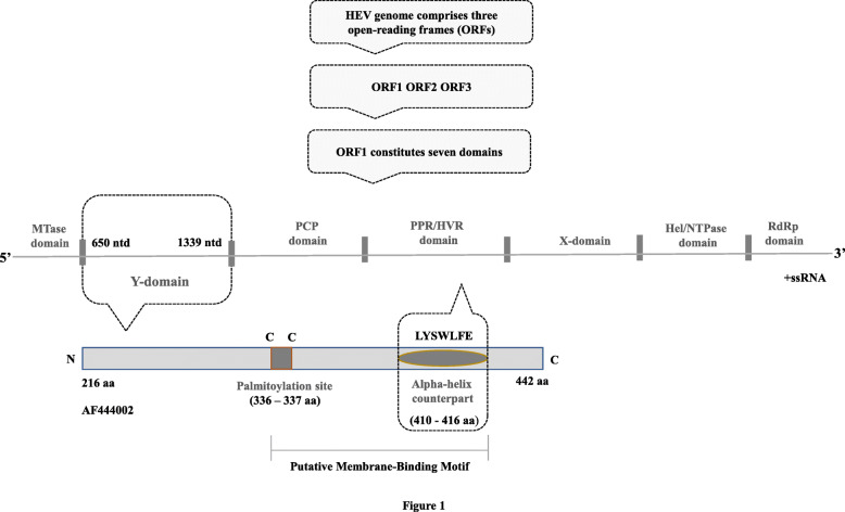
Background: Hepatitis E is a liver disease caused by the pathogen hepatitis E virus (HEV). The largest polyprotein open reading frame 1 (ORF1) contains a nonstructural Y-domain region (YDR) whose activity in HEV adaptation remains uncharted. The specific role of disordered regions in several nonstructural proteins has been demonstrated to participate in the multiplication and multiple regulatory functions of the viruses. Thus, intrinsic disorder of YDR including its structural and functional annotation was comprehensively studied by exploiting computational methodologies to delineate its role in viral adaptation.

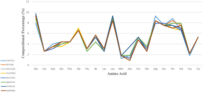
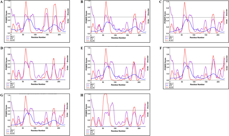
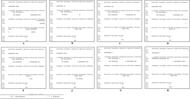
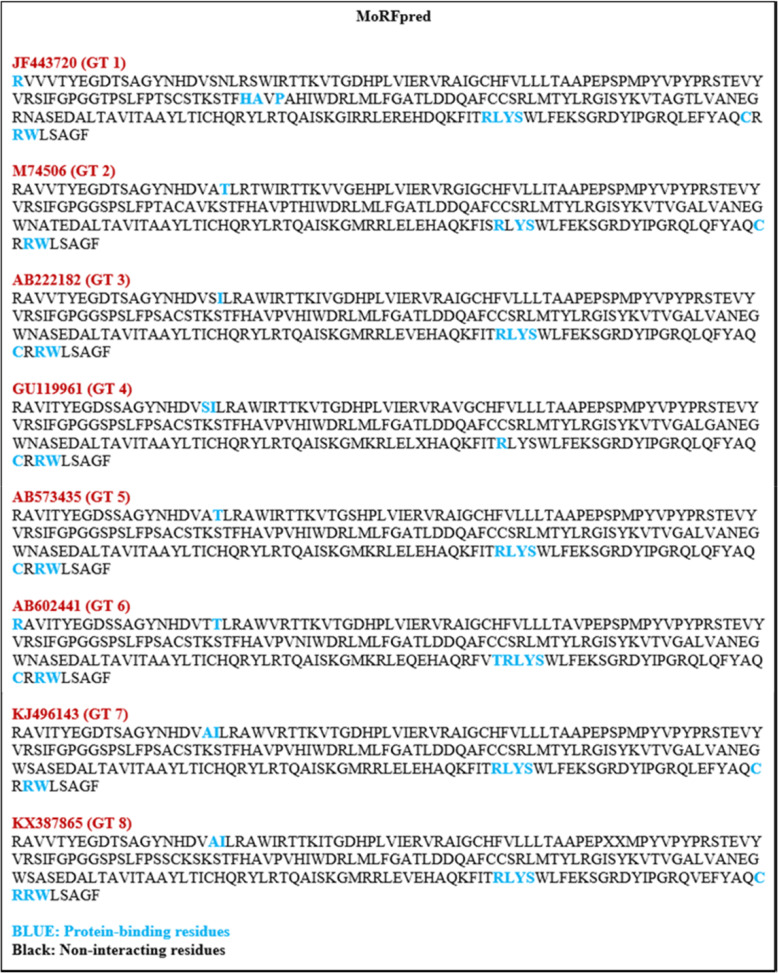
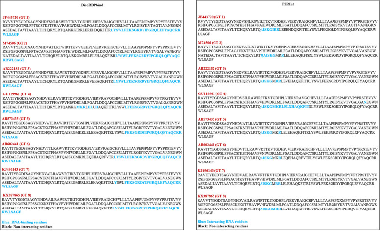
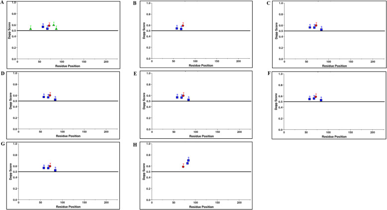
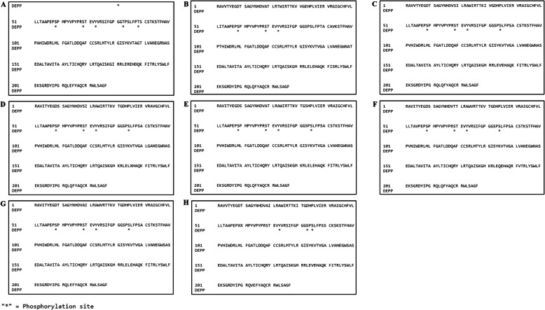
Results: Based on our findings, it was evident that YDR contains significantly higher levels of ordered regions with less prevalence of disordered residues. Sequence-based analysis of YDR revealed it as a "dual personality" (DP) protein due to the presence of both structured and unstructured (intrinsically disordered) regions. The evolution of YDR was shaped by pressures that lead towards predominance of both disordered and regularly folded amino acids (Ala, Arg, Gly, Ile, Leu, Phe, Pro, Ser, Tyr, Val). Additionally, the predominance of characteristic DP residues (Thr, Arg, Gly, and Pro) further showed the order as well as disorder characteristic possessed by YDR. The intrinsic disorder propensity analysis of YDR revealed it as a moderately disordered protein. All the YDR sequences consisted of molecular recognition features (MoRFs), i.e., intrinsic disorder-based protein-protein interaction (PPI) sites, in addition to several nucleotide-binding sites. Thus, the presence of molecular recognition (PPI, RNA binding, and DNA binding) signifies the YDR's interaction with specific partners, host membranes leading to further viral infection. The presence of various disordered-based phosphorylation sites further signifies the role of YDR in various biological processes. Furthermore, functional annotation of YDR revealed it as a multifunctional-associated protein, due to its susceptibility in binding to a wide range of ligands and involvement in various catalytic activities.

Conclusions: As DP are targets for regulation, thus, YDR contributes to cellular signaling processes through PPIs. As YDR is incompletely understood, therefore, our data on disorder-based function could help in better understanding its associated functions. Collectively, our novel data from this comprehensive investigation is the first attempt to delineate YDR role in the regulation and pathogenesis of HEV.

Keywords: Molecular function; Nucleotide-binding propensity; Phosphorylation; Protein disorder; Protein structure; Protein-binding propensity; Y-domain region (YDR).


Conflict of interest statement

The authors declare that they have no competing interests.

Figures

Fig. 1
Fig. 1
Diagrammatic representation of hepatitis E virus nonstructural polyprotein (ORF1) domain, showing the Y-domain. The ORF1 constitutes seven domains, i.e., MTase, methyltransferase; Y, undefined; PCP, papain-like cysteine protease; P/HVR, proline-rich/hypervariable region; X, Macro; Hel/NTPase, helicase/nucleotide triphosphatase; and RdRp, RNA-dependent RNA polymerase. The Y-domain region (YDR) is of 228 amino acids in length (650–1339 nucleotides) and consists of a potential palmitoylation site (C336C337) and an alpha-helix segment (L410Y411S412W413L414F415E416). These segments are found to be indispensable for cytoplasmic membrane binding and are highly conserved within HEV genotypes
Fig. 2
Fig. 2
Depiction of amino acid percentage composition in the YDR sequences considered for the study: (A) JF443720 (GT 1), (B) M74506 (GT 2), (C) AB222182 (GT 3), (D) GU119961 (GT 4), (E) AB573435 (GT 5), (F) AB602441 (GT 6), KJ496143 (GT 7), and (H) KX387865 (GT 8)
Fig. 3
Fig. 3
Analysis of intrinsic disorder predisposition of HEV YDR. (A) JF443720 (GT 1); (B) M74506 (GT 2); (C) AB222182 (GT 3); (D) GU119961 (GT 4); (E) AB573435 (GT 5); (F) AB602441 (GT 6); KJ496143 (GT 7); and (H) KX387865 (GT 8). Graphs AH represent the intrinsic disorder profiles of YDR sequences of HEV. Disorder probability was calculated using three members of the family PONDR (Prediction of Natural Disordered Regions), i.e., VLXT, VL3, and VSL2. A threshold value of 0.5 was set to distinguish between ordered and disordered regions along the genome (dashed line). Regions above the threshold are predicted to be disordered
Fig. 4
Fig. 4
Prediction of disordered residues in HEV YDR. A JF443720 (GT 1); B M74506 (GT 2); C AB222182 (GT 3); D GU119961 (GT 4); E AB573435 (GT 5); F AB602441 (GT 6); KJ496143 (GT 7); and H KX387865 (GT 8). The prediction of disordered residues was carried out using three members of the family PONDR (Prediction of Natural Disordered Regions), i.e., VLXT, VL3, and VSL2. A threshold value of 0.5 was set to distinguish between ordered and disordered regions along the genome (dashed line). Regions above the threshold are predicted to be disordered. The predicted disordered residues are shown with the alphabet “D”
Fig. 5
Fig. 5
Analysis of protein-binding propensity of HEV YDR, i.e., JF443720 (GT 1), M74506 (GT 2), AB222182 (GT 3), GU119961 (GT 4), AB573435 (GT 5), AB602441 (GT 6), KJ496143 (GT 7), and KX387865 (GT 8). The resulting protein-binding profile was calculated using MoRFpred. YDR mainly contains MoRFs at C-terminals. The protein-binding residues are depicted in blue while the non-interacting residues are depicted in black
Fig. 6
Fig. 6
A Analysis of RNA-binding propensity of HEV YDR, i.e., JF443720 (GT 1), M74506 (GT 2), AB222182 (GT 3), GU119961 (GT 4), AB573435 (GT 5), AB602441 (GT 6), KJ496143 (GT 7), and KX387865 (GT 8). The resulting RNA-binding profile was calculated using webservers (A) DisoRDPbind and (B) PPRInt. The RNA-binding residues were situated at the C-terminus of the YDR. The identified RNA-binding residues are depicted in red while the non-interacting residues are depicted in black
Fig. 7
Fig. 7
Analysis of DNA-binding propensity of HEV YDR, i.e., JF443720 (GT 1), M74506 (GT 2), AB222182 (GT 3), GU119961 (GT 4), AB573435 (GT 5), AB602441 (GT 6), KJ496143 (GT 7), and KX387865 (GT 8). The resulting DNA-binding profile was calculated using webservers DRNApred. The DNA-binding residues distributed throughout the polypeptide chains of the YDR sequences
Fig. 8
Fig. 8
Prediction of phosphorylation sites showing the scores of phosphorylated residues (Ser, Thr, Tyr) along with the depicted scores within YDR. A JF443720 (GT 1); B M74506 (GT 2); C AB222182 (GT 3); D GU119961 (GT 4); E AB573435 (GT 5); F AB602441 (GT 6); KJ496143 (GT 7); and H KX387865 (GT 8). Graphs AH represent the phosphorylation patterns of the YDR sequences of HEV. The score was computed using DEPP (Disorder Enhanced Phosphorylation Predictor). A threshold value of 0.5 was set to distinguish between ordered and disordered regions along the genome (line). The predicted phosphorylated residues above the threshold are represented as Ser (S), blue; Thr (T), green; and Tyr (Y), red
Fig. 9
Fig. 9
Depiction of phosphorylated residues within HEV YDR (A) JF443720 (GT 1); (B) M74506 (GT 2); (C) AB222182 (GT 3); (D) GU119961 (GT 4); (E) AB573435 (GT 5); (F) AB602441 (GT 6); KJ496143 (GT 7); and (H) KX387865 (GT 8). The was carried out using DEPP (Disorder Enhanced Phosphorylation Predictor). The predicted phosphorylated residues in the YDR proteins are marked with asterisk (*)

Similar articles

Cited by

References

    1. Khuroo MS. Discovery of hepatitis E: the epidemic non-A, non-B hepatitis 30 years down the memory lane. Virus Res. 2011;161(1):3–14. doi: 10.1016/j.virusres.2011.02.007. - DOI - PubMed
    1. Khuroo MS, Kamili S. Aetiology, clinical course and outcome of sporadic acute viral hepatitis in pregnancy. J Viral Hepat. 2003;10(1):61–69. doi: 10.1046/j.1365-2893.2003.00398.x. - DOI - PubMed
    1. Li P, Liu J, Li Y, Su J, Ma Z, Bramer WM, Cao W, de Man RA, Peppelenbosch MP, Pan Q. The global epidemiology of hepatitis E virus infection: a systematic review and meta-analysis. Liver International. 2020;40(7):1516–1528. doi: 10.1111/liv.14468. - DOI - PMC - PubMed
    1. Teshale EH, Hu DJ, Holmberg SD. The two faces of hepatitis E virus. Clin Infect Dis. 2010;51(3):328–334. doi: 10.1086/653943. - DOI - PubMed
    1. Meng XJ. From barnyard to food table: the omnipresence of hepatitis E virus and risk for zoonotic infection and food safety. Virus Res. 2011;161(1):23–30. doi: 10.1016/j.virusres.2011.01.016. - DOI - PMC - PubMed

LinkOut - more resources