

Altered peripapillary vessel density and nerve fiber layer thickness in thyroid-associated ophthalmopathy using optical coherence tomography angiography
- PMID: 34637060
- PMCID: PMC8917016
- DOI: 10.1007/s10792-021-02051-1
Altered peripapillary vessel density and nerve fiber layer thickness in thyroid-associated ophthalmopathy using optical coherence tomography angiography
Abstract
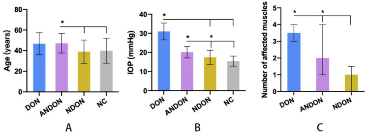
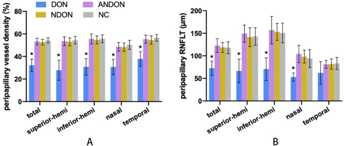
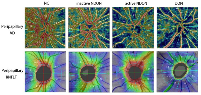
Objective: To measure the peripapillary vessel density (VD) and retinal nerve fiber layer thickness (RNFLT) in thyroid-associated ophthalmopathy (TAO) and dysthyroid optic neuropathy (DON) patients using optical coherence tomography angiography (OCTA), and determine their prognostic relevance.
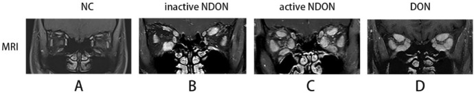
Methods: Forty-three TAO patients with or without DON (82 eyes in total) and 26 healthy subjects (52 eyes) were enrolled. All participants underwent ophthalmology and endocrinology tests. The peripapillary VD in retinal peripapillary capillary layer and RNFLT were analyzed using OCTA images. Multiple linear regression analysis was used to assess the relationship between peripapillary VD /RNFLT and the relevant factors.
Results: The total peripapillary VD and RNFLT were significantly lower in the DON patients compared to the other groups (P < 0.001, 95% confidence intervals), and each quadrant of VD and RNFLT showed similar results except temporal RNFLT. No significant difference was seen between the RNFLT and VD of active non-DON (ANDON), inactive non-DON (NDON) patients and normal control (NC) group. Multivariable linear regression model showed that high IOP is an independent risk factor for lower peripapillary VD and RNFLT (β = -0.465, P < 0.001 and β = -0.343, P = 0.002 respectively).
Conclusion: OCTA parameters are suitable indicators for diagnosing DON. TAO patients with high IOP should be considered at high risk of retinal vessel and nerve fiber layer deterioration. In addition, patients with TAO should be advised to quit smoking since it could affect peripapillary VD and RNFLT.
Keywords: Dysthyroid optic neuropathy; OCTA; Peripapillary vessel density; Retinal nerve fiber layer thickness; Thyroid-associated ophthalmopathy.
© 2021. The Author(s).
Conflict of interest statement
The authors have no conflicts of interest to declare.
Figures
Similar articles
-
Optical coherence tomography angiography in thyroid associated ophthalmopathy: a systematic review.BMC Ophthalmol. 2024 Jul 22;24(1):304. doi: 10.1186/s12886-024-03569-5. BMC Ophthalmol. 2024. PMID: 39039451 Free PMC article.
-
[Peripapillary and macular vessel density in eyes with different phases of thyroid-associated ophthalmopathy].Zhonghua Yan Ke Za Zhi. 2020 Nov 11;56(11):824-831. doi: 10.3760/cma.j.cn112142-20191115-00574. Zhonghua Yan Ke Za Zhi. 2020. PMID: 33152840 Chinese.
-
Peripapillary and Macular Vessel Density in Dysthyroid Optic Neuropathy: An Optical Coherence Tomography Angiography Study.Invest Ophthalmol Vis Sci. 2019 May 1;60(6):1863-1869. doi: 10.1167/iovs.18-25941. Invest Ophthalmol Vis Sci. 2019. PMID: 31042792
-
Evaluation of lamina cribrosa and peripapillary vascular density in thyroid orbitopathy and effect of intravenous methylprednisolone therapy.Can J Ophthalmol. 2024 Oct;59(5):e489-e495. doi: 10.1016/j.jcjo.2023.11.003. Epub 2023 Dec 11. Can J Ophthalmol. 2024. PMID: 38096906
-
Optical coherence tomography angiography (OCT angiography) in anterior ischemic optic neuropathy (AION): A systematic review and meta-analysis.Eur J Ophthalmol. 2023 Jan;33(1):530-545. doi: 10.1177/11206721221113681. Epub 2022 Jul 17. Eur J Ophthalmol. 2023. PMID: 35844139
Cited by
-
Changes in retinal nerve fiber layer, ganglion cell complex, and ganglion cell layer thickness in thyroid eye disease: A systematic review.Taiwan J Ophthalmol. 2023 Feb 20;14(2):217-224. doi: 10.4103/tjo.TJO-D-22-00110. eCollection 2024 Apr-Jun. Taiwan J Ophthalmol. 2023. PMID: 39027065 Free PMC article. Review.
-
Alterations in Spontaneous Neuronal Activity and Microvascular Density of the Optic Nerve Head in Active Thyroid-Associated Ophthalmopathy.Front Endocrinol (Lausanne). 2022 Jul 22;13:895186. doi: 10.3389/fendo.2022.895186. eCollection 2022. Front Endocrinol (Lausanne). 2022. PMID: 35937801 Free PMC article.
-
Optical coherence tomography angiography in thyroid associated ophthalmopathy: a systematic review.BMC Ophthalmol. 2024 Jul 22;24(1):304. doi: 10.1186/s12886-024-03569-5. BMC Ophthalmol. 2024. PMID: 39039451 Free PMC article.
-
Decreased macular choriocapillaris in thyroid-associated ophthalmopathy: focusing on chorioretinal folds with and without optic disc edema.Front Endocrinol (Lausanne). 2023 Apr 21;14:1123820. doi: 10.3389/fendo.2023.1123820. eCollection 2023. Front Endocrinol (Lausanne). 2023. PMID: 37152945 Free PMC article.
-
Alterations in blood flow at the optic nerve head in patients with thyroid eye disease using optic coherence tomography angiography.Front Med (Lausanne). 2025 May 30;12:1585907. doi: 10.3389/fmed.2025.1585907. eCollection 2025. Front Med (Lausanne). 2025. PMID: 40520777 Free PMC article.
References
MeSH terms
Grants and funding
LinkOut - more resources
Full Text Sources
