

Rare primary hepatic carcinosarcoma composed of hepatocellular carcinoma, cholangiocarcinoma, and sarcoma: a case report
- PMID: 34637352
- PMCID: PMC8516393
- DOI: 10.1177/03000605211050539
Rare primary hepatic carcinosarcoma composed of hepatocellular carcinoma, cholangiocarcinoma, and sarcoma: a case report
Abstract
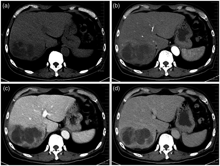
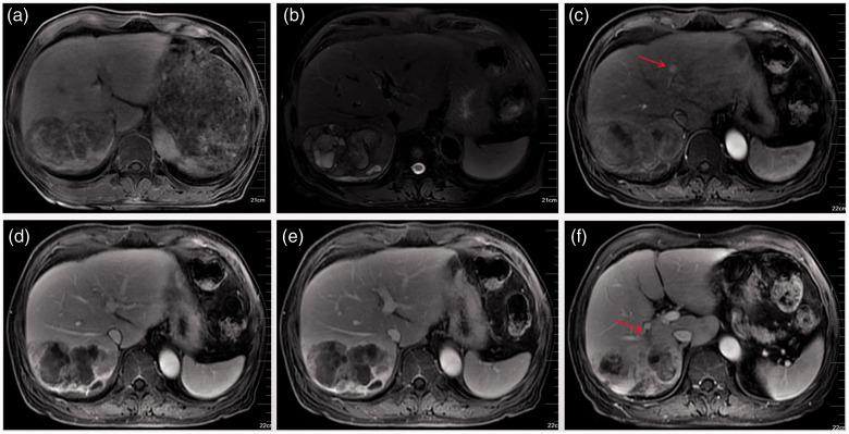
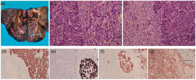
Primary hepatic carcinosarcoma (HCS) is an extremely rare malignant tumor of the liver that contains carcinomatous and sarcomatous components. The diagnosis, treatment, and prognosis of HCS pose great challenges to clinicians. Herein, we present a case of HCS in a 67-year-old man with unique pathological manifestation. Preoperative magnetic resonance imaging showed a malignant lesion in the right liver and a small sub-focus in the left liver. Radical treatment was performed, including excision of the right posterior lobe of the liver, thrombectomy of the right posterior portal vein, and radiofrequency ablation of lesions in the left liver. The specimens were confirmed to be HCS by pathological examinations, which revealed a combination of poorly differentiated hepatocellular carcinoma, moderately differentiated cholangiocellular carcinoma, and spindle cell sarcoma. Transhepatic arterial chemotherapy and embolization was performed after surgery. Unfortunately, pulmonary metastasis occurred 1.5 months later, which meant a poor prognosis. In this report, we discuss the clinicopathological characteristics of this case and factors that affected surgical outcomes, which may add some ideas for the future diagnosis and treatment of HCS patients.
Keywords: Liver; carcinosarcoma; case report; diagnosis; magnetic resonance imaging; tumor.
Conflict of interest statement
Figures
References
-
- Nomura K Aizawa S andUshigome S.. Carcinosarcoma of the liver. Arch Pathol Lab Med. 2000; 124: 888–890. DOI: 10.5858/2000-124-0888-COTL - PubMed
-
- Gagnier JJ, Kienle G, Altman DG, et al. ; CARE Group. The CARE guidelines: Consensus-based clinical case reporting guideline development. Headache. 2013; 53: 1541–1547. DOI: 10.1136/bcr-2013-201554 - PubMed
-
- Wang QB, Cui BK, Weng JM, et al. Clinicopathological characteristics and outcome of primary sarcomatoid carcinoma and carcinosarcoma of the liver. J Gastrointest Surg. 2012; 16: 1715–1726. DOI: 10.1007/s11605-012-1946-y - PubMed
-
- WHO Classification of Tumours Editorial Board. WHO Classification of Tumors: Digestive System Tumours. 5th ed. Lyon, France: International Agency for Research on Cancer; 2019.
Publication types
MeSH terms
LinkOut - more resources
Full Text Sources
Medical
