. 2021 Oct 28;184(22):5608-5621.e18.
doi: 10.1016/j.cell.2021.09.023. Epub 2021 Oct 11.

Mechanoreceptor synapses in the brainstem shape the central representation of touch

Affiliations

Mechanoreceptor synapses in the brainstem shape the central representation of touch

Brendan P Lehnert et al. Cell. .

Abstract

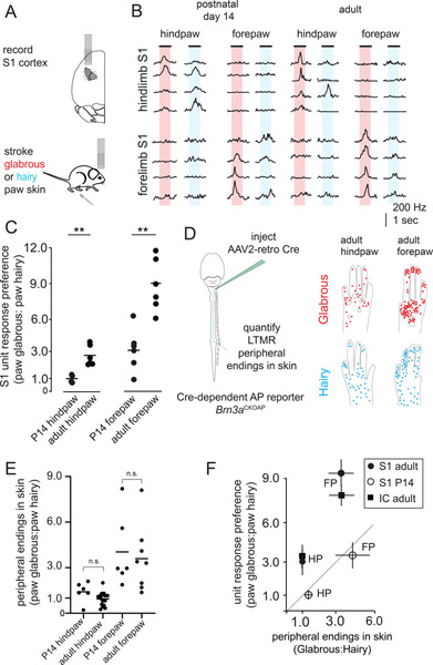
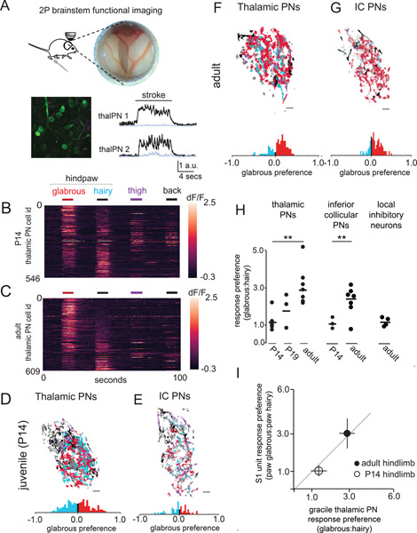
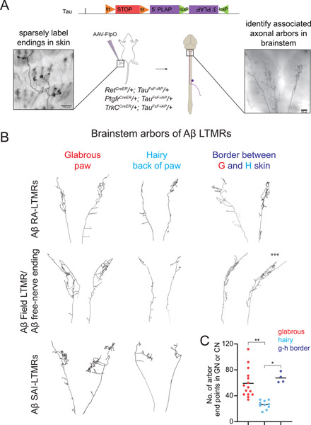
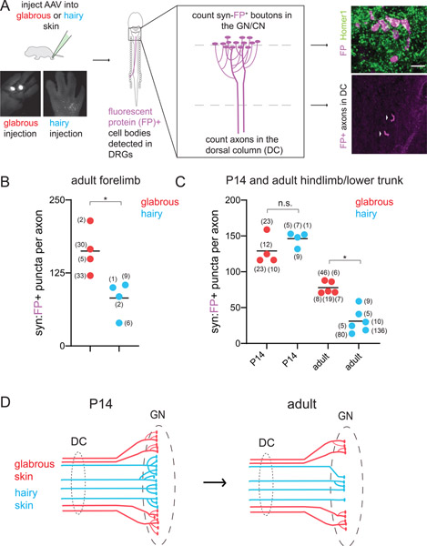
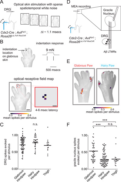
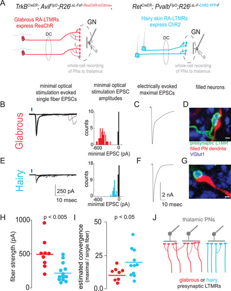
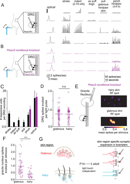
Mammals use glabrous (hairless) skin of their hands and feet to navigate and manipulate their environment. Cortical maps of the body surface across species contain disproportionately large numbers of neurons dedicated to glabrous skin sensation, in part reflecting a higher density of mechanoreceptors that innervate these skin regions. Here, we find that disproportionate representation of glabrous skin emerges over postnatal development at the first synapse between peripheral mechanoreceptors and their central targets in the brainstem. Mechanoreceptor synapses undergo developmental refinement that depends on proximity of their terminals to glabrous skin, such that those innervating glabrous skin make synaptic connections that expand their central representation. In mice incapable of sensing gentle touch, mechanoreceptors innervating glabrous skin still make more powerful synapses in the brainstem. We propose that the skin region a mechanoreceptor innervates controls the developmental refinement of its central synapses to shape the representation of touch in the brain.

Keywords: LTMR; brainstem; disproportionate representation; homunculus; mechanoreceptor; neural activity; peripheral nervous system; piezo2; somatosensation; synaptic expansion; touch.


Conflict of interest statement

Declaration of interests The authors declare no competing interests.

Figures

Figure 1.
Figure 1.. Central representation of glabrous skin expands over postnatal development while receptor density is stable
A. Hindlimb and forelimb S1 regions were stereotactically targeted with multiple multielectrode array (MEA) recordings in adult and postnatal day 14 (P14) mice. B. MEA recordings from S1 at two developmental time points. Representative trial-averaged responses to touch of glabrous and hairy skin for hindpaw (top) and forepaw (bottom) regions of somatosensory cortex reveal a preference for glabrous skin stimulation that emerges over development. C. The ratio of neurons preferentially responding to gentle stroke of glabrous or hairy skin for forepaw S1 and hindpaw S1. An increase in the proportion of neurons preferring glabrous skin stroke occurs over development (n = 4 for P14 hindpaw S1, 6 for adult hindpaw S1, n=6 for P14 forepaw S1, n=6 for adult hindpaw S1, one-way ANOVA, p < 0.01, ** Bonferroni corrected p < 0.01, Mann-Whitney U test). D. (left) AAV2-retro-Cre virus was injected into the dorsal column nuclei to retrogradely label Aβ-LTMRs in Brn3APCKOAP reporter mice. Aβ-LTMR terminals were identified in skin to make estimates of relative innervation density. (right) Compilation of all endings identified in the paws of adult animals that received dorsal column injections, super-imposed on reference images of paws (n=121 neurons from 6 animals for hindpaw skin, n=209 neurons from 7 animals for forepaw skin). Each dot represents a single Aβ LTMR terminal ending in skin. E. The relative number of Aβ LTMR endings in glabrous or hairy forepaw or hindpaw skin is stable from P14 into adulthood (n=6 animals for P14 hindpaw and forepaw, n=12 animals for adult hindpaw, n=7 animals for adult forepaw, p > 0.025, Bonferroni corrected Mann-Whitney U test). For each paw, glabrous:hairy innervation density ratios were calculated by dividing the number of endings per square mm in paw glabrous skin by the number of endings per square mm in paw hairy skin. F. (left) At P14, glabrous skin preference ratios for both forepaw (FP) S1 and hindpaw (HP) S1 are equivalent to innervation density. In adults, a greater proportion of S1 neurons respond to glabrous skin stimulation than predicted by innervation density, and a similar effect is seen in the inferior colliculus, a second major tactile area (right). Together, this demonstrates a developmental expansion of glabrous skin representation in multiple brain regions that is independent of peripheral receptor density.
Figure 2.
Figure 2.. Disproportionate representation of glabrous skin emerges in the brainstem over postnatal development
A. (top) A preparation allowing in vivo multiphoton imaging of the gracile nucleus (GN) of the brainstem. (bottom) A representative, motion-corrected image of thalamic PNs expressing jGCaMP7, and corresponding raw fluorescence time series extracted from ROIs centered on two example neurons (black, 100 μm scale bar) and surrounding neuropil fluorescence (gray) during skin stroke. B. Touch-evoked responses of thalamic projection neurons functionally imaged in the GN. Calcium signals were evoked by stroking equivalent skin areas of the hindpaw glabrous skin (red), hindpaw hairy skin (blue), thigh (purple), and back (black) in P14 mice, data displayed is pooled across n=3 mice (100 μm scale bar). C. Representative response to touch of the body in adult mice, similar to B. D. Representative map of the body within thalamic projection neurons of the GN. A glabrous skin preference index, (glabrous − hairy) / (glabrous + hairy), was computed for all neurons responding to hindpaw skin stimulation in this and other experiments (bottom). At P14, there are roughly equal proportions of glabrous preferring and hairy preferring cells. E. Representative map of the body within inferior collicular projection neurons in the GN, similar to D. F-G. Representative maps of the body in adulthood for thalamic projecting neurons (F) and inferior collicular projecting neurons (G). H. The proportion of thalamic or inferior collicular projecting neurons in the GN that preferentially respond to hindpaw glabrous skin touch expands over developmental time (p < 0.01, Mann-Whitney U test). I. The relationship between glabrous preference in the GN of the brainstem and glabrous preference in hindpaw S1 at two developmental timepoints.
Figure 3.
Figure 3.. Target dependence of Aβ-LTMR central arbor morphologies.
A. Generation of a dual recombinase-dependent AP reporter mouse using the Tau (Mapt) locus (TauFSF-iAP) for intersectional genetic labeling of Aβ-LTMR types with specific CreER lines and AAV2/1-hSyn-FlpO virus injection to hairy or glabrous skin. Animals with sparse labeling are analyzed to relate peripheral arbor morphologies (left) to central arbor anatomies in the GN or CN (right). B. Representative examples of reconstructed brainstem arbors of Aβ RA-LTMRs (top), Aβ field/free nerve ending-LTMRs (middle), or Aβ SAI-LTMRs (bottom) that innervate glabrous or hairy paw skin. Across genetic and functional classes, glabrous skin innervating neurons form more elaborate axonal arbors in the GN or CN than most hairy skin innervating neurons. Neurons innervating hair follicles near glabrous skin (<0.5 mm) form complex arbors similar to those formed by glabrous skin innervating neurons (right). A neuron that spanned both glabrous and hairy skin is indicated with asterisks (***). C. Aβ-LTMRs innervating paw glabrous skin or hairy follicles adjacent to glabrous skin form arbors with more end points in the GN or CN compared to Aβ-LTMRs innervating paw hairy skin that are more than 0.5 mm from glabrous skin (p<0.01 Kruskal-Wallis one-way ANOVA, **p<0.0001 or *p<0.01, Bonferroni corrected Mann-Whitney U test), n=15 neurons from 14 mice for paw glabrous, 10 neurons from 7 mice for paw hairy, 4 neurons from 4 mice for neurons at the edge of paw/glabrous-hairy border.
Figure 4.
Figure 4.. Relationship between skin target identity and Aβ-LTMR brainstem synapses over development
A. (left) AAV virus injection for skin type-specific labeling of Aβ-LTMR central projections with synaptophysin-tdTomato or synaptophysin-mScarlet, hereafter referred to as synaptophysin-FP (fluorescent protein). The GN and CN were sectioned in their entirety and stained for fluorescent proteins and synaptic markers. (right) The number of FP+ pre-synaptic boutons and axons ascending the dorsal column were identified in each sample. Representative example of labeled boutons apposed to the excitatory postsynaptic marker Homer1 in the GN (top). Both hairy and glabrous skin-innervating Aβ-LTMRs form syn-FP+ boutons apposed to Homer1+ puncta in the GN or CN. Representative example of labeled axons (arrows) ascending the dorsal column, in a transverse section of the cervical spinal cord (bottom). Arrows point to individual axons. B-C. Quantification of the average number of FP+ pre-synaptic boutons formed by Aβ-LTMRs innervating different skin targets. Each data point represents one animal; the number of axons identified in each animal is shown in parentheses. B. Adult forepaw glabrous skin-innervating Aβ-LTMRs form more synapses than forepaw hairy skin-innervating Aβ-LTMRs in the CN (*p<0.05, Mann-Whitney U test). C. At P14, hindpaw glabrous skin-innervating Aβ-LTMRs and hindlimb hairy skin-innervating Aβ-LTMRs form similar numbers of pre-synaptic boutons in the GN. At adult time points, paw glabrous skin Aβ-LTMRs form significantly more pre-synaptic boutons in the GN than paw or trunk hairy skin Aβ-LTMRs (p<0.01 Kruskal-Wallis one-way ANOVA, *p<0.01, Mann-Whitney U test with Bonferroni correction for multiple comparisons). D. Summary of skin type-dependent synaptic refinement in Aβ-LTMRs.
Figure 5.
Figure 5.. Aβ-LTMRs that innervate glabrous skin more powerfully excite central tactile neurons in the gracile nucleus than Aβ-LTMRs that innervate hairy skin.
A. Simultaneous in vivo recording from LTMRs with cell bodies in L4 DRG during optical stimulation of skin with sparse spatiotemporal white noise. Single pulses of focused laser light (300 microseconds, 20 mW) were directed to pseudorandom skin locations every 1.1 msec to excite ReaChR present on the terminals of cutaneous sensory neurons. Spatiotemporal optogenetic receptive fields for each unit were estimated through reverse correlation. B. Representative single LTMR unit recorded in vivo with a MEA in the L4 DRG. (top) Spiking response to mechanical indentation for an Aβ RA-LTMR. (bottom) The optogenetic spatiotemporal receptive field of the same RA-LTMR computed through reverse correlation. LTMRs have spatiotemporally simple receptive fields in skin that align with mechanical receptive fields. Scale bars are 1 mm and 200 μm. C. The mean number of spikes evoked by a stimulus to the center of a LTMR’s optogenetic receptive field, computed by integrating the conditional probability of a spike over the 3–6 msec following a light pulse. An equivalent number of LTMR spikes were evoked by stimulating optogenetic RFs in glabrous hindpaw skin, hairy hindpaw skin, and thigh skin (following hair removal), one-way ANOVA p=0.20. D. Strategy for exploring the functional connectivity of the GN at single LTMR resolution. Optogenetic receptive fields of touch sensitive neurons in the GN postsynaptic to Aβ LTMRs were computed similar to A-C. E. Two representative units recorded from the GN with receptive fields on the glabrous (left) or hairy hindpaw skin (right). GN units are composed of spatially and temporally separable “hotspots” in their RFs, indicating convergence of multiple LTMRs onto central touch neurons in the GN. F. The number of spikes evoked in a GN neuron by a single optical pulse to “hotspots” comprising their RFs. Optical stimulation of hindpaw glabrous skin innervating sensory neurons more effectively drives spiking in postsynaptic GN neurons than hindpaw and thigh hairy skin optical stimulation (one-way ANOVA, p < 10−13, *** indicates Bonferroni corrected p < 10−6 via Mann Whitney U-Test).
Figure 6:
Figure 6:. Synaptic strength and functional convergence of glabrous skin- and hairy skin-innervating Aβ-LTMR synapses onto thalamic projection neurons in the brainstem.
A. Genetic strategies for assessing synaptic connections between glabrous skin- or hairy skin-innervating LTMRs and thalamic projection neurons in acute brainstem slices. (left) Glabrous skin Aβ RA-LTMRs that send ascending projections through the dorsal column and synapse in the GN express ReaChR in TrkBCreER; AvilFlpO; R26LsL-FsF-ReaChR::mCitrine mice. (right) Hairy skin-innervating Aβ RA-LTMRs selectively express ChR2 under a RetCreER ON, PvalbFlpO OFF strategy. B. Representative whole-cell recording from a retrogradely labeled thalamic projection neuron in the GN. (left) Minimal optical stimulation of glabrous Aβ RA-LTMRs axons in the dorsal column (1–4 mm from the GN) isolated putative single fiber-evoked EPSCs as stereotyped currents accompanied by failures. (right) Histograms of EPSC amplitudes for these representative recordings are consistent with single fiber stimulation. C. EPSC evoked by maximal stimulation of axons ascending the dorsal column, from the same recording as B. D. The biocytin fill of a recorded cell (red) shows apposition to VGlut1+ (blue), ReaChR::mCitrine+ (green) presynaptic boutons, consistent with a direct synaptic connection (scale bar: 1 μm). E. Representative whole-cell recording from a retrogradely labeled thalamic projection neuron in the GN postsynaptic to hairy skin innervating Aβ RA-LTMRs, similar to B. F. EPSC evoked by maximal stimulation of all axons ascending the dorsal column in the same recording as E. G. Biocytin filled dendrites apposed to ChR2:YFP+ (green) presynaptic axons, similar to D (scale bar: 1 μm). H. Optogenetic activation of individual glabrous skin innervating Aβ RA-LTMRs neurons evokes a larger EPSC than activation of individual hairy skin-innervating Aβ RA-LTMRs (p < .005, Mann Whitney U-test). I. The estimated heterotypic convergence for thalamic projection neurons receiving synaptic input from glabrous skin- (red) and hairy skin-innervating (blue) Aβ RA-LTMRs computed by dividing the maximal EPSC evoked by electrical stimulation of the dorsal column by the genotype-specific single fiber strength. Hairy skin Aβ RA-LTMRs synaptic connections exhibit a greater estimated convergence rate than glabrous skin Aβ RA-LTMRs (p < .05, Mann-Whitney U-test). J. A model of differential synaptic expansion consistent with synaptic strength and convergence measurements presented in (A-I). Glabrous skin LTMR circuits thus have a lower functional convergence ratio while maintaining comparable synaptic input to thalamic PNs, resulting in synaptic expansion that enlarges the representation of glabrous skin relative to hairy skin.
Figure 7:
Figure 7:. Disproportionate glabrous skin representation in the brainstem is maintained in mice that do not sense gentle touch
A. (left) MEA recordings from L4 DRGs in wild-type mice expressing ReaChR in all sensory neurons. (center) Representative units responded to optical stimulation of the skin with short-latency spikes. (right) Mechanical stimulation with stroke, indentation, air puff to the thigh, glabrous skin pull, and strong pinch to the hindpaw. Stroke and indentation were delivered at multiple points on the paw and thigh, and the maximal responses are displayed and quantified. B. MEA recordings from L4 DRG in Piezo2 conditional knockout animals that express ReaChR in all sensory neurons (Cdx2-Cre; Piezo2flox/flox; AvilFlpO; R26FsF-ReaChR). Optical stimulation of the skin produces short-latency spikes (center), as seen in control mice (A), while responses to innocuous mechanical stimuli are largely absent (right). Responses to strong pinch to the hindpaw were preserved. C. The number of DRG units responsive to mechanical and optical stimulation in wildtype and conditional Piezo2 knockout mice (n=6 wt, n=5/7 Piezo2 cKO with mechanical/optical stimulation). Equivalent numbers of Piezo2 cKO and wildtype mouse DRG respond to noxious pinch and optical stimulation, but responses to one or more innocuous mechanical stimuli are reduced in the Piezo2 ckO mutant (one-way ANOVA p < .005, p = 0.33, p=0.46, p < .005, respectively, Mann-Whitney U test). D. Optical stimulation of the receptive fields of neurons innervating glabrous or hairy skin evoked equivalent numbers of spikes in Piezo2 cKO DRG neurons (p=0.49, Mann-Whitney U test). E. (left) Optogenetic receptive fields were computed for neurons in the GN of Piezo2 cKO mice. (right) Representative receptive field spots of GN neurons computed after hairy skin stimulation (top) or glabrous skin stimulation (bottom). F. The number spikes evoked in units recorded from the GN following optical stimulation of receptive field spots in hindpaw glabrous skin, hindpaw hairy skin, and thigh skin (after hair removal). Glabrous skin optical stimulation evoked more spikes in GN units than hairy skin stimulation (p < .001, Mann-Whitney U-test). G. Disproportionate expansion of glabrous skin representation in the brain reflects the skin region innervated by sensory neurons independent of touch-evoked activity.

Similar articles

Cited by

References

    1. Abraira VE, and Ginty DD (2013). The sensory neurons of touch. Neuron 79, 618–639. - PMC - PubMed
    1. Allievi AG, Arichi T, Tusor N, Kimpton J, Arulkumaran S, Counsell SJ, Edwards AD, and Burdet E (2016). Maturation of Sensori-Motor Functional Responses in the Preterm Brain. Cereb Cortex 26, 402–413. - PMC - PubMed
    1. Azzopardi P, and Cowey A (1993). Preferential representation of the fovea in the primary visual cortex. Nature 361, 719–721. - PubMed
    1. Badea TC, Cahill H, Ecker J, Hattar S, and Nathans J (2009a). Distinct roles of transcription factors brn3a and brn3b in controlling the development, morphology, and function of retinal ganglion cells. Neuron 61, 852–864. - PMC - PubMed
    1. Badea TC, Hua ZL, Smallwood PM, Williams J, Rotolo T, Ye X, and Nathans J (2009b). New mouse lines for the analysis of neuronal morphology using CreER(T)/loxP-directed sparse labeling. PLoS One 4, e7859. - PMC - PubMed

Publication types

LinkOut - more resources