. 2021 Nov 18;81(22):4663-4676.e8.
doi: 10.1016/j.molcel.2021.09.020. Epub 2021 Oct 11.

NRF1 association with AUTS2-Polycomb mediates specific gene activation in the brain

Affiliations

NRF1 association with AUTS2-Polycomb mediates specific gene activation in the brain

Sanxiong Liu et al. Mol Cell. .

Erratum in

  • NRF1 association with AUTS2-Polycomb mediates specific gene activation in the brain.
    Liu S, Aldinger KA, Cheng CV, Kiyama T, Dave M, McNamara HK, Zhao W, Stafford JM, Descostes N, Lee P, Caraffi SG, Ivanovski I, Errichiello E, Zweier C, Zuffardi O, Schneider M, Papavasiliou AS, Perry MS, Humberson J, Cho MT, Weber A, Swale A, Badea TC, Mao CA, Garavelli L, Dobyns WB, Reinberg D. Liu S, et al. Mol Cell. 2021 Nov 18;81(22):4757. doi: 10.1016/j.molcel.2021.10.023. Mol Cell. 2021. PMID: 34798045 No abstract available.

Abstract

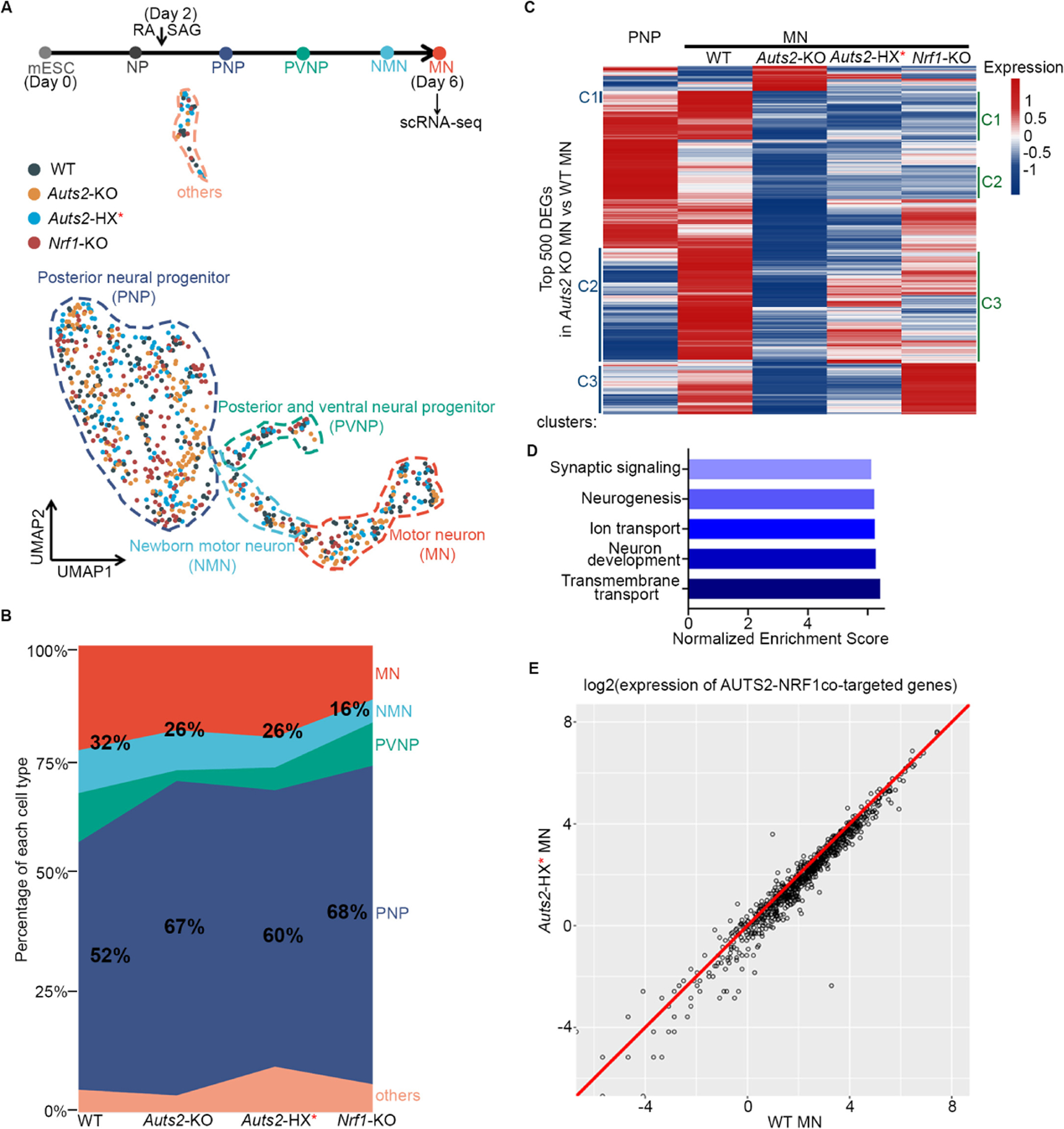
The heterogeneous family of complexes comprising Polycomb repressive complex 1 (PRC1) is instrumental for establishing facultative heterochromatin that is repressive to transcription. However, two PRC1 species, ncPRC1.3 and ncPRC1.5, are known to comprise novel components, AUTS2, P300, and CK2, that convert this repressive function to that of transcription activation. Here, we report that individuals harboring mutations in the HX repeat domain of AUTS2 exhibit defects in AUTS2 and P300 interaction as well as a developmental disorder reflective of Rubinstein-Taybi syndrome, which is mainly associated with a heterozygous pathogenic variant in CREBBP/EP300. Moreover, the absence of AUTS2 or mutation in its HX repeat domain gives rise to misregulation of a subset of developmental genes and curtails motor neuron differentiation of mouse embryonic stem cells. The transcription factor nuclear respiratory factor 1 (NRF1) has a novel and integral role in this neurodevelopmental process, being required for ncPRC1.3 recruitment to chromatin.

Keywords: AUTS2; NRF1; P300; RSTS; active transcription; brain development; ncPRC1.3; polycomb.


Conflict of interest statement

Declaration of interests D.R. is a cofounder of Constellation and Fulcrum Pharmaceuticals.

Figures

Figure 1.
Figure 1.. AUTS2-ncPRC1.3 targets active genes in mouse brain
(A) Schematic showing the mouse Auts2 gene structure and its two major transcripts in the mouse brain. Red arrows indicate the translational start codons used for each AUTS2 isoform. (B) Schematic showing the domains of the long and short isoforms of mouse AUTS2 protein. PR, proline-rich region; PY, PPPY motif; HX, HX repeat motif comprising alternating HQ (x6) or HT (x3) residues; His-rich, eight histidine repeats. (C) Expression of AUTS2 and core ncPRC1.3/1.5 components (PCGF3 and PCGF5, respectively, and RING1B) in the mouse brain. Immunoblotting was performed with whole brain extracts at various developmental stages, as indicated. (D) Bar graphs showing the value of transcripts per kilobase million (TPM) for Pcgf3 and Pcgf5 revealed by RNA-Seq from whole brain lysate at postnatal day 1 (P1). (E) Proteomic mass spectrometry results of immunoprecipitation (IP) using AUTS2 antibody in whole brain lysate at P1. (F) IGV browser views showing ChIP-seq for input, AUTS2, RING1B, RYBP, PCGF3, P300, H3K27ac, H3K4me3, H3K27me3, H2AK119ub1, and RNA Polymerase II (PolII) at the representative loci. ChIP-seq was performed in whole brain lysate at P1. (G) Heatmap showing AUTS2, RING1B, RYBP, PCGF3, P300, H3K27ac, H3K4me3, H3K27me3, H2AK119ub1, and PolII ChIP-seq signals centered on AUTS2 bound regions (±5 kb). ChIP-seq was performed in whole brain lysate at P1.
Figure 2.
Figure 2.. Patients with mutations in the AUTS2 HX repeat have features overlapping those of Rubinstein-Taybi syndrome.
(A) Schematic illustrating mutations in the AUTS2 gene from individual patients as identified through trio-based exome sequencing. Mutations resulting in similar clinical features are labeled with same color. PY, PPPY motif; HX, HX repeat motif comprising alternating HQ (x6) or HT (x3) residues. (B) Clinical features of individuals with mutations in AUTS2 PY motif and HX repeat domain. Frontal photos of our original proband (LR05–007 with p.Thr534Pro mutation) at ~1 (i) and 13 (ii) years show reflexive eye closure with smiling, as well as a low hanging columella, features often seen in RSTS (i-ii). Photo of his right hand shows severe symphalangism of the right third finger (iii). Frontal (iv) and profile (v) photos of a boy (LR15–003 with p.His535_Thr542 del) at 17 years show thick horizontal eyebrows with synophrys, prominent (high) nasal bridge, broad nose with mildly low columella, and posteriorly-rotated left ear. Photos of his feet show mildly broad halluces (vi). Frontal (vii) and profile (viii) photos of a girl (LR15–004 with p.Pro517Leu) show a normal facial appearance. (C) Magnetic resonance images from 5 individuals with missense variants in AUTS2 exon 9 and a normal control. The three midline sagittal images in the top row from a normal control (i), a girl with a missense variant (p. Pro517Leu) (ii), and a boy with the recurrent INDEL (p.His535_Thr542 del) (iii) respectively, all show normal midline structures. The three midline sagittal images in the bottom row come from a boy with a missense mutation (p.Thr534Pro) (iv), and two boys with the recurrent INDEL (p.His535_Thr542 del) (v-vi). All three show thin and dysplastic corpus callosum and small cerebellar vermis (arrows and asterisks, respectively, in iv-vi). The horizontal white or black lines mark the level of the obex, the usual lower extent of the vermis.
Figure 3.
Figure 3.. The HX repeat domain and ncPRC1.3/1.5 core components are required for efficient P300 recruitment and transcriptional activation.
(A) Schematic showing human AUTS2 variants constructed and expressed in 293 T-REx cells. (B-C) Western blots show co-IP results from nuclear extract of 293 T-REx cells expressing Flag–AUTS2, either WT or mutant versions as indicated, using Flag antibody (B) or reciprocal IP using P300 antibody (C). (D) Schematic of the reporter construct for the luciferase assay in the context of GAL4-AUTS2, either WT or mutant versions as indicated. (E) Luciferase activity in cells expressing GAL4-AUTS2, either WT or mutant versions, before and after doxycycline treatment. (F-G) Western blots show co-IP results from nuclear extract of MN differentiated from WT and Auts2 HX mutant (T534P and 535–542 aa deletion, respectively) mESC as indicated, using AUTS2 antibody (F) or reciprocal IP using P300 antibody (G). (H) Luciferase activity in cells expressing GAL4-AUTS2, either in the presence or absence of PCGF3 and PCGF5, before and after doxycycline treatment. (I) ChIP-qPCR at the UAS element using the antibodies as indicated in cells expressing GAL4-AUTS2, either in the presence or absence of PCGF3 and PCGF5, before and after doxycycline treatment.
Figure 4.
Figure 4.. Transcription factor NRF1 colocalizes with AUTS2 on chromatin and physically interacts with AUTS2 in mouse brain
(A) Top 5 enriched motifs identified in AUTS2-bound regions in mouse brain using HOMER. (B) Heatmap showing AUTS2 and NRF1 ChIP-seq signals centered on AUTS2-bound regions (±5 kb) with two replicates. ChIP-seq was performed in whole brain lysate at P1. (C) Venn diagram showing the extent of overlap for AUTS2- and NRF1-bound regions revealed by ChIP-seq from (B). (D) Reciprocal co-IP and western blot analyses demonstrating AUTS2 and NRF1 interaction in whole brain lysate at P1. (E) Expression of CBP, P300, AUTS2, and NRF1 in mouse brain. Immunoblotting was performed with whole brain extracts at various developmental stages, as indicated.
Figure 5.
Figure 5.. NRF1 is crucial for AUTS2-ncPRC1.3 associated active transcription in MN
(A) The schematic at top depicts the protocol for differentiation of mouse embryonic stem cells (mESC) to motor neurons (MN) using retinoic acid (RA) and smoothened agonist (SAG). EB, embryoid bodies. Below is a western blot showing the expression of AUTS2, NRF1, PCGF3, and RING1B in mESC and MN. (B) Heatmap showing AUTS2, NRF1 and RNA Pol II ChIP-seq signals centered on AUTS2-bound regions identified in WT MN (±5 kb). (C) k-means clustering of RING1B, H2AK119ub1, H3K27me3, H3K27ac, H3K4me3, RNA Pol II, RYBP, AUTS2, and NRF1 ChIP-seq signals from WT MN centered on RING1B-bound regions identified in WT MN (±5 kb). (D) Violin plot of the log2(TPM) of genes assigned to each cluster [as indicated in (C)], quantified from RNA-seq in WT MN. (E) Average density profiles (top) and heatmap (bottom) showing AUTS2 ChIP-seq signals from WT, Auts2-KO, and Nrf1-KO MN centered on AUTS2-bound regions identified in WT MN (±5 kb). (F) Average density profiles (top) and heatmap (bottom) showing NRF1 ChIP-seq signals from WT, Nrf1-KO, and Auts2-KO MN centered on NRF1-bound regions identified in WT MN (±5 kb). (G) Venn diagram showing the extent of overlap for RING1B-bound regions in WT MN, RING1B cluster 3 regions [as indicated in (C)], and RING1B-bound regions in Nrf1-KO MN revealed by ChIP-seq. (H) k-means clustering of RING1B, H2AK119ub1, H3K27me3, H3K27ac, AUTS2, and NRF1 ChIP-seq signals from Nrf1-KO MN centered on RING1B-bound regions identified in Nrf1-KO MN (±5 kb). (I) Violin plot of the log2(normalized counts), quantified from H2AK119ub, H3K27me3, and H3K27ac ChIP-Seq signal at RING1B cluster 3 regions [as indicated in (C)] in WT and Nrf1-KO MN.
Figure 6.
Figure 6.. Defect in PNP to MN differentiation under Auts2-KO, mutation in Auts2 HX repeat or Nrf1-KO as revealed by scRNA-Seq
(A) The schematic at top depicts cell lineage transitions from mESC to MN. MN (day 6) differentiated from WT, Auts2 knockout, Auts2 HX mutant (535–542 aa deletion) and Nrf1 knockout ESC were harvested for scRNA-Seq. Below is the dimensionality reduction (UMAP) of 816 cells from WT, Auts2-KO, Auts2 HX mutant, and Nrf1-KO samples, sequenced with the Smart-seq3 technique and colored by sample identity (WT, 228 cells; Auts2-KO, 221 cells; Auts2-HX*, 184 cells; Nrf1-KO, 183 cells). Five cell classes revealed by unsupervised clustering of cellular transcriptomics are represented by color-coded circles of dashed-lines and annotated based on marker gene expression. Colors of dashed-lines match those for the cell types shown at top. (B) Proportional stacked area graph showing the abundances of each cell type in MN differentiated from WT, Auts2-KO, Auts2 HX mutant (535–542 aa deletion), and Nrf1-KO ESC. The percentage of MN (MN+NMN) and PNP in each sample are labelled. (C) Top 500 DEGs were identified by comparing Auts2-KO MN versus WT MN. Expression of these DEGs across WT PNP, WT MN, Auts2-KO MN, Auts2 HX mutant MN, and Nrf1-KO MN is shown by heatmap. Color scale represents the averaged and scaled expression values from each cell population. Labelling of clusters (C1, C2, C3) on left is based on gene expression differences in WT PNP, WT MN, and Auts2-KO MN. Labelling of clusters (C1, C2, C3) on right is based on gene expression differences in WT MN, Auts2-KO MN, and Nrf1-KO MN. (D) Bar plot summarizing results of GO analysis for genes downregulated in Auts2 KO MN versus WT MN. (E) Scatter plot of log2(normalized expression of AUTS2-NRF1 co-targeted genes) in WT and Auts2 HX mutant MN from scRNA-Seq.
Figure 7.
Figure 7.. Model: AUTS2-ncPRC1.3 activates its targeted genes for brain development through NRF1-mediated recruitment and HX repeat-mediated P300 interaction.
Mutations or deletions of CBP or P300 impede neuronal differentiation during early brain development and result in the malformation of several brain regions, including the corpus collosum and cerebellum in RSTS patients. As shown here, mutations in the AUTS2 HX repeat domain impair AUTS2 interaction with P300 within the context of ncPRC1.3, effectively disabling transcription activation. We speculate such disruptions to appropriate gene activation lead to a defect in brain development that overlaps with RSTS.

References

    1. Ajmone PF, Avignone S, Gervasini C, Giacobbe A, Monti F, Costantino A, Esposito S, Marchisio P, Triulzi F, and Milani D (2018). Rubinstein-Taybi syndrome: New neuroradiological and neuropsychiatric insights from a multidisciplinary approach. Am J Med Genet B Neuropsychiatr Genet 177, 406–415. - PubMed
    1. Aldinger KA, Timms AE, Thomson Z, Mirzaa GM, Bennett JT, Rosenberg AB, Roco CM, Hirano M, Abidi F, Haldipur P, et al. (2019). Redefining the Etiologic Landscape of Cerebellar Malformations. The American Journal of Human Genetics 105, 606–615. - PMC - PubMed
    1. Aloia L, Stefano BD, and Croce LD (2013). Polycomb complexes in stem cells and embryonic development. Development 140, 2525–2534. - PubMed
    1. Badea TC, Cahill H, Ecker J, Hattar S, and Nathans J (2009). Distinct Roles of Transcription Factors Brn3a and Brn3b in Controlling the Development, Morphology, and Function of Retinal Ganglion Cells. Neuron 61, 852–864. - PMC - PubMed
    1. Bannister AJ, and Kouzarides T (1996). The CBP co-activator is a histone acetyltransferase. Nature 384, 641–643. - PubMed

Publication types

MeSH terms