. 2021 Sep 23;13(19):4761.
doi: 10.3390/cancers13194761.

Fibroblasts Influence Metastatic Melanoma Cell Sensitivity to Combined BRAF and MEK Inhibition

Affiliations

Fibroblasts Influence Metastatic Melanoma Cell Sensitivity to Combined BRAF and MEK Inhibition

Delphine Morales et al. Cancers (Basel). .

Abstract

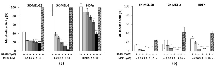
The sensitivity of melanoma cells to targeted therapy compounds depends on the tumor microenvironment. Three-dimensional (3D) in vitro coculture systems better reflect the native structural architecture of tissues and are ideal for investigating cellular interactions modulating cell sensitivity to drugs. Metastatic melanoma (MM) cells (SK-MEL-28 BRAF V600E mutant and SK-MEL-2 BRAF wt) were cultured as a monolayer (2D) or cocultured on 3D dermal equivalents (with fibroblasts) and treated with a BRAFi (vemurafenib) combined with a MEK inhibitor (MEKi, cobimetinib). The drug combination efficiently inhibited 2D and 3D MM cell proliferation and survival regardless of their BRAF status. Two-dimensional and three-dimensional cancer-associated fibroblasts (CAFs), isolated from a cutaneous MM biopsy, were also sensitive to the targeted therapy. Conditioned media obtained from healthy dermal fibroblasts or CAFs modulated the MM cell's response differently to the treatment: while supernatants from healthy fibroblasts potentialized the efficiency of drugs on MM, those from CAFs tended to increase cell survival. Our data indicate that the secretory profiles of fibroblasts influence MM sensitivity to the combined vemurafenib and cobimetinib treatment and highlight the need for 3D in vitro cocultures representing the complex crosstalk between melanoma and CAFs during preclinical studies of drugs.

Keywords: 3D human melanoma model; BRAFi + MEKi efficiency; cancer-associated fibroblasts; targeted therapy; tissue engineering.


Conflict of interest statement

The authors declare no conflict of interest.

Figures

Figure A1
Figure A1
CAF characterization. (A) Observation of CAF morphology; (B) F-actin staining using phalloidin-FITC (FP AZ0130, Fluoprobes) in isolated CAFs. Fibroblast nuclei were stained blue with 4′,6-diamidino-2-phenylindole); (C) CAFs were negative for Melan-A expression; (D) SK-MEL-2 cells were used as positive control for Mela-A expression; (E) CAFs strongly expressed vimentin protein (red staining); (F) CAFs expressed the PDGFRβ marker (red staining).
Figure A2
Figure A2
SK-MEL-28 and SK-MEL-2 cell response to combined BRAFi + MEKi or to MEKi single treatment (from 0.2 to 10 µM). Cells were cultured as a monolayer, treated with vemurafenib + cobimetinib or cobimetinib alone during 48 h, and metabolic activity was quantified using MTS assay. n = 6; * p < 0.05 and *** p < 0.001 indicate significant difference between cells treated with BRAFi + MEKi vs. cells treated with MEKi alone (Mann–Whitney test).
Figure A3
Figure A3
EdU-labeled SK-MEL-28 cells cultured as a monolayer (2D) or in the dermal equivalent (3D) during BRAFi (2 µM) and MEKi treatment (2 µM).For the 3D coculture model, EdU positive melanoma cells are indicated with white arrows and fibroblasts with dashed arrows. The polycarbonate membranes from the inserts are indicated with dashed lines. Scale bar = 250 µm.
Figure A4
Figure A4
TUNEL-labeled SK-MEL-2 cells cultured as a monolayer (2D) or in the dermal equivalent (3D) during BRAFi (2 µM) and MEKi treatment (2 µM). For the 3D coculture model, TUNEL positive melanoma cells are indicated with white arrows and fibroblasts with dashed arrows. The polycarbonate membranes from the inserts are indicated with dashed lines. Scale bar = 250 µm (2D) or 100 µm (3D).
Figure A5
Figure A5
IL-6 release measured in the cell culture media of HDFn and CAF dermal equivalents treated with BRAFi and MEKi for 48 h. IL-6 concentration was quantified by an ELISA assay (Affymetrix eBioscience), n = 1.
Figure 1
Figure 1
Characterization of the effect of BRAFi and MEKi on MM cells and healthy fibroblasts cultured as a monolayer. Melanoma cells (SK-MEL-28, SK-MEL-2) and human dermal fibroblasts (HDFn) were treated with vemurafenib (2 µM) combined with different concentrations of cobimetinib for 48 h. Cell proliferation relative to untreated controls was assessed by means of (a) cell metabolic activity (MTS assay) and (b) DNA synthesis (EdU assay). Results are means ±  s.d. of three experiments performed in triplicate: * p < 0.05; ** p < 0.01; *** p < 0.001 indicate significant difference vs. untreated cells.
Figure 2
Figure 2
Histological images of 3D coculture models upon BRAFi and MEKi treatment. Dermal equivalents were obtained by seeding HDFn cells in a type I collagen gel. Fibroblasts were cultured in 3D for 7 days. Melanoma cells (SK-MEL-28 or SK-MEL-2) were then added for 24 h. The coculture models were treated with 2 µM of BRAFi and 2 µM of MEKi for 48 h and stained with hematoxylin-eosin-saffron. The melanoma cells on the dermal equivalent are indicated with black arrows. Fibroblasts (dashed arrows) were found inside the tissue and aligned with the collagen fibers. Inserts show a higher magnification of the pictures. Scale bar = 100 µm (200×).
Figure 3
Figure 3
Effect of BRAFi and MEKi on MM cell proliferation in the 3D HDFn dermal equivalent. Coculture models were treated with 2 µM of BRAFi and different concentrations of MEKi for 48 h. Total cell proliferation (MM + fibroblasts) relative to untreated controls was assessed by means of DNA synthesis (EdU assay). Results are means ±  s.d. of three experiments carried out in triplicate: * p < 0.05; *** p < 0.001 indicate significant difference vs. untreated cells.
Figure 4
Figure 4
Characterization of the effect of BRAFi and MEKi on cancer-associated fibroblasts (CAFs) cultured as a monolayer or as dermal equivalents. CAFs were treated with BRAFi (2 µM) combined with different concentrations of MEKi for 48 h. Cell proliferation relative to untreated controls was assessed by means of (a) cell metabolic activity (MTS assay) and (b) cell counting. Results are means ± s.d. of three experiments carried out in triplicate: * p < 0.05; ** p < 0.01; *** p < 0.001 indicate significant difference vs. untreated cells.
Figure 5
Figure 5
Effect of HDFn- and CAF-conditioned media on 2D SK-MEL-2 sensitivity to BRAFi + MEKi. Conditioned media obtained from (a) HDFn or (b) CAFs were used for SK-MEL-2 cells cultured as a monolayer and treated with BRAFi + MEKi. SK-MEL-2 cell proliferation relative to the untreated control medium was assessed by cell metabolic activity (MTS assay). Results are means ±  s.d. of three triplicate experiments: * p < 0.05; ** p < 0.01 indicate significant difference vs. untreated cells with control medium.

References

    1. Donia M., Ellebaek E., Øllegaard T.H., Duval L., Aaby J.B., Hoejberg L., Køhler U.H., Schmidt H., Bastholt L., Svane I.M. The real-world impact of modern treatments on the survival of patients with metastatic melanoma. Eur. J. Cancer. 2019;108:25–32. doi: 10.1016/j.ejca.2018.12.002. - DOI - PubMed
    1. Larkin J., Ascierto P.A., Dréno B., Atkinson V., Liszkay G., Maio M., Mandalà M., Demidov L., Stroyakovskiy D., Thomas L., et al. Combined Vemurafenib and Cobimetinib in BRAF-mutated melanoma. N. Engl. J. Med. 2014;371:1867–1876. doi: 10.1056/NEJMoa1408868. - DOI - PubMed
    1. Long G.V., Stroyakovskiy D., Gogas H., Levchenko E., de Braud F., Larkin J., Garbe C., Jouary T., Hauschild A., Grob J.J., et al. Combined BRAF and MEK Inhibition versus BRAF Inhibition Alone in Melanoma. N. Engl. J. Med. 2014;371:1877–1888. doi: 10.1056/NEJMoa1406037. - DOI - PubMed
    1. Robert C., Karaszewska B., Schachter J., Rutkowski P., Mackiewicz A., Stroiakovski D., Lichinitser M., Dummer R., Grange F., Mortier L., et al. Improved overall survival in melanoma with combined Dabrafenib and Trametinib. N. Engl. J. Med. 2015;372:30–39. doi: 10.1056/NEJMoa1412690. - DOI - PubMed
    1. Flaherty K.T., Puzanov I., Kim K.B., Ribas A., McArthur G.A., Sosman J.A., O’Dwyer P.J., Lee R.J., Grippo J.F., Nolop K., et al. Inhibition of mutated, activated BRAF in metastatic melanoma. N. Engl. J. Med. 2010;363:809–819. doi: 10.1056/NEJMoa1002011. - DOI - PMC - PubMed