. 2021 Oct 4;13(19):4983.
doi: 10.3390/cancers13194983.

Mir-21 Suppression Promotes Mouse Hepatocarcinogenesis

Affiliations

Mir-21 Suppression Promotes Mouse Hepatocarcinogenesis

Marta Correia de Sousa et al. Cancers (Basel). .

Abstract

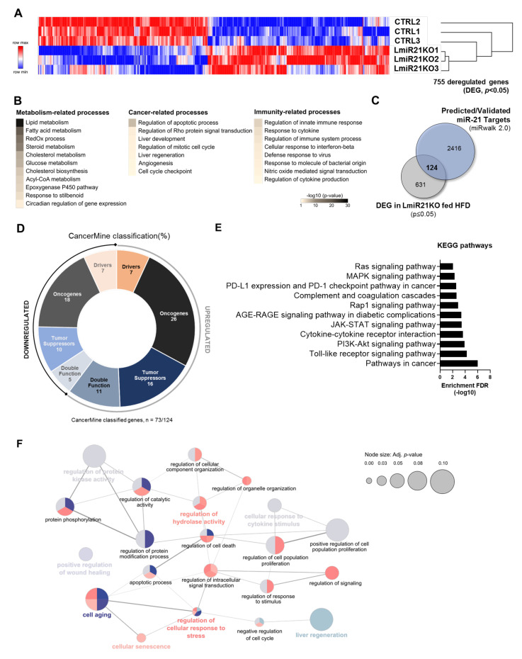
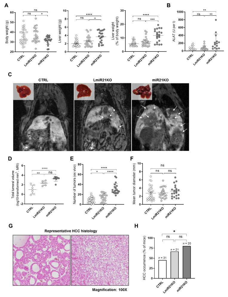
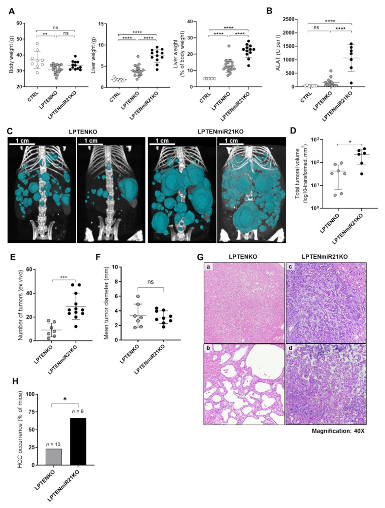
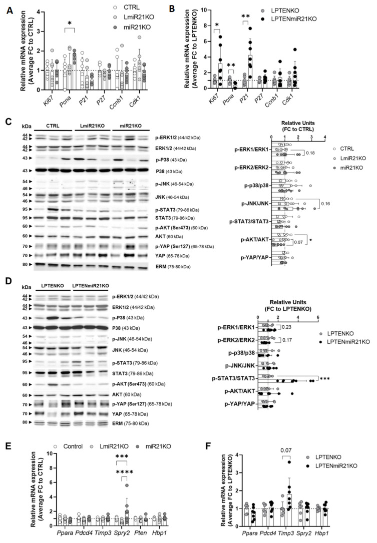
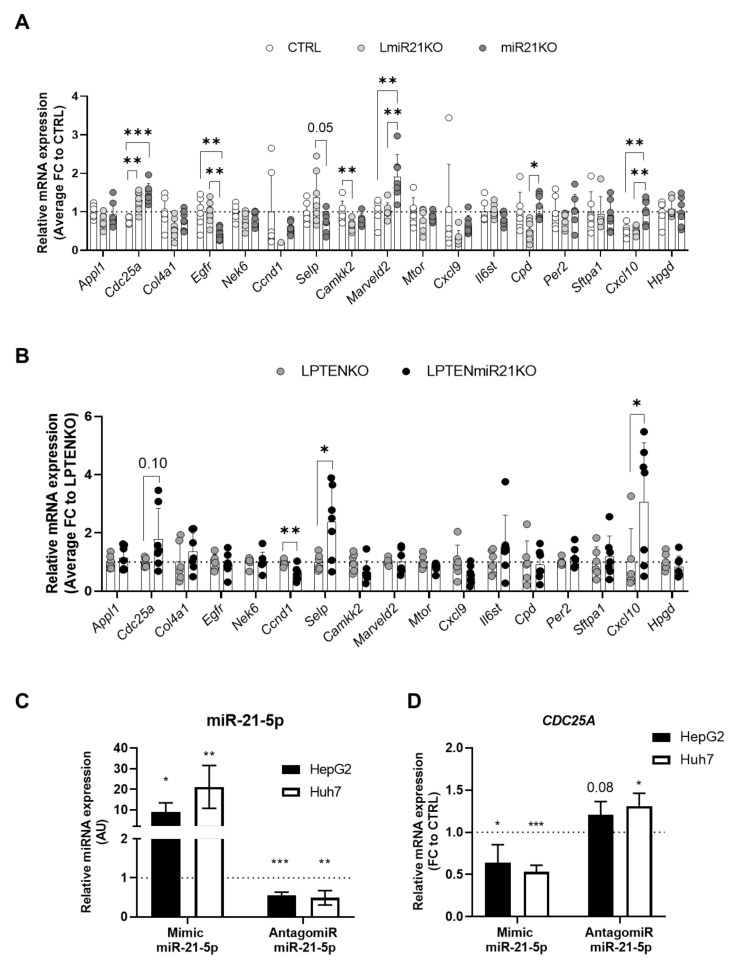
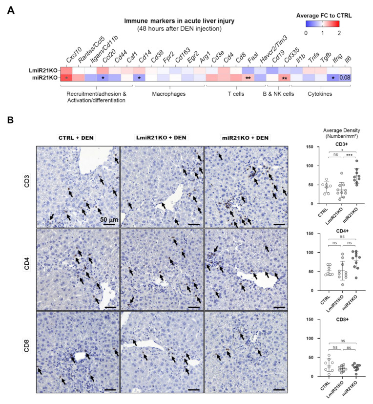
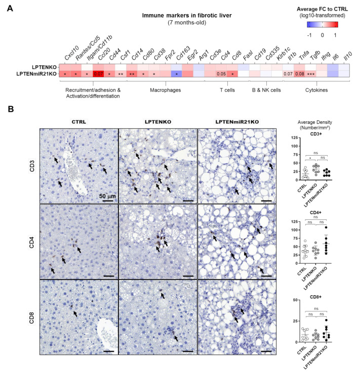
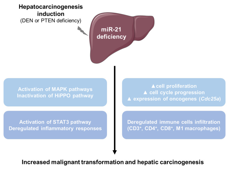
The microRNA 21 (miR-21) is upregulated in almost all known human cancers and is considered a highly potent oncogene and potential therapeutic target for cancer treatment. In the liver, miR-21 was reported to promote hepatic steatosis and inflammation, but whether miR-21 also drives hepatocarcinogenesis remains poorly investigated in vivo. Here we show using both carcinogen (Diethylnitrosamine, DEN) or genetically (PTEN deficiency)-induced mouse models of hepatocellular carcinoma (HCC), total or hepatocyte-specific genetic deletion of this microRNA fosters HCC development-contrasting the expected oncogenic role of miR-21. Gene and protein expression analyses of mouse liver tissues further indicate that total or hepatocyte-specific miR-21 deficiency is associated with an increased expression of oncogenes such as Cdc25a, subtle deregulations of the MAPK, HiPPO, and STAT3 signaling pathways, as well as alterations of the inflammatory/immune anti-tumoral responses in the liver. Together, our data show that miR-21 deficiency promotes a pro-tumoral microenvironment, which over time fosters HCC development via pleiotropic and complex mechanisms. These results question the current dogma of miR-21 being a potent oncomiR in the liver and call for cautiousness when considering miR-21 inhibition for therapeutic purposes in HCC.

Keywords: HCC; PTEN; fibrosis; immune cells; inflammation; microRNA 21; oncogenes; tumor suppressors.


Conflict of interest statement

The authors declare no conflict of interest.

Figures

Figure 1
Figure 1
Transcriptomic profile of cancer-related factors altered in hepatic tissues from LmiR-21KO livers. (A) Heatmap representation of differentially expressed genes (DEG, p-value < 0.05, |log10FC| > 0.5) identified through RNAseq analysis of explanted liver tissues from control (CTRL) and hepatocyte-specific miR-21 knockout mice fed obesogenic diet (LmiR21KO, HFD 60% of fat, high-fat diet) for 3 weeks (n = 3 per group, upregulated and downregulated genes represented in red and blue, respectively). (B) Gene Ontology (GO) enrichment analysis of DEG using DAVID database ((https://david.ncifcrf.gov/, accessed on 27 March 2020) retrieved metabolism-, cancer-, and immunity-related processes. (C) Potential miR-21 targets of interest were identified by cross-comparison of DEG identified in LmiR21KO with a list of predicted/validated targets of miR-21-5p/-3p extracted from the miRwalk 2.0 database (accessed on 11 June 2020). (D) Literature screening and the CancerMine database (accessed on 07/06/2021) identified candidates as oncogenes (ONC—black and dark grey for upregulated and downregulated genes, respectively), tumor suppressors (TS—dark blue and light blue for upregulated and downregulated genes, respectively), or drivers (D—orange and light orange for upregulated and downregulated genes, respectively) of carcinogenesis. Genes identified as both ONC and TS were classified as having double function (grey-blue and light grey for upregulated and downregulated genes, respectively). (E) Gene Ontology (GO) enrichment analysis of potential miR-21 targets upregulated under stress conditions identified key pathways involved in hepatocarcinogenesis. GO enrichment analysis based on Kyoto Encyclopedia of Genes and Genomes (KEGG) pathways was performed on upregulated genes identified in panels CE. Only significant enrichments are represented. The detailed results of these analyses are presented in Figure S3. (F) ClueGO+CluePedia (Cytoscape) analysis of upregulated candidates identified numerous functionally grouped network of biological processes (clusters of terms) with putative interactions between them based on their kappa score level (0.45). Node size represents enrichment significance of each cluster (adjusted p-value ≤ 0.10). Each cluster of terms is represented by a specific color (detailed in Figure S2). Functionally related terms that overlap in different clusters due to multiple functions of single genes are represented as well. The most representative term of each cluster is highlighted in bold with the respective color attributed to the cluster.
Figure 2
Figure 2
MiR-21 deficiency fosters DEN-induced HCC development. (A) Body weight, liver weight, and liver-to-body weight ratio of 11-month-old control (CTRL—miR21fx, n = 32), hepatocyte-specific and total miR-21 knockout (LmiR21KO, n = 21, and miR21KO n = 20, respectively) mice injected with diethylnitrosamine (DEN, 25 mg/kg of body weight) intraperitoneally at day 15 post-birth. (B) Plasma levels of alanine-aminotransferase (ALAT) in CTRL (n = 27), LmiR21KO (n = 21), and miR21KO (n = 19) mice. (C) Representative pictures of explanted livers post-sacrifice and magnetic resonance imaging (MRI) prior to sacrifice of CTRL, LmiR21KO, and miR21KO mice. Tumoral masses are indicated by white arrows in the MRI scans. (D) Volume of the total tumoral mass quantified by MRI (values log10-transformed) in CTRL (n = 8), LmiR21KO (n = 8), and miR21KO (n = 8) mice. (E) Number and (F) mean diameter of isolated tumors from explanted liver in CTRL (n = 32), LmiR21KO (n = 21), and miR21KO (n = 20) mice. (G) Representative histopathological sections (hematoxylin-eosin staining, original magnification ×100) of typical HCC tumors found indiscriminately in HCC in either CTRL, LmiR21KO, or miR21KO mice. Left panel shows HCC in miR21KO with a pseudo-glandular pattern and eosinophilic cells. Right panel shows HCC in miR21KO with trabecular architectures and clear cell changes. (H) Percentage of mice in the respective cohorts developing hepatocellular carcinoma (HCC) as determined by histopathological analyses of H&E stained sections (CTRL, n = 31; LmiR21KO, n = 21, miR21KO, n = 20). Data that failed normality tests was submitted to log10-transformation prior to statistical analysis. An outliers test was performed using the ROUT method (Q = 1%). One-way ANOVA with Holm–Sidak’s correction was used for comparison between more than two groups. Chi-square test was used to evaluate the independence of categorical variables (percentage of samples with detected expression). ns, not significant, * p-value < 0.05, ** p-value < 0.01, *** p-value < 0.001, **** p-value < 0.0001.
Figure 3
Figure 3
Hepatocyte-specific miR-21 deletion promotes exacerbates hepatocarcinogenesis induced by PTEN deficiency. (A) Body weight, liver weight, and liver-to-body weight ratio of control (CTRL—floxed mice not expressing the Alb-Cre recombinase, n = 9), hepatocyte-specific PTEN knockout (LPTENKO (n = 17), and LPTENmiR21KO (n = 12), respectively) sacrificed at 12 months of age. (B) Plasma levels of alanine-aminotransferase (ALAT) in CTRL (n = 7), LPTENKO (n = 14), and LPTENmiR21KO (n = 7) mice. Data is represented as mean ± SD. (C) Representative three-dimensional reconstructions of computed tomography (CT) scans performed prior to sacrifice. Tumoral masses are represented in blue. (D) Volume of the total tumoral mass (values log10 transformed) quantified by CT scanning (LPTENKO, n = 7; LPTENmiR21KO, n = 6). Data is represented as mean ± SD. (E) Number and (F) mean diameter of isolated tumors from explanted liver in LPTENKO (n = 7 mice) and LPTENmiR21KO (n = 12 mice). Data is represented as mean ± SD. Outliers were removed. (G) Histopathological characterization (hematoxylin-eosin staining, magnification 40×) of tumoral nodules in the livers of LPTENKO mice (a) well-differentiated hepatocellular adenoma (HCA) and (b) cholangioma) and LPTENmiR21KO mice (c) hepatobiliary tumors of mixed origin and (d) HCC). (H) Percentage of mice in the respective cohorts developing hepatocellular carcinoma (HCC) as determined by histopathological analyses of H&E stained sections (LPTENKO, n = 13; LPTENmiR21KO, n = 9). Data that failed normality tests was submitted to log10-transformation prior to statistical analysis. An outliers test was performed using the ROUT method (Q = 1%). Unpaired t-test with Welch’s correction was used for comparison between two groups. One-way ANOVA with Holm–Sidak’s correction was used for comparison between more than two groups. Chi-square test was used to evaluate the independence of categorical variables (percentage of samples with detected expression). ns, not significant, * p-value < 0.05, ** p-value < 0.01, *** p-value < 0.001, **** p-value < 0.0001.
Figure 4
Figure 4
Impact of miR-21 deficiency on cell proliferation, oncogenic signaling pathways, and previously identified miR-21 cancer-related targets. Ki67, Pcna, p21, p27, Ccnb1, Cdk1 expression in hepatic tissues of (A) 11-month-old control (CTRL, n = 6), hepatocyte-specific (LmiR21KO, n = 7), and total miR-21 (miR21KO n = 7) knockout mice injected with DEN and (B) 12-month-old hepatocyte-specific PTEN knockout (LPTENKO, n = 6) and PTEN/miR-21 knockout (LPTENmiR21KO, n=7) mice. (C) Representative Western blot (left panel, 3 mice per group) and quantifications (right panel) of phosphorylated over total expression ratio of ERK1, ERK2, p38, JNK, STAT3, AKT, and YAP in hepatic tissues of 11-month-old control, hepatocyte-specific, and total miR-21 knockout mice injected with DEN. Quantifications were performed on Western blot analysis of n = 6 CTRL mice, n = 6 LmiR21KO, and n = 8 miR21KO. (D) Representative Western blot (left panel, 3 mice per group) and quantifications (right panel) of phosphorylated over total expression ratio of ERK1, ERK2, p38, JNK, STAT3, AKT, and YAP in hepatic tissues of 12-month-old hepatocyte-specific PTEN knockout and PTEN/miR-21 knockout mice. Quantifications were performed on Western blot analysis of n=5 LPTENKO and n = 8 LPTENmiR21KO. ERM expression was assessed as a loading control for WB. RT-qPCR analysis of Ppara, Pdcd4, Timp3, Spry2, Pten, and Hbp1 expression in the livers of (E) 11-month-old control (CTRL—miR21fx, n = 6), hepatocyte-specific, and total miR-21 knockout (LmiR21KO, n = 7, and miR21KO n =7, respectively) mice injected with DEN and (F) in the livers of hepatocyte-specific PTEN knockout (LPTENKO, n = 6) and PTEN-miR21 knockout (LPTENmiR21KO, n = 7) mice sacrificed at 12 months of age. Data from DEN-injected miR-21 KO mice is represented as mean± SD fold-change (FC) to CTRL mice (miR21fx treated with DEN). Data from LPTENmiR21KO is represented as mean ± SD FC to LPTENKO mice. Data that failed normality tests was submitted to log10-transformation prior to statistical analysis. An outliers test was performed using the ROUT method (Q = 1%). Unpaired t-test with Welch’s correction was used for comparison between two groups. One-way ANOVA with Holm–Sidak’s correction was used for comparison between more than two groups. * p-value < 0.05, ** p-value < 0.01, *** p-value < 0.001, **** p-value < 0.0001.
Figure 5
Figure 5
miR-21 deficiency in vivo is associated with Cdc25a overexpression and deregulated expression of immunity-related factors in the liver. RT-qPCR analysis of Appl1, Cdc25a, Col4a1, Egfr, Nek6, Ccnd1, Selp, Camkk2, Marveld2, Mtor, Cxcl9, Il6st, Cpd, Per2, Sftpa1, Cxcl10, and Hpgd expression in hepatic tissues of (A) 11-month-old control (CTRL, n = 6), hepatocyte-specific (LmiR21KO, n = 7), and total miR-21 (miR21KO n = 7) knockout mice injected with DEN and (B) 12-month-old hepatocyte-specific PTEN knockout (LPTENKO, n = 6) and PTEN/miR-21 knockout (LPTENmiR21KO, n = 7) mice. Data from DEN-injected miR-21 KO mice is represented as mean ± SD fold-change (FC) to DEN-treated CTRL mice. Data from LPTENmiR21KO is represented as mean ± SD FC to LPTENKO mice. (C) miR-21-5p expression and (D) CDC25A expression in HepG2 and Huh7 hepatic cancer cell lines, transfected for 48 h under basal culturing conditions with either miR-21-5p mimicking oligonucleotides (mimic) or miR-21-5p inhibiting oligonucleotides (antagomir). CDC25A expression is represented as mean ± SD FC to Control Mimic or Control Antagomir. Data that failed normality tests was submitted to log10-transformation prior to statistical analysis. An outliers test was performed using the ROUT method (Q=1%). Unpaired t-test with Welch’s correction was used for comparison between two groups. One-way ANOVA with Holm–Sidak’s correction was used for comparison between more than two groups. * p-value < 0.05, ** p-value < 0.01, *** p-value < 0.001.
Figure 6
Figure 6
MiR-21 deficiency alters DEN-induced acute inflammatory responses and immune cells recruitment in the liver prior to tumor initiation. (A) Heatmap representation of relative mRNA expression of inflammatory markers involved in immune cell recruitment, adhesion, activation, and differentiation; of macrophages, T cells, B cells and NK cells; and of pro- and anti-inflammatory cytokines in non-tumoral hepatic tissues from 2-month-old mice 48 h post-DEN injection (100 mg/kg of body weight; CTRL, n = 9 mice, LmiR21KO, n=10 mice; miR21KO mice, n = 10 mice). Data are represented by mean fold-change (FC) to CTRL (miR21fx treated with DEN). (B) Representative immunohistochemical staining of CD3, CD4 and CD8 in liver sections (left) and quantification of positive cells (number of positive cells per mm2, right) in DEN-injected mice (CTRL, n = 8; LmiR21KO, n = 5–10, miR21KO, n = 7–9). Data is represented as mean ± SD. Data that failed normality tests was submitted to log10-transformation prior to statistical analysis. An outliers test was performed using the ROUT method (Q = 1%). One-way ANOVA with Holm–Sidak’s correction was used for comparison between more than two groups with one variable. * p-value <0.05, ** p-value < 0.01, *** p-value < 0.001.
Figure 7
Figure 7
Loss of miR-21 in PTEN-deficient hepatocytes prevents macrophages and increases T-cell recruitment in pre-tumoral hepatic tissues. (A) Heatmap representation of relative mRNA expression of inflammatory markers involved in immune cell recruitment, adhesion, activation, and differentiation; of macrophages, T cells, B cells and NK cells; and of pro- and anti-inflammatory cytokines in pre-tumoral hepatic tissues of 7-month-old mice (CTRL, n = 9; LPTENKO, n = 8; LPTENmiR21KO, n = 12). Data were submitted to log10-transformation of the fold-change (FC) to CTRL and is represented as means. (B) Representative immunohistochemical staining of CD3, CD4, and CD8 in liver sections (left) and quantification of positive cells (number of positive cells per mm2, right) in CTRL (n = 9), LPTENKO (n = 8), and LPTENmiR21KO (n = 7–8) mice. Data is represented as mean ± SD. Data that failed normality tests was submitted to log10-transformation prior to statistical analysis. An outliers test was performed using the ROUT method (Q = 1%). Unpaired t-test with Welch’s correction was used for comparison between two groups. One-way ANOVA with Holm–Sidak’s correction was used for comparison between more than two groups. * p-value < 0.05, ** p-value < 0.01, *** p-value < 0.001.
Figure 8
Figure 8
miR-21 deficiency promotes hepatocarcinogenesis in two distinct HCC animal models through pleiotropic mechanisms. These mechanisms involve subtle alterations of pro-oncogenic (MAPK, HiPPO pathways) and pro-inflammatory signaling pathways (STAT3 pathways), increased cell proliferation and cell cycle progression, increased expression of oncogenes (e.g., Cdc25a), and deregulated immune cells’ infiltration.

References

    1. Villanueva A. Hepatocellular Carcinoma. N. Engl. J. Med. 2019;380:1450–1462. doi: 10.1056/NEJMra1713263. - DOI - PubMed
    1. Llovet J.M., Zucman-Rossi J., Pikarsky E., Sangro B., Scwhartz M., Sherman M., Gores G. Hepatocellular carcinoma. Nat. Rev. Dis. Primers. 2016;2:16018. doi: 10.1038/nrdp.2016.18. - DOI - PubMed
    1. Younossi Z., Stepanova M., Ong J.P., Jacobson I.M., Bugianesi E., Duseja A., Eguchi Y., Wong V.W., Negro F., Yilmaz Y., et al. Nonalcoholic Steatohepatitis Is the Fastest Growing Cause of Hepatocellular Carcinoma in Liver Transplant Candidates. Clin. Gastroenterol. Hepatol. 2018;17:748–755. doi: 10.1016/j.cgh.2018.05.057. - DOI - PubMed
    1. Marrone A.K., Shpyleva S., Chappel G., Tryndyak V., Uehara T., Tsuchiya M., Beland F.A., Rusyn I., Pogribny I.P. Differentially Expressed MicroRNAs Provide Mechanistic Insight into Fibrosis-Associated Liver Carcinogenesis in Mice. Mol. Carcinog. 2015;55:808–817. doi: 10.1002/mc.22323. - DOI - PubMed
    1. Dolicka D., Sobolewski C., Correia de Sousa M., Gjorgjieva M., Foti M. mRNA Post-Transcriptional Regulation by AU-Rich Element-Binding Proteins in Liver Inflammation and Cancer. Int. J. Mol. Sci. 2020;21:6648. doi: 10.3390/ijms21186648. - DOI - PMC - PubMed