. 2021 Sep 22;22(19):10204.
doi: 10.3390/ijms221910204.

Selective Extracellular Signal-Regulated Kinase 1/2 (ERK1/2) Inhibition by the SCH772984 Compound Attenuates In Vitro and In Vivo Inflammatory Responses and Prolongs Survival in Murine Sepsis Models

Affiliations

Selective Extracellular Signal-Regulated Kinase 1/2 (ERK1/2) Inhibition by the SCH772984 Compound Attenuates In Vitro and In Vivo Inflammatory Responses and Prolongs Survival in Murine Sepsis Models

Michal Kopczynski et al. Int J Mol Sci. .

Abstract

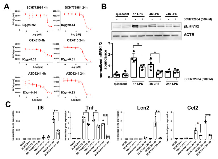
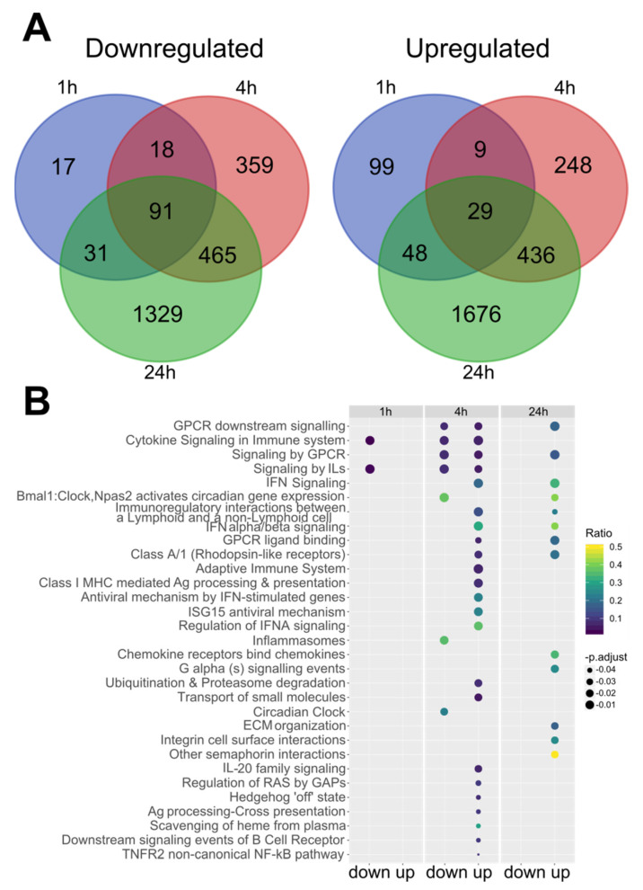
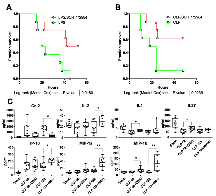
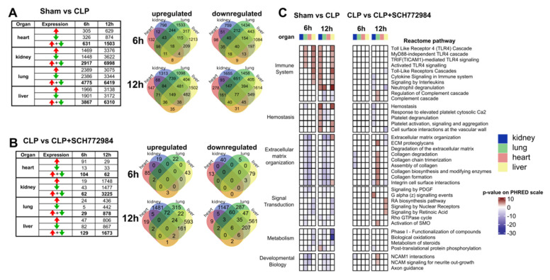
Sepsis is the leading cause of death in intensive care units worldwide. Current treatments of sepsis are largely supportive and clinical trials using specific pharmacotherapy for sepsis have failed to improve outcomes. Here, we used the lipopolysaccharide (LPS)-stimulated mouse RAW264.7 cell line and AlphaLisa assay for TNFa as a readout to perform a supervised drug repurposing screen for sepsis treatment with compounds targeting epigenetic enzymes, including kinases. We identified the SCH772984 compound, an extracellular signal-regulated kinase (ERK) 1/2 inhibitor, as an effective blocker of TNFa production in vitro. RNA-Seq of the SCH772984-treated RAW264.7 cells at 1, 4, and 24 h time points of LPS challenge followed by functional annotation of differentially expressed genes highlighted the suppression of cellular pathways related to the immune system. SCH772984 treatment improved survival in the LPS-induced lethal endotoxemia and cecal ligation and puncture (CLP) mouse models of sepsis, and reduced plasma levels of Ccl2/Mcp1. Functional analyses of RNA-seq datasets for kidney, lung, liver, and heart tissues from SCH772984-treated animals collected at 6 h and 12 h post-CLP revealed a significant downregulation of pathways related to the immune response and platelets activation but upregulation of the extracellular matrix organization and retinoic acid signaling pathways. Thus, this study defined transcriptome signatures of SCH772984 action in vitro and in vivo, an agent that has the potential to improve sepsis outcome.

Keywords: ERK1/2; SCH772984; cecal ligation and puncture; drugs repurposing; sepsis.


Conflict of interest statement

The authors declare no conflict of interest. The funders had no role in the design of the study; in the collection, analyses, or interpretation of data; in the writing of the manuscript, or in the decision to publish the results.

Figures

Figure 1
Figure 1
SCH772984 significantly attenuates LPS-induced TNFa production and inflammatory genes expression in the RAW 264.7 cell line. (A) IC50 values of TNFa production inhibition for the most effective drugs at 4 h and 24 h post-LPS challenge. (B) Representative Western blot (upper panel) showing the levels of ERK1/2 phosphorylation following SCH772984 treatment upon LPS time course challenge. The graph (lower panel) shows the averaged pERK1/2 abundance normalized to Actb by densitometry analysis of Western blotting bands. Values derived from four independent experiments and their means (+/− SD) are presented. * p ≤ 0.05, as determined by unpaired t-test. (C) qPCR results showing the mRNA expression of inflammatory genes following SCH772984 treatment upon LPS time course. Data from three independent experiments and their means and +/− SD are presented. * p ≤ 0.05, ** p < 0.01, *** p < 0.001 as determined by unpaired t-test.
Figure 2
Figure 2
SCH772984 changes RAW 264.7 cells transcriptomes by altering gene expression pathways related to immune system pathways upon LPS challenge. (A) Venn diagram illustrating the number of up and downregulated DEGs (adj. p. value < 0.05) and Reactome pathways (B) altered upon SCH772984 treatment during the LPS challenge time points. IL—interleukin; IFN—interferon; Ag—antigen; ECM—extracellular matrix.
Figure 3
Figure 3
SCH772984 improves survival in mouse models of sepsis and reduces plasma levels of Ccl2/Mcp1 chemokine. Kaplan–Meier survival curves of (A) LPS-treated C57BL/6W mice (20 mg/kg, i.p., n = 8 per group) or mice subjected to cecal ligation puncture (CLP) procedure (B) that were both injected i.p. with solvent control or 10 mg/kg of SCH772984 2 h after LPS administration/CLP procedure. (C) Plasma levels of indicated cytokines were measured with the 26-Plex Mouse ProcartaPlex immunoassay in CLP, SCH772984 (ERKi)-treated CLP, and sham-operated animals (n ≥ 6) at 6 and 12 h endpoint. ** p < 0.01, * p < 0.05 as determined by unpaired t-test. Only seven cytokines with significant alternations in plasma levels following SCH772984 are shown.
Figure 4
Figure 4
SCH772984 downregulates transcripts associated with the immune response and hemostasis in kidney and liver tissues, and positively regulates ECM and retinoic acid pathways in the lungs and liver, respectively, in a CLP-induced mouse model of sepsis. The numbers of differentially expressed genes (adj. p-value < 0.05) across the organs and two time points in the sham vs. CLP groups (A) and upon SCH772984 (10 mg/kg) treatment (B). Red and green arrows indicate up- and downregulated genes, respectively. Venn diagrams show the overlapping numbers of DEGs for organs at a given time point post-CLP for up-and downregulated genes. (C) Significantly altered (adj. p-value < 0.05) recurrent Reactome terms across the organs and time points in the sham vs. CLP and the CLP vs. CLP+ SCH772984 groups. The Reactome pathways with a differential gene ratio higher than 10% and recurrently altered in more than two types of samples are shown. The p-value is shown on the PHRED scale and is reversed for downregulated pathways.

References

    1. Taeb A.M., Hooper M.H., Marik P.E. Sepsis: Current Definition, Pathophysiology, Diagnosis, and Management. Nutr. Clin. Pr. 2017;32:296–308. doi: 10.1177/0884533617695243. - DOI - PubMed
    1. Rudd K.E., Johnson S.C., Agesa K.M., Shackelford K.A., Tsoi D., Kievlan D.R., Colombara D.V., Ikuta K.S., Kissoon N., Finfer S., et al. Global, regional, and national sepsis incidence and mortality, 1990–2017: Analysis for the Global Burden of Disease Study. Lancet. 2020;395:200–211. doi: 10.1016/S0140-6736(19)32989-7. - DOI - PMC - PubMed
    1. Heming N., Lamothe L., Ambrosi X., Annane D. Emerging drugs for the treatment of sepsis. Expert Opin. Emerg. Drugs. 2016;21:27–37. doi: 10.1517/14728214.2016.1132700. - DOI - PubMed
    1. Singer M., Deutschman C.S., Seymour C.C., Shankar-Hari M., Annane D., Bauer M., Bellomo R., Bernard G.R., Chiche J.-D., Coopersmith C.C.M., et al. The Third International Consensus Definitions for Sepsis and Septic Shock (Sepsis-3) JAMA. 2016;315:801–810. doi: 10.1001/jama.2016.0287. - DOI - PMC - PubMed
    1. Nguyen H.B., Smith D. Sepsis in the 21st century: Recent definitions and therapeutic advances. Am. J. Emerg. Med. 2007;25:564–571. doi: 10.1016/j.ajem.2006.08.015. - DOI - PubMed

MeSH terms