. 2021 Sep 23;22(19):10255.
doi: 10.3390/ijms221910255.

Difluoromethylornithine Induces Apoptosis through Regulation of AP-1 Signaling via JNK Phosphorylation in Epithelial Ovarian Cancer

Affiliations

Difluoromethylornithine Induces Apoptosis through Regulation of AP-1 Signaling via JNK Phosphorylation in Epithelial Ovarian Cancer

Woo Yeon Hwang et al. Int J Mol Sci. .

Abstract

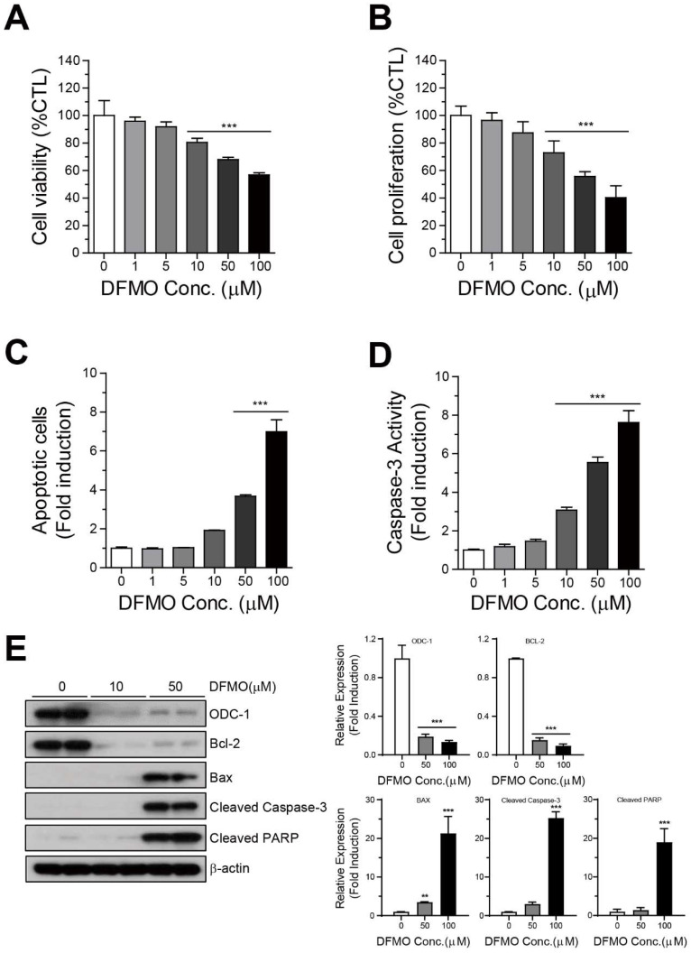
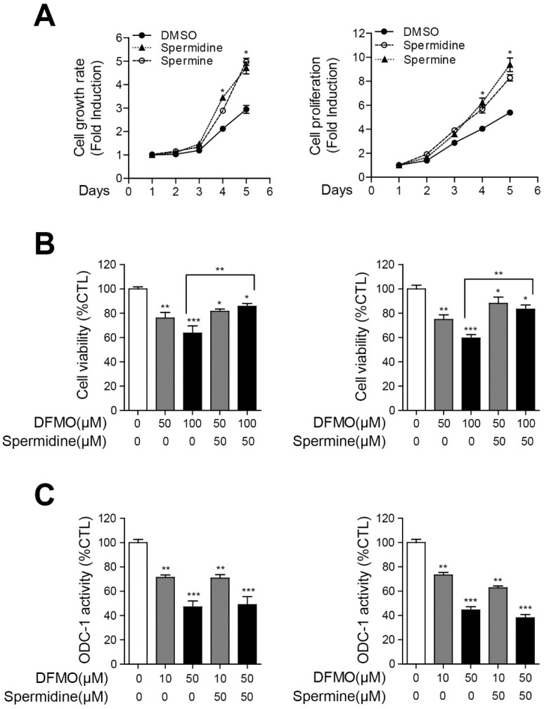
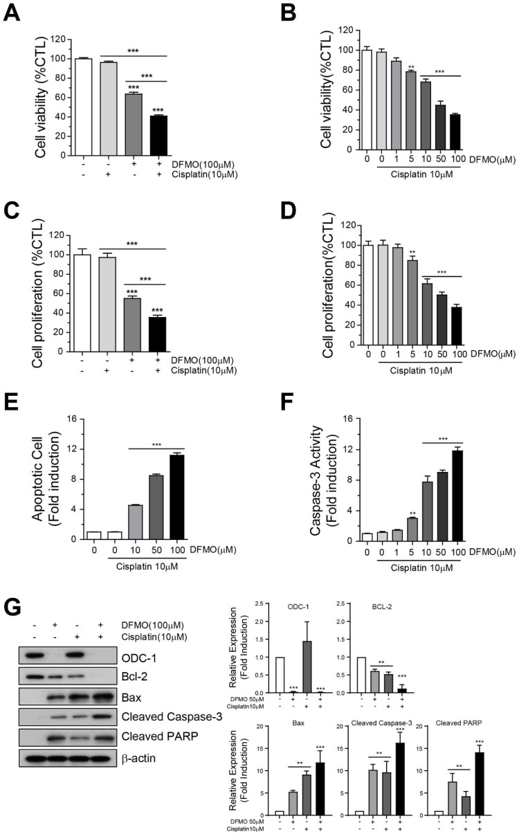
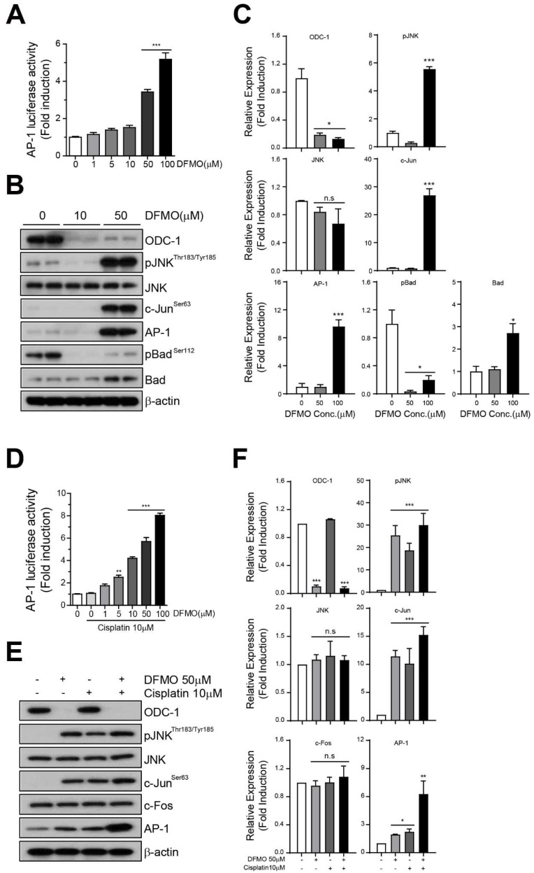
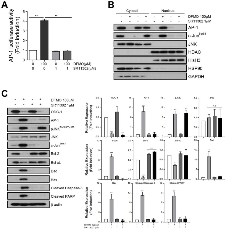
Difluoromethylornithine (DFMO), an irreversible inhibitor of ornithine decarboxylase (ODC), has promising activity against various cancers and a tolerable safety profile for long-term use as a chemopreventive agent. However, the anti-tumor effects of DFMO in ovarian cancer cells have not been entirely understood. Our study aimed to identify the effects and mechanism of DFMO in epithelial ovarian cancer cells using SKOV-3 cells. Treatment with DFMO resulted in a significantly reduced cell viability in a time- and dose-dependent manner. DFMO treatment inhibited the activity and downregulated the expression of ODC in ovarian cancer cells. The reduction in cell viability was reversed using polyamines, suggesting that polyamine depletion plays an important role in the anti-tumor activity of DFMO. Additionally, significant changes in Bcl-2, Bcl-xL, Bax protein levels, activation of caspase-3, and cleavage of poly (ADP-ribose) polymerase were observed, indicating the apoptotic effects of DFMO. We also found that the effect of DFMO was mediated by AP-1 through the activation of upstream JNK via phosphorylation. Moreover, DFMO enhanced the effect of cisplatin, thus showing a possibility of a synergistic effect in treatment. In conclusion, treatment with DFMO alone, or in combination with cisplatin, could be a promising treatment for ovarian cancer.

Keywords: AP-1; DFMO; JNK; apoptosis; ovarian cancer; polyamines.


Conflict of interest statement

The authors declare no conflict of interest.

Figures

Figure 1
Figure 1
Inhibition of cell survival and induction of apoptotic cell death by DFMO in SKOV-3 cells. (A) SKOV-3 cells were cultured with varying concentrations of DFMO (from 0 to 100 μM) for 48 h, and cell viability was detected using the PrestoBlue assay. (B) Cell proliferation was determined by analyzing ATP content in DFMO-treated SKOV-3 cells using the CellTiter-Glo assay. (C) Flow cytometric analysis of Annexin V-FITC/PI-stained SKOV-3 cells treated with the control (0.1% DMSO) or 0–100 μM DFMO was used to determine the apoptotic rates of SKOV-3 cells under various concentrations of DFMO. (D) Caspase-3 activity in DFMO-treated SKOV-3 cells was measured using the Luciferase Assay (Caspase-Glo 3/7 Assay system). (E) Western blot and quantification analysis for the apoptotic proteins Bcl-2, Bcl-xL, Bax, cleaved caspase-3, and cleaved PARP in cells treated with DFMO. The controls were treated with 0.1% DMSO. Data are expressed as mean ± SD. ** p < 0.01, *** p < 0.001 compared with the controls.
Figure 2
Figure 2
Interruption in DFMO-induced cell death by polyamines in SKOV-3 cells. (A) SKOV-3 cells were incubated with polyamines for 1–5 days. Cell growth rate and proliferation were determined using the PrestoBlue and CellTiter-Glo assays, respectively. (B) Polyamines were treated with a concentration of 0 or 50 μM for 48 h in DFMO-treated SKOV-3 cells, and the cell viability in DFMO/polyamines-treated SKOV-3 cells was measured using the PrestoBlue assay. (C) ODC-1 activity in DFMO/polyamines-treated SKOV-3 cells was detected using the Luciferase Assay (Luciferase Assay System). The controls were treated with 0.1% DMSO. Data are expressed as mean ± SD. * p < 0.05, ** p < 0.01, *** p < 0.001 compared with the controls.
Figure 3
Figure 3
Effect of combination therapy using DFMO and cisplatin on SKOV-3 cell survival and apoptotic cell death. Cell viability was determined of cells treated with (A) 10 μM cisplatin, 100 μM DFMO, or (B) 10 μM cisplatin/0–100 μM DFMO for 48 h using the PrestoBlue assay and CellTiter-Glo assay. (C) Proliferation was determined in cells treated with (C) 10 μM cisplatin, 100 μM DFMO, or (D) 10 μM cisplatin/0–100 μM DFMO for 48 h. (E) Flow cytometric analysis of Annexin V-FITC/PI-stained SKOV-3 cells treated with 10 μM cisplatin/0–100 μM DFMO for 48 h was performed to determine the apoptotic rates of SKOV-3 cells under various concentrations of DFMO. (F) Caspase-3 activity in cisplatin/DFMO-treated SKOV-3 cells was measured using the Luciferase Assay (Caspase-Glo 3/7 Assay system). (G) Western blot and quantification analysis for the apoptotic proteins Bcl-2, Bcl-xL, Bax, cleaved caspase-3, and cleaved PARP in cells treated with DFMO. The controls were treated with 0.1% DMSO. Data are expressed as mean ± SD. ** p < 0.01, *** p < 0.001 compared with the controls.
Figure 4
Figure 4
Induction of AP-1 activity and JNK signaling using the combination therapy of DFMO and cisplatin. An AP-1 luciferase plasmid vector was transiently transfected into SKOV-3 cells for 48 h. The transfected cells were seeded in cell culture plates and treated with cisplatin or DFMO for 48 h. The AP-1 luciferase activity of DFMO- or cisplatin/DFMO-treated cells was measured using the Luciferase Assay System (A,D). Western blot and quantification of the proteins of JNK signaling (phospho-JNK, JNK, phospho-cJun, AP-1, c-Fos), and apoptosis (phospho-Bad, Bad) in DFMO- or cisplatin/DFMO-treated cells (B,C,E,F). The controls were treated with 0.1% DMSO. Data are expressed as mean ± SD. * p < 0.05, ** p < 0.01, *** p < 0.001 compared with the controls.
Figure 5
Figure 5
Induction of DFMO-induced apoptotic cell death through the regulation of AP-1 activity. The transfected cells were seeded in cell culture plates and treated with DFMO, SR11302, or DFMO/SR11302 for 48 h. (A) The AP-1 luciferase activity in the treated cells was determined using the Luciferase Assay System. (B) Western blot of organelle extracts (10 μg) was performed using the following markers: nucleus; HDAC, Histone H3/ cytosol: Hsp90, GAPDH. (C) Western blot analysis of the JNK signaling pathway detected JNK signaling proteins (phospho-JNK, JNK, phospho-cJun, AP-1), anti-apoptotic proteins (Bcl-2, Bcl-xL), and proapoptotic proteins (Bad, Bax cleaved caspase-3, cleaved PARP) in DFMO-, SR11302-, or DFMO/SR11302-treated cells, respectively. The controls were treated with 0.1% DMSO. Data are expressed as mean ± SD. * p < 0.05, ** p < 0.01, *** p < 0.001 compared with the controls.

References

    1. Siegel R.L., Miller K.D., Jemal A. Cancer statistics, 2020. CA Cancer J. Clin. 2020;70:7–30. doi: 10.3322/caac.21590. - DOI - PubMed
    1. Tothill R., Tinker A.V., George J., Brown R., Fox S., Lade S., Johnson D.S., Trivett M.K., Etemadmoghadam D., Locandro B., et al. Novel Molecular Subtypes of Serous and Endometrioid Ovarian Cancer Linked to Clinical Outcome. Clin. Cancer Res. 2008;14:5198–5208. doi: 10.1158/1078-0432.CCR-08-0196. - DOI - PubMed
    1. Alexiou G.A., Lianos G.D., Ragos V., Galani V., Kyritsis A.P. Difluoromethylornithine in cancer: New advances. Futur. Oncol. 2017;13:809–819. doi: 10.2217/fon-2016-0266. - DOI - PubMed
    1. Soda K. The mechanisms by which polyamines accelerate tumor spread. J. Exp. Clin. Cancer Res. 2011;30:95. doi: 10.1186/1756-9966-30-95. - DOI - PMC - PubMed
    1. Murray-Stewart T., Ferrari E., Xie Y., Yu F., Marton L.J., Oupicky D., Casero R.A., Jr. Biochemical evaluation of the anticancer potential of the polyamine-based nanocarrier Nano11047. PLoS ONE. 2017;12:e0175917. - PMC - PubMed

MeSH terms