. 2021 Sep 24;22(19):10282.
doi: 10.3390/ijms221910282.

Structure-Based Understanding of ABCA3 Variants

Affiliations

Structure-Based Understanding of ABCA3 Variants

Marion Onnée et al. Int J Mol Sci. .

Abstract

ABCA3 is a crucial protein of pulmonary surfactant biosynthesis, associated with recessive pulmonary disorders such as neonatal respiratory distress and interstitial lung disease. Mutations are mostly private, and accurate interpretation of variants is mandatory for genetic counseling and patient care. We used 3D structure information to complete the set of available bioinformatics tools dedicated to medical decision. Using the experimental structure of human ABCA4, we modeled at atomic resolution the human ABCA3 3D structure including transmembrane domains (TMDs), nucleotide-binding domains (NBDs), and regulatory domains (RDs) in an ATP-bound conformation. We focused and mapped known pathogenic missense variants on this model. We pinpointed amino-acids within the NBDs, the RDs and within the interfaces between the NBDs and TMDs intracellular helices (IHs), which are predicted to play key roles in the structure and/or the function of the ABCA3 transporter. This theoretical study also highlighted the possible impact of ABCA3 variants in the cytosolic part of the protein, such as the well-known p.Glu292Val and p.Arg288Lys variants.

Keywords: 3D modeling; ABCA3; mutation; nucleotide-binding domain (NBD); regulatory domain (RD).


Conflict of interest statement

The authors declare no conflict of interest.

Figures

Figure 1
Figure 1
(A) Schematic 2D representation of the human ABCA3. (B) Ribbon 3D representation of the human ABCA3 3D structure. C-alpha positions of amino-acids with mutations discussed in this study are depicted with balls (orange: mutations listed in the Table 1, pink: mutations listed in Table S2). ATP molecules are colored in yellow.
Figure 2
Figure 2
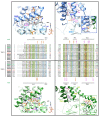
The IHs and interfaces with NBDs. Alignment of the sequences of the IHs in the ABCA family and mapping of critical amino-acids on the model of the 3D structure of human ABCA3. Sequences from human proteins designated with their UniProt identifiers (UniProt accession numbers: ABC1: O95477, ABCA2: Q9BZC7, ABCA4: P78363, ABCA7: Q8IZY2, ABCAC (ABCA12): Q86UK0, ABCAD (ABCA13): Q86UQ4; ABCA3: Q99758, ABCA5: Q8WW27, ABCA6: Q8N139, ABCA8: Q94911, ABCA9: Q8IUA7, ABCAA (ABCA10): Q8WWZ4. IH1 is not present in the ABCA10 sequence. The secondary structures, as observed in the experimental 3D structure of human ABCA4 (PDB 7l kz) are reported above and below the alignments. Positions conserved along the two equivalent IHs in TMD1 and TMD2 are colored according to their properties (and related): green: hydrophobic, orange: aromatic, blue: basic, pink: acidic, yellow: small. Positions of mutated amino-acids are shown with stars (orange: mutations listed in the Table 1, pink: mutations listed in Table S2 and some mutations in TMD parts in contact with IHs) on the alignment and depicted with the same colors on the 3D structure views, together with other amino-acids predicted to play important roles in these regions.
Figure 3
Figure 3
ATP-binding sites in the NBDs. (A) Alignment of the sequences of the NBDs in the ABCA family. (B) Mapping of critical amino-acids of the ATP-binding sites on the model of the 3D structure of humanABCA3. ATP molecules are in white. Important amino-acids positions are written in blue (NBD1) or green (NBD2) and are highlighted in colors according to the motif they belong to.
Figure 4
Figure 4
The RDs. (A) Alignment of the sequences of the RDs in the ABCA family. (B) Mapping of critical amino-acids on the model of the 3D structure of human ABCA3. The two highly conserved phenylalanine of the pinning helices (PH) are also shown in atomic details, highlighting interactions with the NBDs cores.

References

    1. Guagliardo R., Pérez-Gil J., De Smedt S., Raemdonck K. Pulmonary Surfactant and Drug Delivery: Focusing on the Role of Surfactant Proteins. J. Control. Release. 2018;291:116–126. doi: 10.1016/j.jconrel.2018.10.012. - DOI - PubMed
    1. Beers M.F., Mulugeta S. The Biology of the ABCA3 Lipid Transporter in Lung Health and Disease. Cell Tissue Res. 2017;367:481–493. doi: 10.1007/s00441-016-2554-z. - DOI - PMC - PubMed
    1. Wambach J.A., Casey A.M., Fishman M.P., Wegner D.J., Wert S.E., Cole F.S., Hamvas A., Nogee L.M. Genotype–Phenotype Correlations for Infants and Children with ABCA3 Deficiency. Am. J. Respir. Crit. Care Med. 2014;189:1538–1543. doi: 10.1164/rccm.201402-0342OC. - DOI - PMC - PubMed
    1. Shulenin S., Nogee L.M., Annilo T., Wert S.E., Whitsett J.A., Dean M. ABCA3 Gene Mutations in Newborns with Fatal Surfactant Deficiency. N. Engl. J. Med. 2004;350:1296–1303. doi: 10.1056/NEJMoa032178. - DOI - PubMed
    1. Klay D., Platenburg M.G.J.P., van Rijswijk R.H.N.A.J., Grutters J.C., van Moorsel C.H.M. ABCA3 Mutations in Adult Pulmonary Fibrosis Patients: A Case Series and Review of Literature. Curr. Opin. Pulm. Med. 2020;26:293–301. doi: 10.1097/MCP.0000000000000680. - DOI - PubMed

MeSH terms

Substances

LinkOut - more resources