Clinical Trial
. 2021 Sep 27;22(19):10397.
doi: 10.3390/ijms221910397.

Interleukin-9 Facilitates Osteoclastogenesis in Rheumatoid Arthritis

Affiliations
Clinical Trial

Interleukin-9 Facilitates Osteoclastogenesis in Rheumatoid Arthritis

Santanu Kar et al. Int J Mol Sci. .

Abstract

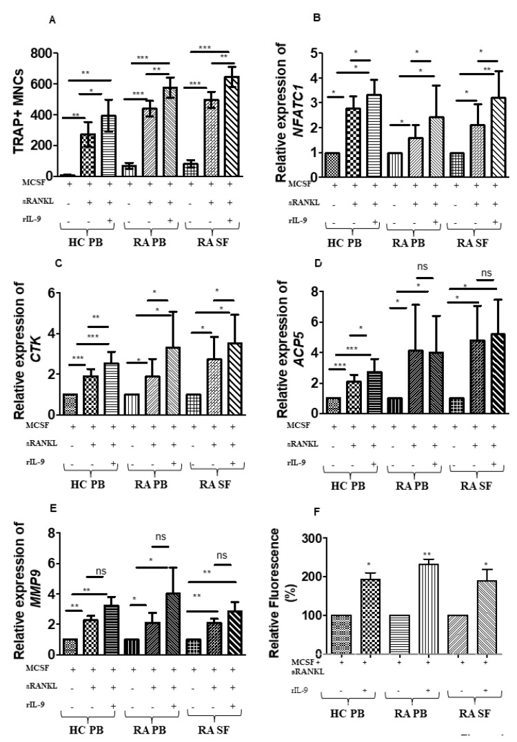
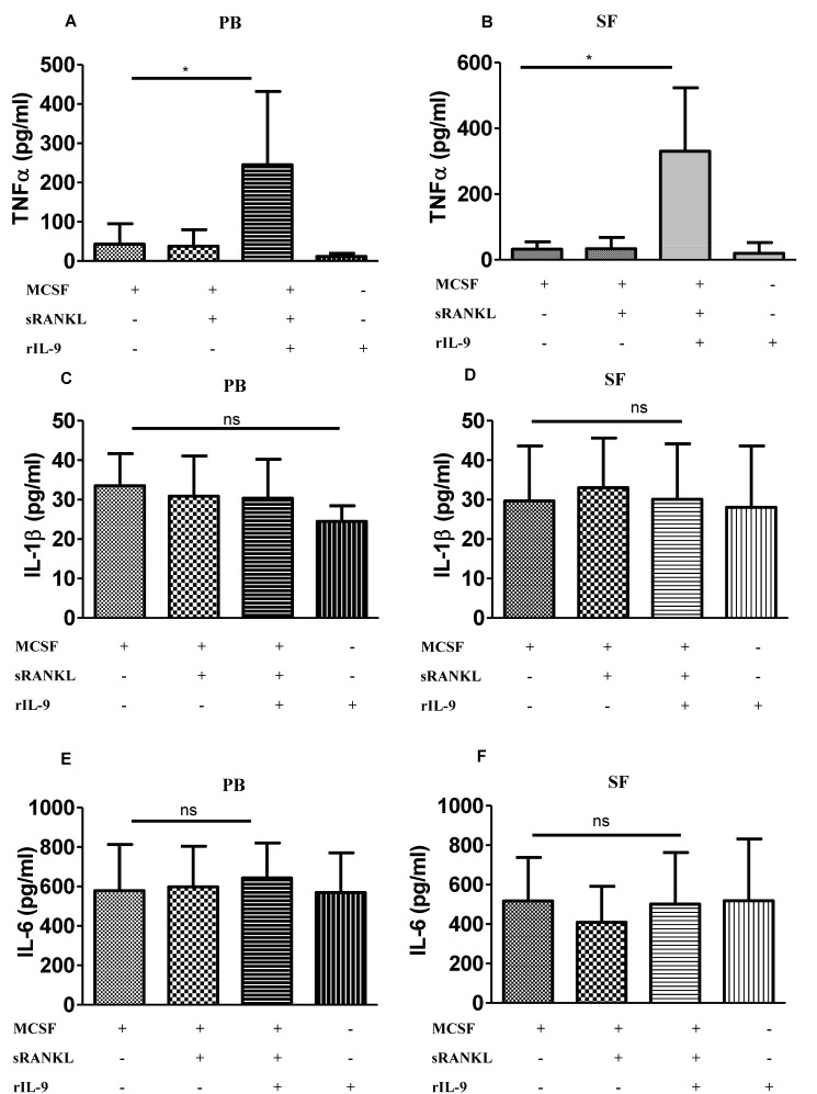
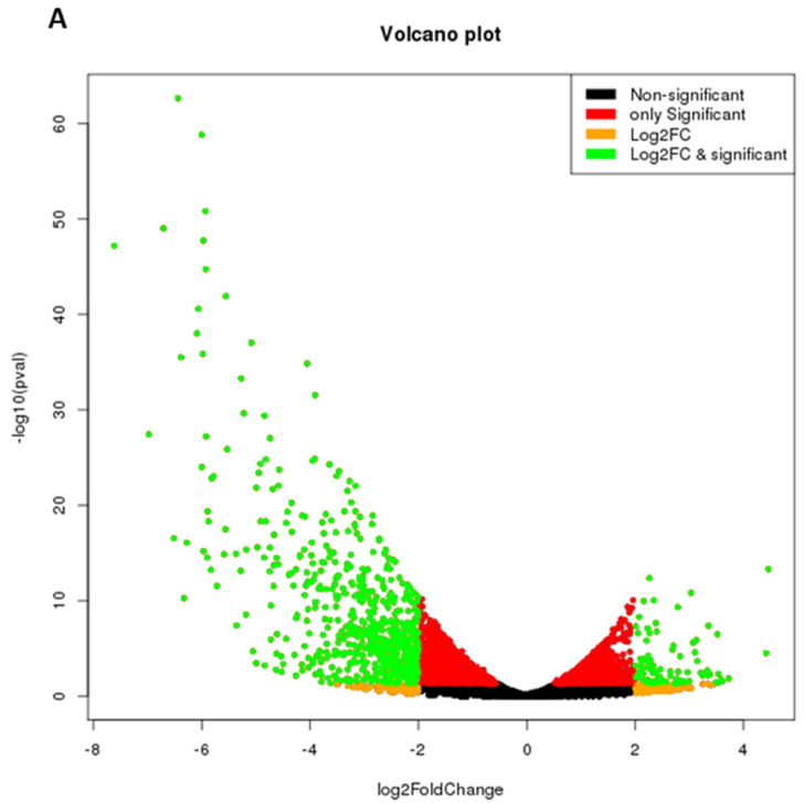
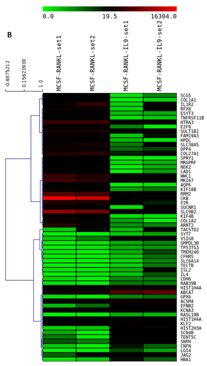
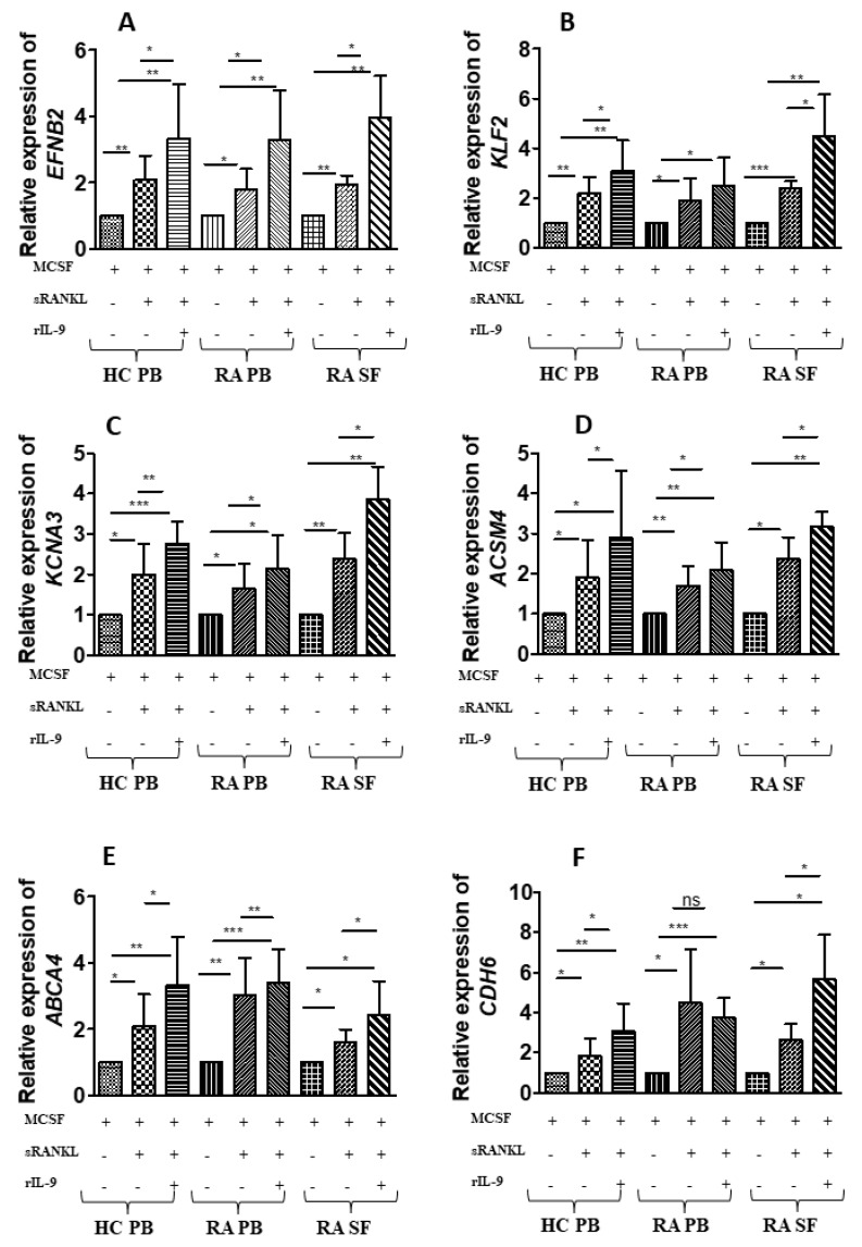
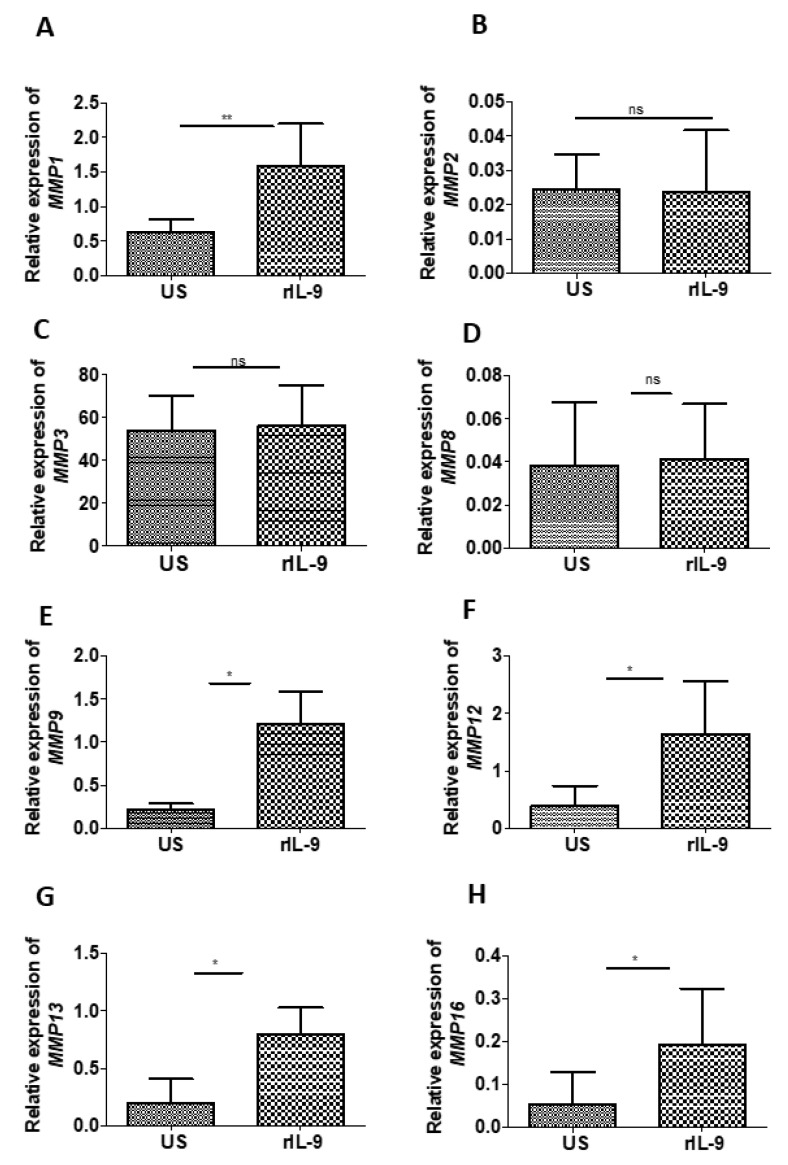
In rheumatoid arthritis (RA), inflammatory cytokines play a pivotal role in triggering abnormal osteoclastogenesis leading to articular destruction. Recent studies have demonstrated enhanced levels of interleukin-9 (IL-9) in the serum and synovial fluid of patients with RA. In RA, strong correlation has been observed between tissue inflammation and IL-9 expression in synovial tissue. Therefore, we investigated whether IL-9 influences osteoclastogenesis in patients with RA. We conducted the study in active RA patients. For inducing osteoclast differentiation, mononuclear cells were stimulated with soluble receptor activator of NF-kB ligand (sRANKL) and macrophage-colony-stimulating factor (M-CSF) in the presence or absence of recombinant (r) IL-9. IL-9 stimulation significantly enhanced M-CSF/sRANKL-mediated osteoclast formation and function. Transcriptome analysis revealed differential gene expression induced with IL-9 stimulation in the process of osteoclast differentiation. IL-9 mainly modulates the expression of genes, which are involved in the metabolic pathway. Moreover, we observed that IL-9 modulates the expression of matrix metalloproteinases (MMPs), which are critical players in bone degradation. Our results indicate that IL-9 has the potential to influence the structural damage in the RA by promoting osteoclastogenesis and modulating the expression of MMPs. Thus, blocking IL-9 pathways might be an attractive immunotherapeutic target for preventing bone degradation in RA.

Keywords: differential gene expression; interleukin-9; matrix metalloproteinases; osteoclast; osteoclastogenesis; rheumatoid arthritis.


Conflict of interest statement

The authors declare no conflict of interest. The funders had no role in the design of the study; in the collection, analyses, or interpretation of data; in the writing of the manuscript, or in the decision to publish the results.

Figures

Figure 1
Figure 1
Interleukin (IL)-9 enhances osteoclast formation and function in cells derived from RA and HC. (A) Cells derived from peripheral blood (PB) of healthy control (HC); cells derived from PB and synovial fluid (SF) of patients with RA were treated as indicated with macrophage colony-stimulating factor (M-CSF; 25 ng/mL), soluble receptor activator of nuclear factor κB ligand (sRANKL; 50 ng/mL), or IL-9 (100 ng/mL) for 21 days. Half of the culture medium was replenished with fresh culture medium containing stimulating factors (M-CSF, sRANKL, and rIL-9) at 4-day intervals. Cells were then fixed and stained for tartrate-resistant acid phosphatase (TRAP). Using a light microscope, multinucleated (≥3 nuclei) TRAP+ cells were counted manually. Graph shows TRAP+ multinucleated cells (MNCs) (mean  ±  SD; n  =  5). Statistical analysis was performed using paired Student’s t-test comparing M-CSF/sRANKL/rIL-9-treated cells to M-CSF/sRANKL, M-CSF, or M-CSF/sRANKL-treated cells to M-CSF (*: p  ≤  0.05; **: p  ≤  0.005; ***: p  ≤  0.0005). (BE) Cells derived from PB of HC, PB, and SF of patients with RA were stimulated as indicated with M-CSF (25 ng/mL), sRANKL (50 ng/mL), or IL-9 (100 ng/mL) for 3 days. Cells were then lysed followed by RNA extraction and cDNA preparation. Quantitative real-time PCR (RT-PCR) was performed for osteoclast-specific marker genes, nuclear factor of activated T-cells, cytoplasmic 1 (NFATc1), cathepsin K (CTSK), acid phosphatase 5, tartrate-resistant (ACP5), and matrix metalloproteinase-9 (MMP-9). The graphs represent the relative expression of NFATc1, CTSK, ACP5, and MMP-9 (mean  ±  SD; n  =  8). Statistical analysis was performed using paired Student’s t-test comparing M-CSF/sRANKL/rIL-9-treated cells to M-CSF/sRANKL-, M-CSF-, or M-CSF/sRANKL-treated cells to M-CSF (*: p  ≤  0.05; **: p  ≤  0.005; ***: p  ≤  0.0005; ns –non significant). (F) Cells derived from PB of HC, PB, and SF of patients with RA were stimulated with M-CSF (25 ng/mL), sRANKL (100 ng/mL), or IL-9 (100 ng/mL) as indicated for 24 days. Half of the culture medium was replenished with fresh culture medium containing stimulating factors (M-CSF, sRANKL, and rIL-9) at 4-day intervals. Using a fluorometer, fluorescence intensity of the culture supernatant was measured. The graph represents the relative fluorescence intensity (mean  ±  SD; n  =  3). Statistical analysis was performed using paired Student’s t-test comparing M-CSF/sRANKL/rIL-9-treated cells to M-CSF/sRANKL-treated cells. (*: p  ≤  0.05; **: p  ≤  0.005).
Figure 2
Figure 2
Interleukin (IL)-9 enhances TNF-α production during osteoclastogenesis in RA. (AF) Cells derived from peripheral blood (PB) and synovial fluid (SF) of patients with RA were treated as indicated with macrophage colony-stimulating factor (M-CSF; 25 ng/mL), soluble receptor activator of nuclear factor κB ligand (sRANKL; 50 ng/mL), or IL-9 (100 ng/mL) for 4 days. Cell culture supernatants were analyzed for TNF-α (A,B), IL-1β (C,D), and IL-6 (E,F) using ELISA (mean  ±  SD; n = 8). Statistical analysis was performed using paired Student’s t-test comparing M-CSF/sRANKL/rIL-9-treated cells to M-CSF/sRANKL- or M-CSF-treated cells (*: p  ≤  0.05; ns–non significant).
Figure 3
Figure 3
IL-9 mediated differential gene expression profile during osteoclastogenesis in RA. Cells derived from synovial fluid (SF) of patients with RA were treated with macrophage colony-stimulating factor (M-CSF; 25 ng/mL) and soluble receptor activator of nuclear factor κB ligand (sRANKL; 50 ng/mL) in the presence or absence of rIL-9 (100 ng/mL) for 4 days. After that RNA sequencing was performed with the treated cells as described in Materials and Methods. (A) Volcano plot of differentially expressed genes between M-CSF + sRANKL-treated cells and M-CSF + sRANKL + rIL-9-treated cells. (B) Heatmap of hierarchical cluster analysis of differentially expressed genes between M-CSF + sRANKL-treated cells and M-CSF + sRANKL + rIL-9-treated cells.
Figure 3
Figure 3
IL-9 mediated differential gene expression profile during osteoclastogenesis in RA. Cells derived from synovial fluid (SF) of patients with RA were treated with macrophage colony-stimulating factor (M-CSF; 25 ng/mL) and soluble receptor activator of nuclear factor κB ligand (sRANKL; 50 ng/mL) in the presence or absence of rIL-9 (100 ng/mL) for 4 days. After that RNA sequencing was performed with the treated cells as described in Materials and Methods. (A) Volcano plot of differentially expressed genes between M-CSF + sRANKL-treated cells and M-CSF + sRANKL + rIL-9-treated cells. (B) Heatmap of hierarchical cluster analysis of differentially expressed genes between M-CSF + sRANKL-treated cells and M-CSF + sRANKL + rIL-9-treated cells.
Figure 4
Figure 4
Validation that IL-9 induced six differentially expressed genes during osteoclast differentiation. (AF) Cells derived from peripheral blood (PB) of healthy control (HC) and cells derived from PB and synovial fluid (SF) of patients with RA were treated as indicated with macrophage colony-stimulating factor (M-CSF; 25 ng/mL), soluble receptor activator of nuclear factor κB ligand (sRANKL; 50 ng/mL), or IL-9 (100 ng/mL) for 4 days. Cells were then lysed followed by RNA extraction and cDNA preparation from an equal quantity of RNA. Glyceraldehyde phosphate dehydrogenease (GAPDH) was used as an internal control for validation of the differentially expressed genes, as the expression of GAPDH showed least variation with stimulation. Quantitative real-time PCR (RT-PCR) was performed for ephrinB2 (EFNB2), ATP-binding cassette subfamily A member 7 (ABCA7), Krueppel-like factor 2 (KLF2), cadherin 6 (CDH6), acyl-coenzyme A synthetase (ACSM4), and potassium voltage-gated channel subfamily A member 3 (KCNA3). The graphs represent the relative expression of these genes (mean  ±  SD; n  =  8). Statistical analysis was performed using paired Student’s t-test comparing M-CSF/sRANKL/rIL-9-treated cells to M-CSF/sRANKL-, M-CSF-, and M-CSF/sRANKL-treated cells to M-CSF (*: p  ≤  0.05; **: p  ≤  0.005; ***: p  ≤  0.0005; ns–non significant).
Figure 5
Figure 5
Effect of IL-9 on the expression of matrix metalloproteinases (MMPs). (AH) Bone explant culture was stimulated with and without rIL-9 (100 ng/mL) for 5 days. Cells were then lysed and homogenized, followed by RNA extraction and cDNA preparation from an equal quantity of RNA. Glyceraldehyde phosphate dehydrogenease (GAPDH) was used as an internal control as the expression of GAPDH showed the least variation with stimulation. Quantitative real-time PCR (RT-PCR) was performed for MMP-1 (A), MMP-2 (B), MMP-3 (C), MMP-8 (D), MMP-9 (E), MMP-12 (F), MMP-13 (G), and MMP-16 (H). The graphs represent the relative expression of these genes (mean  ±  SD; n  =  6). Statistical analysis was performed using paired Student’s t-test comparing treated cells to untreated cells (*: p  ≤  0.05; **: p  ≤  0.005; ns—non significant).

References

    1. Feldmann M., Brennan F.M., Maini R.N. Rheumatoid arthritis. Cell. 1996;85:307–310. doi: 10.1016/S0092-8674(00)81109-5. - DOI - PubMed
    1. Hoes J.N., Bultink I.E.M., Lems W.F. Management of osteoporosis in rheumatoid arthritis patients. Expert Opin. Pharm. 2015;16:559–571. doi: 10.1517/14656566.2015.997709. - DOI - PubMed
    1. Chen B., Cheng G., Wang H., Feng Y. Increased risk of vertebral fracture in patients with rheumatoid arthritis: A meta-analysis. Medicine. 2016;95:e5262. doi: 10.1097/MD.0000000000005262. - DOI - PMC - PubMed
    1. Schett G. Cells of the synovium in rheumatoid arthritis. Osteoclasts. Arthritis Res. Ther. 2007;9:203. doi: 10.1186/ar2110. - DOI - PMC - PubMed
    1. Walsh N.C., Reinwald S., Manning C.A., Condon K.W., Iwata K., Burr D.B., Gravallese E.M. Osteoblast function is compromised at sites of focal bone erosion in inflammatory arthritis. J. Bone Miner. Res. 2009;24:1572–1585. doi: 10.1359/jbmr.090320. - DOI - PubMed

Publication types