. 2021 Sep 28;22(19):10489.
doi: 10.3390/ijms221910489.

Establishment of Bovine-Induced Pluripotent Stem Cells

Affiliations

Establishment of Bovine-Induced Pluripotent Stem Cells

Yue Su et al. Int J Mol Sci. .

Abstract

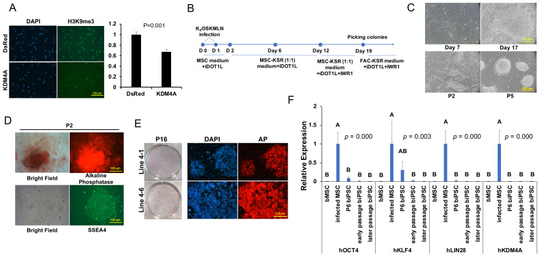
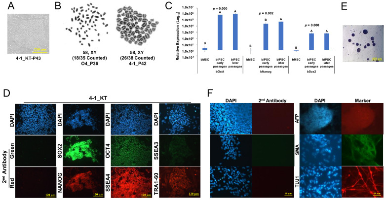
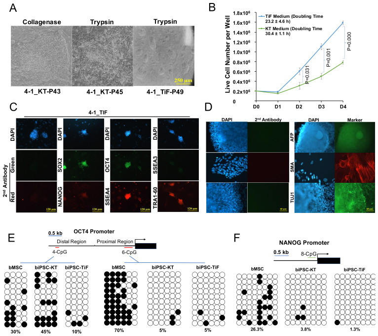
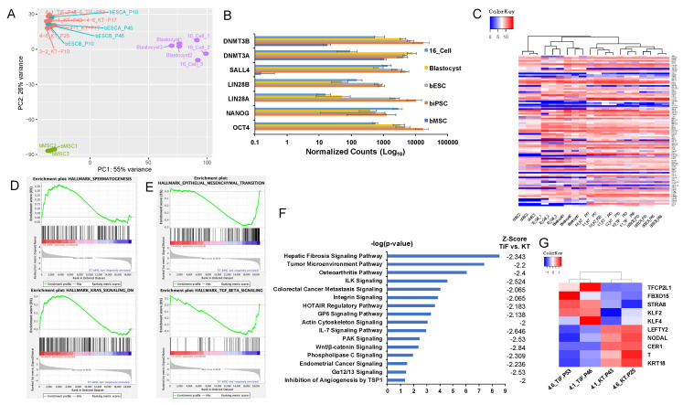
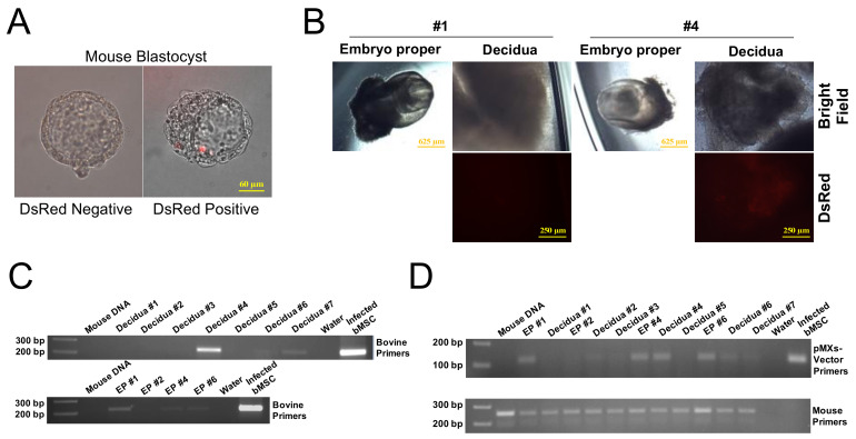
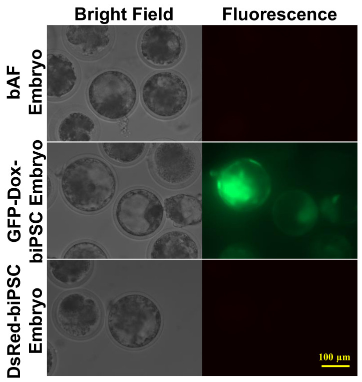
Pluripotent stem cells (PSCs) have been successfully developed in many species. However, the establishment of bovine-induced pluripotent stem cells (biPSCs) has been challenging. Here we report the generation of biPSCs from bovine mesenchymal stem cells (bMSCs) by overexpression of lysine-specific demethylase 4A (KDM4A) and the other reprogramming factors OCT4, SOX2, KLF4, cMYC, LIN28, and NANOG (KdOSKMLN). These biPSCs exhibited silenced transgene expression at passage 10, and had prolonged self-renewal capacity for over 70 passages. The biPSCs have flat, primed-like PSC colony morphology in combined media of knockout serum replacement (KSR) and mTeSR, but switched to dome-shaped, naïve-like PSC colony morphology in mTeSR medium and 2i/LIF with single cell colonization capacity. These cells have comparable proliferation rate to the reported primed- or naïve-state human PSCs, with three-germ layer differentiation capacity and normal karyotype. Transcriptome analysis revealed a high similarity of biPSCs to reported bovine embryonic stem cells (ESCs) and embryos. The naïve-like biPSCs can be incorporated into mouse embryos, with the extended capacity of integration into extra-embryonic tissues. Finally, at least 24.5% cloning efficiency could be obtained in nuclear transfer (NT) experiment using late passage biPSCs as nuclear donors. Our report represents a significant advance in the establishment of bovine PSCs.

Keywords: bovine; differentiation; embryo aggregation; induced pluripotent stem cells (iPSCs); nuclear transfer (NT); pluripotency; reprogramming.


Conflict of interest statement

The authors declare no conflict of interest.

Figures

Figure 1
Figure 1
Induction of biPSCs. (A): Left Panel: Immunostaining of H3K9me3 expression in bMSCs infected with retroviral vector control or KDM4A. Bar = 120 µm. Right Panel: Relative fluorescence intensity for H3K9me3 in bMSCs. Bar = mean ± sd, n = 3. Student’s t-test was used for data analysis. (B): Scheme of reprogramming of the bovine MSCs. (C): Upper panel: Development of iPSC colonies at day 7 and 17 of retroviral KdOSKMLN infection of bMSCs, bar = 120 µm. Lower panel: Picked biPSC colonies at P2 and P5, bar = 250 µm. (D): Expression of AP (upper panel) and SSEA4 (lower panel) in P2 biPSC colonies, bar = 120 µm. (E): AP staining of biPSCs expanded in KT medium at P16. Left: Two AP-stained biPSC lines from one well of a 6-well plate. Right: AP-fluorescence from 2 biPSC lines under the microscope. Bar = 120 µm. (F): qRT-PCR for transgene expression in four lines of biPSCs at passage 6 (P6), early passages (Lines 4-1 at P17, 4-6 at P17, 3-2 at P10), and later passages (Lines 4-1 at P43, 4-6 at P25, 3-2 at P29, O4 at P40). Transgene infected bMSCs at 48 h were used as the positive control. Bar = mean ± sd, n = 4. One way–ANOVA with Tukey’s post hoc multiple comparison test was used for data analysis.
Figure 2
Figure 2
Characterization of primed-like bovine iPSCs. (A): Flat colony morphology of 4-1 in KT medium. Bar = 250 µm. (B): Karyotype for O4 and 4-1 biPSC lines at passage 36 and 42, respectively. (C): qRT-PCR for endogenous expression of pluripotent genes in four lines of biPSCs at early passages (4-1 at P17, 4-6 at P17, 3-2 at P10) and later passages (4-1 at P43, 4-6 at P25, 3-2 at P29, O4 at P40). Bar = mean ± sd, n = 4. One way–ANOVA with Tukey’s post hoc multiple comparison test was used for data analysis. (D): Representative immunostaining images of biPSCs in KT medium for OCT4, NANOG, SOX2, SSEA3, SSEA4, TRA-1-60. Bar = 120 µm. (E): EBs formed from 4-1_KT, bar = 625 µm. (F): Immunostaining of differentiated cells for the three-germ layer markers (AFP for endoderm, SMA for mesoderm, and TUJ1 for ectoderm) after passaging of the EBs. bar = 120 µm.
Figure 3
Figure 3
Characterization of naïve-like bovine iPSCs. (A): Flat, monolayered primed-like 4-1 biPSC colonies cultured in KT medium and passaged with collagenase (left), deteriorated colony morphology upon trypsinization (middle), and dome-shaped, naïve-like biPSCs cultured in TiF medium and passaged with trypsin (right), bar = 250 µm. (B): Cell proliferation difference between naïve and primed-like biPSCs. Mean ± sd, n = 3. Student’s t-test was used for data comparison. (C): immunostaining of naïve-like 4-1 biPSCs in TiF medium with OCT4, NANOG, SOX2, SSEA3, SSEA4, TRA-1-60 antibodies. Bar = 120 µm. (D): Immunostaining of EBs derived from naïve-like biPSCs for three-germ layer markers (AFP for endoderm, SMA for mesoderm, and TUJ1 for ectoderm) after passaging of the EBs, bar = 120 µm. (E): Bisulfite sequencing results of bovine OCT4 distal enhancer (Left Panel) and proximal promoter (Right Panel) genomic regions. Open and closed circles represent unmethylated and methylated CpGs, respectively. The percentage of methylated CpG is shown at the bottom of each sample. (F): Bisulfite sequencing results of bovine NANOG promoter genomic region. Open and closed circles represent unmethylated and methylated CpGs, respectively. The percentage of methylated CpG is shown at the bottom of each sample.
Figure 4
Figure 4
Transcription analysis of biPSCs. (A): PCA analysis of RNA-seq data from biPSCs in KT and TiF media, bESCs, and bovine embryos at the 16-cell and blastocyst stages. (B): Comparison of pluripotent gene expression between biPSCs, bESCs, bovine embryos, and bMSCs based on RNA-seq data. (C): Heatmap2 clustering analysis on 97 bovine pluripotent genes detected from RNA-seq data. (D): Biological processes enriched in naïve-like biPSCs by GSEA analysis. (E): Biological processes enriched in primed-like biPSCs by GSEA analysis. (F): Significantly inhibited signaling pathways in naïve-like biPSCs (in TiF medium) compared with primed-like biPSCs (in KT medium). (G): Heatmap2 analysis on naïve- and primed- markers for biPSCs in TiF and KT media based on RNA-seq data.
Figure 5
Figure 5
Mouse embryo chimerism analysis of biPSCs. (A): DsRed-biPSCs incorporation of mouse blastocyst (right) at 24 h after embryo aggregation. Bar = 60 µm. (B): DsRed-biPSCs contribution to the decidua of #4 (right) but not #1 (left) E8.5 mouse embryo. Bar = 625 or 250 µm as indicated. (C): PCR analysis of DsRed-biPSC contribution in E8.5 mouse decidual tissue (upper panel) and embryo proper (EP, lower panel) using bovine-specific primers. (D): PCR analysis of DsRed-biPSC contribution on E8.5 embryos using pMXs-vector primers (upper panel). PCR using mouse-specific primers (lower panel) served as reference.
Figure 6
Figure 6
Blastocysts cloned from biPSCs. Blastocysts using bAF as nuclear donors (upper panel); GFP-Dox-biPSC as nuclear donors (middle panel), Dox was added into the embryo culture medium 6 days after activation of cloned embryos to induce the GFP expression; and DsRed-biPSC as nuclear donors (lower panel). Bar = 100 µm.

References

    1. Okita K., Ichisaka T., Yamanaka S. Generation of germline-competent induced pluripotent stem cells. Nat. Cell Biol. 2007;448:313–317. doi: 10.1038/nature05934. - DOI - PubMed
    1. Takahashi K., Yamanaka S. Induction of Pluripotent Stem Cells from Mouse Embryonic and Adult Fibroblast Cultures by Defined Factors. Cell. 2006;126:663–676. doi: 10.1016/j.cell.2006.07.024. - DOI - PubMed
    1. Yu J., Vodyanik M.A., Smuga-Otto K., Antosiewicz-Bourget J., Frane J.L., Tian S., Nie J., Jonsdottir G.A., Ruotti V., Stewart R., et al. Induced pluripotent stem cell lines derived from human somatic cells. Science. 2007;318:1917–1920. doi: 10.1126/science.1151526. - DOI - PubMed
    1. Stice S.L., Strelchenko N.S., Keefer C., Matthews L. Pluripotent bovine embryonic cell lines direct embryonic development following nuclear transfer. Biol. Reprod. 1996;54:100–110. doi: 10.1095/biolreprod54.1.100. - DOI - PubMed
    1. Gong G., Roach M.L., Jiang L., Yang X., Tian X.C. Culture Conditions and Enzymatic Passaging of Bovine ESC-Like Cells. Cell. Reprogram. 2010;12:151–160. doi: 10.1089/cell.2009.0049. - DOI - PubMed

MeSH terms