. 2021 Sep 29;22(19):10520.
doi: 10.3390/ijms221910520.

Effects of Substitution on Cytotoxicity of Diphenyl Ditelluride in Cultured Vascular Endothelial Cells

Affiliations

Effects of Substitution on Cytotoxicity of Diphenyl Ditelluride in Cultured Vascular Endothelial Cells

Takato Hara et al. Int J Mol Sci. .

Abstract

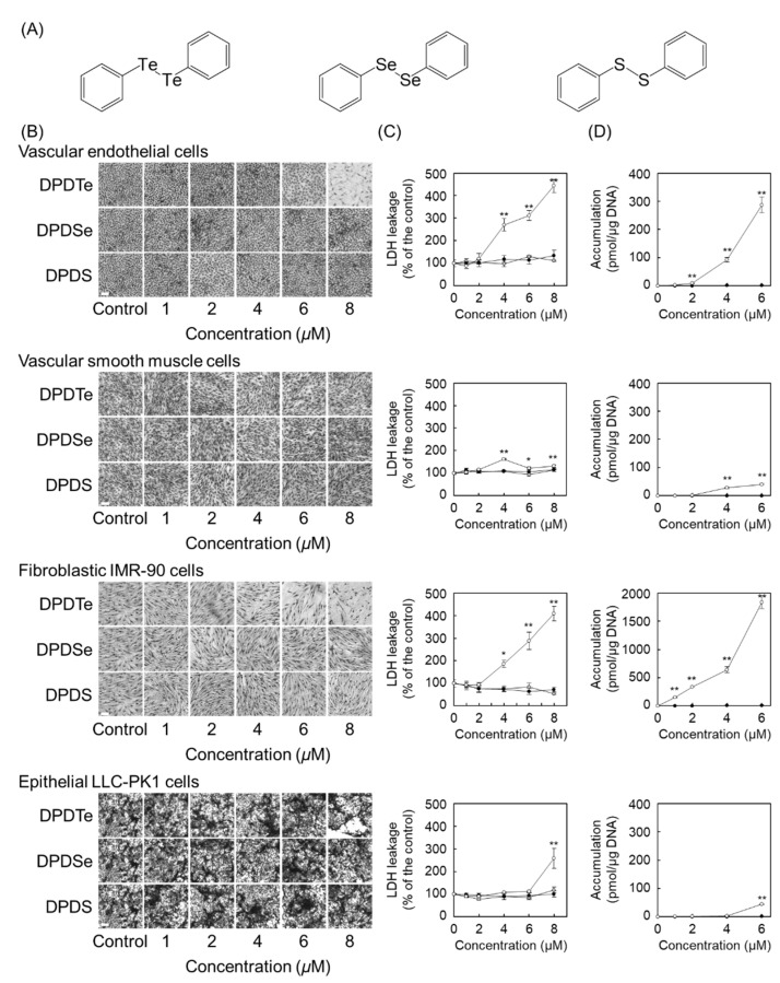
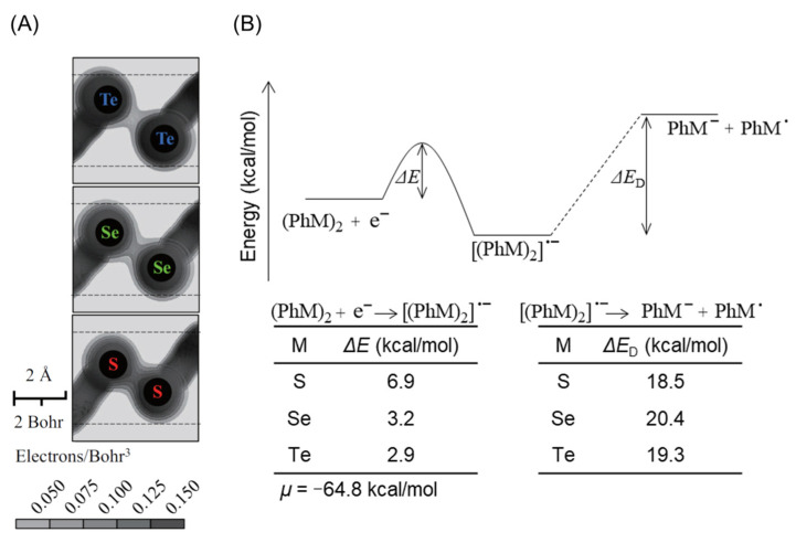
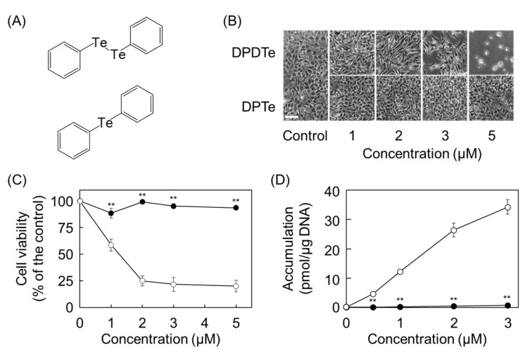
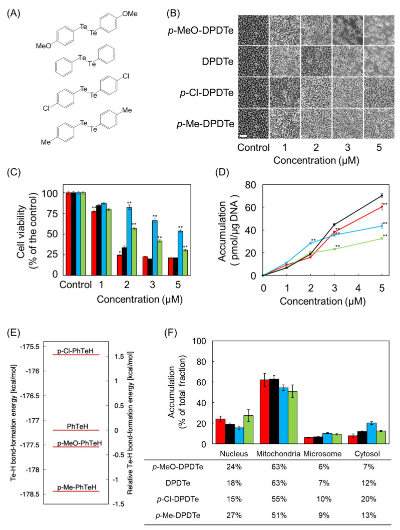
Among organic-inorganic hybrid molecules consisting of organic structure(s) and metal(s), only few studies are available on the cytotoxicity of nucleophilic molecules. In the present study, we investigated the cytotoxicity of a nucleophilic organotellurium compound, diphenyl ditelluride (DPDTe), using a cell culture system. DPDTe exhibited strong cytotoxicity against vascular endothelial cells and fibroblasts along with high intracellular accumulation but showed no cytotoxicity and had less accumulation in vascular smooth muscle cells and renal epithelial cells. The cytotoxicity of DPDTe decreased when intramolecular tellurium atoms were replaced with selenium or sulfur atoms. Electronic state analysis revealed that the electron density between tellurium atoms in DPDTe was much lower than those between selenium atoms of diphenyl diselenide and sulfur atoms of diphenyl disulfide. Moreover, diphenyl telluride did not accumulate and exhibit cytotoxicity. The cytotoxicity of DPDTe was also affected by substitution. p-Dimethoxy-DPDTe showed higher cytotoxicity, but p-dichloro-DPDTe and p-methyl-DPDTe showed lower cytotoxicity than that of DPDTe. The subcellular distribution of the compounds revealed that the compounds with stronger cytotoxicity showed higher accumulation rates in the mitochondria. Our findings suggest that the electronic state of tellurium atoms in DPDTe play an important role in accumulation and distribution of DPDTe in cultured cells. The present study supports the hypothesis that nucleophilic organometallic compounds, as well as electrophilic organometallic compounds, exhibit cytotoxicity by particular mechanisms.

Keywords: bio-organometallics; cytotoxicity; organoselenium compound; organotellurium compound.


Conflict of interest statement

The authors declare no conflict of interest.

Figures

Figure 1
Figure 1
Cytotoxicity and intracellular accumulation of diphenyl ditelluride (DPDTe), and compounds, in which tellurium in DPDTe was replaced by selenium and sulfur atoms (DPDSe and DPDS). Vascular endothelial cells, vascular smooth muscle cells, fibroblastic IMR-90 cells, and epithelial LLC-PK1 cells were treated with DPDTe, DPDSe, or DPDS (1, 2, 4, 6, or 8 µM) for 24 h. (A) Structure of DPDTe (left), DPDSe (middle), and DPDS (right). (B) Morphological observation (bar = 50 µm) and (C) lactate dehydrogenase (LDH) leakage from the cells treated with DPDTe (white), DPDSe (black), or DPDS (gray). (D) Intracellular accumulation of DPDTe (white) and DPDSe (black). Values are expressed as the mean ± standard error of three replicates. * p < 0.05, ** p < 0.01 compared with the corresponding control.
Figure 2
Figure 2
Estimated cleavage process of DPDTe, DPDSe, and DPDS. (A) Electron density of DPDTe, DPDSe, and DPDS determined by ab initio quantum chemical calculations. The darker the black, the higher the electron density. (B) Energy diagram of DPDTe, DPDSe, and DPDS in heterolytic cleavage process.
Figure 3
Figure 3
Assessment of contribution of the Te–Te bond to cytotoxicity of DPDTe. (A) Structure of DPDTe (top) and DPTe (bottom). (B) Morphological observation (bar = 50 µm), (C) viability of vascular endothelial cells treated with DPDTe (white) and DPTe (black), and (D) intracellular accumulation of DPDTe (white) and DPTe (black) in vascular endothelial cells after treatment (0.5, 1, 2, 3, or 5 µM) for 24 h. Values are expressed as the mean ± standard error of three replicates. ** p < 0.01 compared with the corresponding DPDTe treatment.
Figure 4
Figure 4
Effect of substitution on cytotoxicity and intracellular accumulation of DPDTe. (A) Structure of p-MeO-DPDTe, DPDTe, p-Cl-DPDTe, and p-Me-DPDTe. (B) Morphological observation (bar = 50 µm), (C) viability of vascular endothelial cells, and (D) intracellular accumulation of p-MeO-DPDTe, DPDTe, p-Cl-DPDTe, and p-Me-DPDTe. Vascular endothelial cells were treated with p-MeO-DPDTe (red), DPDTe (black), p-Cl-DPDTe (blue), and p-Me-DPDTe (green) (1, 2, 3, or 5 µM) for 24 h. (E) Te–H bond formation energies in p-MeO-PhTeH, PhTeH, p-Cl-PhTeH, and p-Me-PhTeH. (F) Accumulation of p-MeO-DPDTe, DPDTe, p-Cl-DPDTe, and p-Me-DPDTe in cellular fractions. Vascular endothelial cells were treated with 2 µM of p-MeO-DPDTe (red), DPDTe (black), p-Cl-DPDTe (blue), and p-Me-DPDTe (green) for 24 h. Values are expressed as the mean ± standard error of three replicates. * p < 0.05, ** p < 0.01 compared with the corresponding DPDTe treatment.
Figure 5
Figure 5
Summary of this study. DPDTe acquires electrons and forms radical anionic molecules. Heterolytic cleavage of the Te–Te bond occurs and produces PhTe. PhTe is taken up through cell-type dependent mechanisms and translocated to mitochondria. Accumulated PhTe in the mitochondria induces cell dysfunction and cytotoxicity.

Similar articles

Cited by

References

    1. Grignard V. Regarding some new organometallic combinations of magnesium and their application to alcohol synthesis and hydrocarbides. CR Acad. Sci. 1900;130:1322–1324.
    1. Fujie T., Hara T., Kaji T. Toxicology of organic-inorganic hybrid molecules: Bio-organometallics and its toxicology. J. Toxicol. Sci. 2016;41:SP81–SP88. doi: 10.2131/jts.41.SP81. - DOI - PubMed
    1. Hara T., Matsuzaki H., Nakamura T., Yoshida E., Ohkubo T., Maruyama H., Yamamoto C., Saito S., Kaji T. Cytotoxicity of zinc, copper and rhodium complexes with 1, 10-phenanthroline or 2, 9-dimethyl-1, 10-phenanthroline in cultured vascular endothelial cells. Fundam. Toxicol. Sci. 2016;3:109–113. doi: 10.2131/fts.3.109. - DOI
    1. Hara T., Nakano S., Kitamura Y., Yamamoto C., Yasuike S., Kaji T. Intracellular accumulation-independent cytotoxicity of pentavalent organoantimony compounds in cultured vascular endothelial cells. J. Toxicol. Sci. 2019;44:845–848. doi: 10.2131/jts.44.845. - DOI - PubMed
    1. Hara T., Nonaka Y., Yasuike S., Kaji T., Yamamoto C. Structure-activity relationship of [1,5]azastibocines in cytotoxicity to vascular endothelial cells. J. Toxicol. Sci. 2018;43:735–740. doi: 10.2131/jts.43.735. - DOI - PubMed

MeSH terms

LinkOut - more resources