Clinical Trial
. 2021 Sep 29;22(19):10541.
doi: 10.3390/ijms221910541.

A Distinctive NAFLD Signature in Adipose Tissue from Women with Severe Obesity

Affiliations
Clinical Trial

A Distinctive NAFLD Signature in Adipose Tissue from Women with Severe Obesity

Óscar Osorio-Conles et al. Int J Mol Sci. .

Abstract

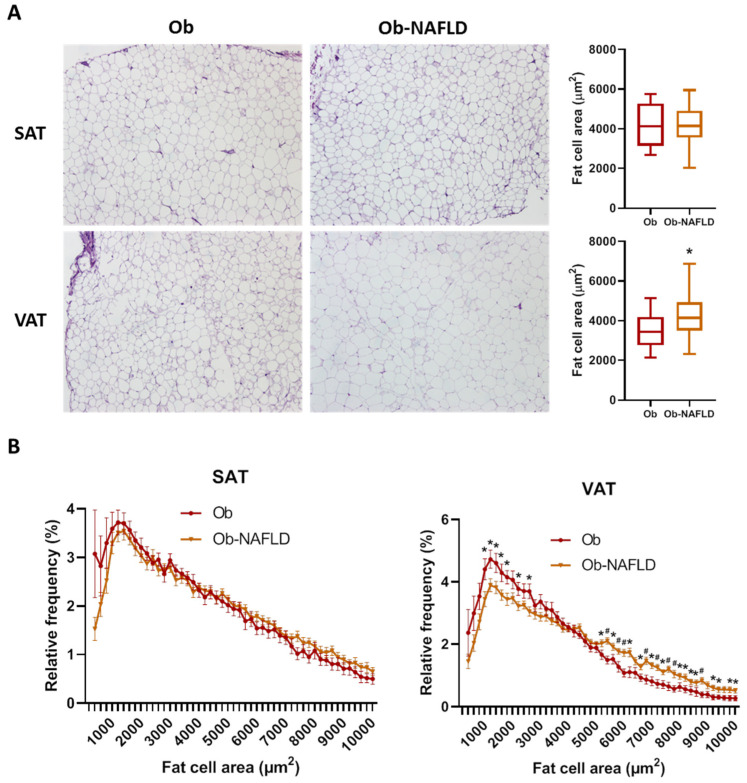
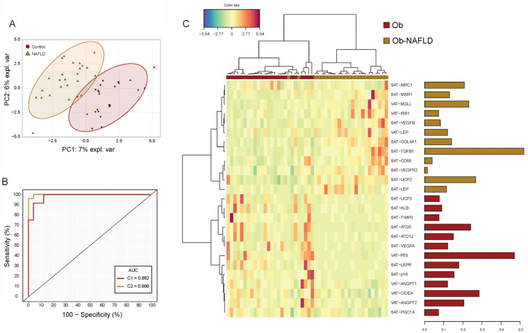
Development and severity of nonalcoholic fatty liver disease (NAFLD) have been linked to obesity and white adipose tissue (WAT) dysfunction plays a key role in this relation. We compared the main features of subcutaneous (SAT) and visceral WAT (VAT) tissue dysfunction in 48 obese women without (Ob) and with NAFLD (Ob-NAFLD) undergoing bariatric surgery and matched for age, BMI and T2D status. Fat cell area, adipocyte size distribution, the degree of histological fibrosis and the mRNA expression of adipokines and genes implicated in inflammation, adipogenesis, angiogenesis, metabolism and extracellular matrix remodeling were measured by RT-qPCR in both fat depots. Ob-NAFLD group showed higher TG and lower HDL circulating levels, increased VAT fat cell area and similar WAT fibrosis in comparison with Ob group. A sPLS-DA was performed in order to identify the set of genes that better characterize the presence of NAFLD. Finally, we build a multinomial logistic model including seven genes that explained 100% of the variance in NAFLD and correctly predicted 100% of cases. Our data support the existence of distinctive NAFLD signatures in WAT from women with severe obesity. A better understanding of these pathways may help in future strategies for the prevention and treatment of NAFLD.

Keywords: NAFLD; adipose tissue; obesity.


Conflict of interest statement

The authors declare no conflict of interest.

Figures

Figure 1
Figure 1
Fat cell size distribution. (A) Comparison of adipocyte mean cell surface area and representative images of SAT and VAT samples from Ob and Ob-NAFLD individuals. (B) Frequency distribution analysis of fat cell areas divided by size into bin intervals of 200 µm2. Data are presented as mean ± SD frequencies of cells within each bin and compared by Mann-Whitney U test. SAT, subcutaneous adipose tissue; VAT, visceral adipose tissue. * = p < 0.05, # = p < 0.01.
Figure 2
Figure 2
Subcutaneous and visceral fat gene expression signature model for NAFLD. (A) Sparse partial least square discriminant analysis (sPLSDA) individual scatter score plot (X-axis: component 1 = 25 genes; Y-axis: component 2 = 25 genes). Ellipses shades indicate 95% CI. (B) Receiver operating characteristic (ROC) analysis of the gene expression signature model. Red line corresponds to the accuracy classification performance of component 1-genes whereas yellow line to the model composed by genes (n = 50) of both components. (C) Hierarchical dendrogram-heatmap of the genes conforming sPLSDA Component 1. Log2 transformation is performed on the mRNA raw values of each gene. Complete linkage clustering method with Euclidean distance are used. The contribution of each gene for the model is represented in the barplot where each bar length corresponds to the loading weight (importance) of each variable. Bar color indicates the group in which the mean gene expression is greater.

References

    1. Younossi Z.M., Koenig A.B., Abdelatif D., Fazel Y., Henry L., Wymer M. Global epidemiology of nonalcoholic fatty liver disease—Meta-analytic assessment of prevalence, incidence, and outcomes. Hepatology. 2016;64:73–84. doi: 10.1002/hep.28431. - DOI - PubMed
    1. Marchesini G., Bugianesi E., Forlani G., Cerrelli F., Lenzi M., Manini R., Natale S., Vanni E., Villanova N., Melchionda N., et al. Nonalcoholic fatty liver, steatohepatitis, and the metabolic syndrome. Hepatology. 2003;37:917–923. doi: 10.1053/jhep.2003.50161. - DOI - PubMed
    1. Tilg H., Moschen A.R. Evolution of inflammation in nonalcoholic fatty liver disease: The multiple parallel hits hypothesis. Hepatology. 2010;52:1836–1846. doi: 10.1002/hep.24001. - DOI - PubMed
    1. Adams L.A., Anstee Q.M., Tilg H., Targher G. Non-Alcoholic fatty liver disease and its relationship with cardiovascular disease and other extrahepatic diseases. Gut. 2017;66:1138–1153. doi: 10.1136/gutjnl-2017-313884. - DOI - PubMed
    1. Polyzos S.A., Kountouras J., Mantzoros C.S. Obesity and nonalcoholic fatty liver disease: From pathophysiology to therapeutics. Metabolism. 2019;92:82–97. doi: 10.1016/j.metabol.2018.11.014. - DOI - PubMed

MeSH terms