Review
. 2021 Oct 2;22(19):10701.
doi: 10.3390/ijms221910701.

Chronic and Cycling Hypoxia: Drivers of Cancer Chronic Inflammation through HIF-1 and NF-κB Activation: A Review of the Molecular Mechanisms

Affiliations
Review

Chronic and Cycling Hypoxia: Drivers of Cancer Chronic Inflammation through HIF-1 and NF-κB Activation: A Review of the Molecular Mechanisms

Jan Korbecki et al. Int J Mol Sci. .

Abstract

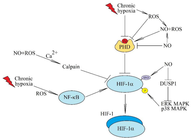
Chronic (continuous, non-interrupted) hypoxia and cycling (intermittent, transient) hypoxia are two types of hypoxia occurring in malignant tumors. They are both associated with the activation of hypoxia-inducible factor-1 (HIF-1) and nuclear factor κB (NF-κB), which induce changes in gene expression. This paper discusses in detail the mechanisms of activation of these two transcription factors in chronic and cycling hypoxia and the crosstalk between both signaling pathways. In particular, it focuses on the importance of reactive oxygen species (ROS), reactive nitrogen species (RNS) together with nitric oxide synthase, acetylation of HIF-1, and the action of MAPK cascades. The paper also discusses the importance of hypoxia in the formation of chronic low-grade inflammation in cancerous tumors. Finally, we discuss the effects of cycling hypoxia on the tumor microenvironment, in particular on the expression of VEGF-A, CCL2/MCP-1, CXCL1/GRO-α, CXCL8/IL-8, and COX-2 together with PGE2. These factors induce angiogenesis and recruit various cells into the tumor niche, including neutrophils and monocytes which, in the tumor, are transformed into tumor-associated neutrophils (TAN) and tumor-associated macrophages (TAM) that participate in tumorigenesis.

Keywords: HIF-1α; HIF-1β; NF-κB; cancer; cycling hypoxia; hypoxia-inducible factor; low-grade inflammation; tumor.


Conflict of interest statement

The authors declare no conflict of interest.

Figures

Figure 1
Figure 1
The effects of ROS and NO on the activation of HIF-1 in chronic hypoxia. Chronic hypoxia there is associated with an increase in the level of ROS which inactivate FIH and PHD. This increases the activation of HIF-1. ROS are also involved in the activation of NF-κB, a transcription factor important in the full activation of HIF-1. HIF-1 activation can also be induced by NO, especially at sites of inflammatory reactions. NO causes the S-nitrosylation of HIF-1α, which increases the stability of this protein. Another post-translational modification of HIF-1α induced by NO is phosphorylation associated with the inactivation of DUSP1. NO can also bind to the iron atom in PHDs and thus inactivate these enzymes. However, in combination with ROS, NO can restore activity of PHDs in chronic hypoxia. It can also increase calcium ion levels in the cytoplasm which activates calpain—a protease that degrades HIF-1α independently of the 26S proteasome.
Figure 2
Figure 2
The importance of MAPK cascades for the HIF-1 activation pathway. During chronic hypoxia, MAPK cascades, in particular ERK MAPK and p38 MAPK, are activated by ROS and increased calcium ions. These kinases cause phosphorylation of HIF-1α and consequently increase the stability and transcriptional activity of HIF-1. p38 MAPK can also cause the activation of SIAH2, which results in the ubiquitination and degradation of PHD3. Important in this model of HIF-1 activation are also phosphatases, in particular DUSP1 and DUSP2—enzymes that catalyze a reaction reverse to ERK MAPK and p38 MAPK. In chronic hypoxia, there is a decrease in DUSP2 expression but an increase in DUSP1, which is a mechanism for regulating HIF-1 activation.
Figure 3
Figure 3
The hypoxia-induced mechanism of NF-κB activation. In hypoxia, NF-κB is an important factor in the increase in HIF-1 mRNA expression, which is activated when oxygen concentration is decreased. This process occurs through multiple pathways. Like HIF-1α, IKKβ activation is inhibited by hydroxylation by PHD1. In hypoxia, PHD1 activity is reduced, which enables IKKβ activation. NF-κB activation during hypoxia also involves ROS and calcium ion mobilization into the cytoplasm. These factors cause the ubiquitination of IKKγ/NEMO, which increases IKK activity. IκBα is SUMOylated, which decreases the activity of this inhibitor of the NF-κB activation pathway. Chronic hypoxia is also associated with the activation of kinases such as p38 MAPK, ERK MAPK, and Akt/PKB, which phosphorylate NF-κB and IKKβ, thus activating this transcription factor.
Figure 4
Figure 4
The inhibition of the NF-κB pathway activation by HIF. Chronic hypoxia is associated with NF-κB activation, although there are also mechanisms that silence the proinflammatory response, such as an increase in PHD3 expression, which inhibits IKK activity. Additionally, there is an HIF-1 induced increase in the expression of IκBα, an inhibitor of NF-κB. The simultaneous activation of NF-κB and HIF causes these two transcription factors to compete for the coactivator p300.
Figure 5
Figure 5
Effect of ROS in cycling hypoxia on the activation of HIF-1 and NF-κB. Cycling hypoxia induces the generation of ROS, which cause the activation of HIF-1 and NF-κB. In particular, ROS inactivate FIH and PHD, which results in increased stability of HIF-1α protein. ROS also activate PKA and mTOR, which phosphorylate HIF-1α and thus increase the stability of this protein and its accumulation in the cell. ROS also causes an increase in the expression of Trx1, which enhances the transcriptional activity of HIF-1.
Figure 6
Figure 6
Effect of cycling hypoxia on angiogenesis in cancer. Cycling hypoxia activates HIF-1 and NF-κB in the tumor cell. (a) This leads to increased production of VEGF-A, CCL2/MCP-1, CXCL1/GRO-α, CXCL8/IL-8, and PGE2. (b) Subsequently, CCL2/MCP-1, CXCL1/GRO-α and CXCL8/IL-8 induce recruitment of TAM and TAN to the tumor niche. Cells that possess pro-angiogenic properties. TAN secrete MMP-9 into the tumor microenvironment, whereas TAM secrete MMP-9 and VEGF-A. MMP-9 is a metalloproteinase that releases VEGF-A. PGE2 also increases the expression of proangiogenic factors. (c) VEGF-A, CCL2/MCP-1, CXCL1/GRO-α and CXCL8/IL-8 directly cause angiogenesis.

References

    1. Chanmee T., Ontong P., Konno K., Itano N. Tumor-associated macrophages as major players in the tumor microenvironment. Cancers. 2014;6:1670–1690. doi: 10.3390/cancers6031670. - DOI - PMC - PubMed
    1. Najafi M., Farhood B., Mortezaee K. Contribution of regulatory T cells to cancer: A review. J. Cell Physiol. 2019;234:7983–7993. doi: 10.1002/jcp.27553. - DOI - PubMed
    1. Ligęza J., Ligęza J., Klein A. Growth factor/growth factor receptor loops in autocrine growth regulation of human prostate cancer DU145 cells. Acta Biochim. Pol. 2011;58:391–396. doi: 10.18388/abp.2011_2252. - DOI - PubMed
    1. Sulciner M.L., Gartung A., Gilligan M.M., Serhan C.N., Panigrahy D. Targeting lipid mediators in cancer biology. Cancer Metastasis Rev. 2018;37:557–572. doi: 10.1007/s10555-018-9754-9. - DOI - PubMed
    1. Do H.T.T., Lee C.H., Cho J. Chemokines and their Receptors: Multifaceted Roles in Cancer Progression and Potential Value as Cancer Prognostic Markers. Cancers. 2020;12:287. doi: 10.3390/cancers12020287. - DOI - PMC - PubMed

MeSH terms