. 2021 Sep 27;14(19):5618.
doi: 10.3390/ma14195618.

Hard Tissue Volume Stability Effect beyond the Bony Envelope of a Three-Dimensional Preformed Titanium Mesh with Two Different Collagen Barrier Membranes on Peri-Implant Dehiscence Defects in the Anterior Maxilla: A Randomized Clinical Trial

Affiliations

Hard Tissue Volume Stability Effect beyond the Bony Envelope of a Three-Dimensional Preformed Titanium Mesh with Two Different Collagen Barrier Membranes on Peri-Implant Dehiscence Defects in the Anterior Maxilla: A Randomized Clinical Trial

So-Ra Lee et al. Materials (Basel). .

Abstract

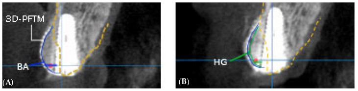
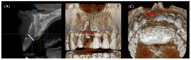
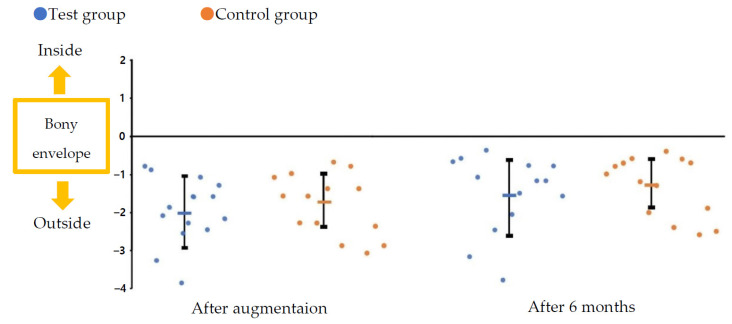
This single-blinded, randomized, controlled study aimed to clinically and radiographically evaluate hard tissue volume stability beyond the bony envelope using three-dimensional preformed titanium mesh (3D-PFTM) for peri-implant dehiscence defects in the anterior maxilla. A total of 28 patients who wished to undergo implant surgery combined with guided bone regeneration (GBR) after extraction of a single maxillary anterior tooth were randomly assigned to two groups depending on the type of collagen membrane used, additionally with the 3D-PFTM-test (n = 14, cross-linked collagen membrane; CCM) and control (n = 14, non-cross-linked collagen membrane; NCCM) groups. Each implant was evaluated radiographically using CBCT at baseline, immediately after surgery, and at 6 months postoperatively. The relative position and distances from the bony envelope to the outlines of the augmented ridge were further determined immediately after GBR and 6 months after healing. At the platform level, the mean horizontal hard tissue gain (HG) at all the sites was 2.35 ± 0.68 mm at 6 months postoperatively. The mean HG rate was 84.25% ± 14.19% in the CCM group and 82.56% ± 13.04% in the NCCM group, but the difference was not significant between the groups. In all cases, HG was maintained beyond the bony envelope even after 6 months of GBR. This study suggests that 3D-PFTM should be considered a valuable option for GBR for peri-implant dehiscence defects in the anterior maxilla. In addition, 3D-PFTM may confer predictable hard tissue volume stability even after the healing period of hard tissue augmented outside the bony envelope by GBR.

Keywords: bone regeneration; clinical study; dental implants; tissue pressure; titanium.


Conflict of interest statement

The authors declare no conflict of interest.

Figures

Figure 1
Figure 1
Surgical procedure stages. (A) Baseline: #11 is missing, (B,C) Implantation: peri-implant dehiscence bony defect is seen, (D) Bone grafts and 3D preformed titanium mesh application, (E) Collagen membrane application, (F) Suturing, (G) Healing state before second implant surgery, (H) Minimal incision and flap elevation: good hard tissue gain is noted, (I) Healing abutment installation and suturing, and (J) Final prosthetic restoration.
Figure 2
Figure 2
Method of radiographic analysis. A line parallel to the long major axis of the implant and a perpendicular line extending buccally at the level of the implant platform are drawn. (A) BA, bone augmentation immediately after surgery; (B) HG, hard tissue gain after 6 months of healing. The gap between the blue and green lines indicates bone resorption, while the red spot indicates the bony envelope. 3D-PFTM, three-dimensional preformed titanium mesh.
Figure 3
Figure 3
(A) CBCT sectional view. Note that the bony envelope appears as a red spot, (B,C) 3D radiographic analysis of bony envelope level.
Figure 4
Figure 4
Flowchart for clinical trial enrollment. 3D-PFTM, three-dimensional preformed titanium mesh; CCM, cross-linked collagen membrane; NCCM, non-cross-linked collagen membrane.
Figure 5
Figure 5
Distance from the boundary line of the bony envelope to the hard tissue profile immediately after augmentation and after 6 months of healing.

References

    1. Breine U., Brånemark P.-I. Reconstruction of alveolar jaw bone. Scand. J. Plast. Reconstr. Surg. 1980;14:23–48. doi: 10.3109/02844318009105733. - DOI - PubMed
    1. Merheb J., Quirynen M., Teughels W. Critical buccal bone dimensions along implants. Periodontology 2000. 2014;66:97–105. doi: 10.1111/prd.12042. - DOI - PubMed
    1. Bassetti M.A., Bassetti R.G., Bosshardt D.D. The alveolar ridge splitting/expansion technique: A systematic review. Clin. Oral Implant. Res. 2016;27:310–324. doi: 10.1111/clr.12537. - DOI - PubMed
    1. Hong J.Y., Baek W.S., Cha J.K., Lim H.C., Lee J.S., Jung U.W. Long-term evaluation of sinus floor elevation using a modified lateral approach in the posterior maxilla. Clin. Oral Implant. Res. 2017;28:946–953. doi: 10.1111/clr.12901. - DOI - PubMed
    1. Lorenzoni M., Pertl C., Polansky R., Wegscheider W. Guided bone regeneration with barrier membranes—A clinical and radiographic follow-up study after 24 months. Clin. Oral Implant. Res. 1999;10:16–23. doi: 10.1034/j.1600-0501.1999.100103.x. - DOI - PubMed

LinkOut - more resources