. 2021 Oct 7;14(19):5857.
doi: 10.3390/ma14195857.

Application of Texture and Fractal Dimension Analysis to Evaluate Subgingival Cement Surfaces in Terms of Biocompatibility

Affiliations

Application of Texture and Fractal Dimension Analysis to Evaluate Subgingival Cement Surfaces in Terms of Biocompatibility

Katarzyna Skośkiewicz-Malinowska et al. Materials (Basel). .

Abstract

Biocompatibility is defined as "the ability of a biomaterial, prosthesis, or medical device to perform with an appropriate host response in a specific application". Biocompatibility is especially important for restorative dentists as they use materials that remain in close contact with living tissues for a long time. The research material involves six types of cement used frequently in the subgingival region: Ketac Fil Plus (3M ESPE, Germany), Riva Self Cure (SDI, Australia) (Glass Ionomer Cements), Breeze (Pentron Clinical, USA) (Resin-based Cement), Adhesor Carbofine (Pentron, Czech Republic), Harvard Polycarboxylat Cement (Harvard Dental, Great Britain) (Zinc polycarboxylate types of cement) and Agatos S (Chema-Elektromet, Poland) (Zinc Phosphate Cement). Texture and fractal dimension analysis was applied. An evaluation of cytotoxicity and cell adhesion was carried out. The fractal dimension of Breeze (Pentron Clinical, USA) differed in each of the tested types of cement. Adhesor Carbofine (Pentron, Czech Republic) cytotoxicity was rated 4 on a 0-4 scale. The Ketac Fil Plus (3M ESPE, Germany) and Riva Self Cure (SDI, Australia) cements showed the most favorable conditions for the adhesion of fibroblasts, despite statistically significant differences in the fractal dimension of their surfaces.

Keywords: cytotoxicity; dental cements; fibroblasts adhesion; fractal dimension analysis; subgingival restoration; texture analysis.


Conflict of interest statement

The authors declare no conflict of interest.

Figures

Figure 1
Figure 1
(A) Triangle (fractal dimension = 2), (B) Sierpinski triangle (fractal dimension ≈ 1.585) (Generated by https://codinglab.huostravelblog.com/math/fractal-generator/ Accessed on 1 October 2021).
Figure 2
Figure 2
A plastic plate constituting the matrix for the production of test material cubes.
Figure 3
Figure 3
Graphical operations. (A) Source image, (B) image after auto-levels, (C) high-pass filter application.
Figure 4
Figure 4
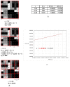
Graphical interpretation of the intensity difference algorithm of the fractal dimension calculation. (A) An example of a grayscale 8-bit image (8 × 8 pixels), the numbers in squares represent the intensity level of each pixel: 0, black, 255, white. The red squares represent the scale, ε. (B) The values of the intensity difference for each step of scale reduction (ε). (C) A straight line drawn through the points from table B on the x–y chart in a natural logarithm scale. The slope factor of this straight line is a value fractal dimension counted using intense difference algorithm.
Figure 5
Figure 5
Examples of light microscope images of the surface of the tested cements subjected to subsequent analysis of textures and the fractal dimension (magnified 18 times).
Figure 6
Figure 6
Morphology of Balb/3T3 cells after 24 h of contact with Agatos S (Chema-Elektromet, Poland), Harvard Polycarboxylat Cement (Harvard Dental, Great Britain) and Breeze (Pentron Clinical, USA); (A) under and (B) near the cement, (C) in area up to 1 cm, (D) in area more than 1 cm. Control culture had no contact with the test materials. Magn. 100×.
Figure 7
Figure 7
Morphology of Balb/3T3 cells after 24 h of contact with Riva Self Cure (SDI, Australia), Ketac Fil Plus (3M ESPE, Germany) and Adhesor Carbofine (Pentron, Czech Republic); (A) under and (B) near the cement, (C) in area up to 1 cm, (D) in area more than 1 cm. Control culture had no contact with the test materials. Magn. 100×.
Figure 8
Figure 8
Adhesion of NHDF cells on cement surface after 48 h and 5 days.
Figure 9
Figure 9
Texture index (TI) calculated for tested cements. The most expanded surface can be seen in Adhesor Carbofine (Pentron, Czech Republic) and Ketac Fil Plus (3M ESPE, Germany) contrary to Agatos S (Chema-Elektromet, Poland) and Breeze (Pentron Clinical, USA) presenting the lowest developed surface (p < 0.05).

References

    1. Anderson J.M. Biological Responses to Materials. Annu. Rev. Mater. Res. 2001;31:81–110. doi: 10.1146/annurev.matsci.31.1.81. - DOI
    1. Wataha J.C. Principles of biocompatibility of dental practicioners. J. Prosthet. Dent. 2001;86:203–209. doi: 10.1067/mpr.2001.117056. - DOI - PubMed
    1. Costa C.A.D.S., Hebling J., Scheffel D.L., Soares D.G., Basso F.G., Ribeiro A.P.D. Methods to evaluate and strategies to improve the biocompatibility of dental materials and operative techniques. Dent. Mater. 2014;30:769–784. doi: 10.1016/j.dental.2014.04.010. - DOI - PubMed
    1. Mallineni S.K., Nuvvula S., Matinlinna J.P., You C.K., King N.M. Biocompatibility of various dental materials in contem-porary dentistry: A narrative insight. J. Investig. Clin. Dent. 2013;4:9–19. doi: 10.1111/j.2041-1626.2012.00140.x. - DOI - PubMed
    1. Bertoldi C., Monari E., Cortellini P., Generali L., Lucchi A., Spinato S., Zaffe D. Clinical and histological reaction of peri-odontal tissues to subgingival resin composite restorations. Clin. Oral Investig. 2020;24:1001–1011. doi: 10.1007/s00784-019-02998-7. - DOI - PubMed

LinkOut - more resources