. 2021 Sep 22;10(19):4295.
doi: 10.3390/jcm10194295.

The Effect of Selective Retina Therapy with Automatic Real-Time Feedback-Controlled Dosimetry for Chronic Central Serous Chorioretinopathy: A Randomized, Open-Label, Controlled Clinical Trial

Affiliations

The Effect of Selective Retina Therapy with Automatic Real-Time Feedback-Controlled Dosimetry for Chronic Central Serous Chorioretinopathy: A Randomized, Open-Label, Controlled Clinical Trial

Ji-Young Lee et al. J Clin Med. .

Abstract

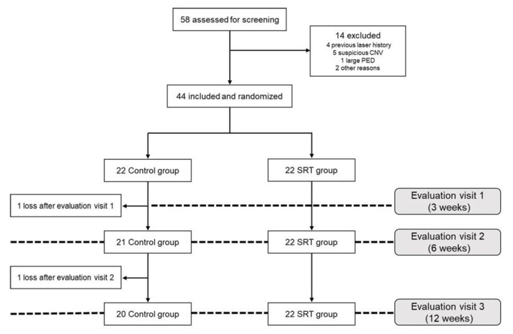
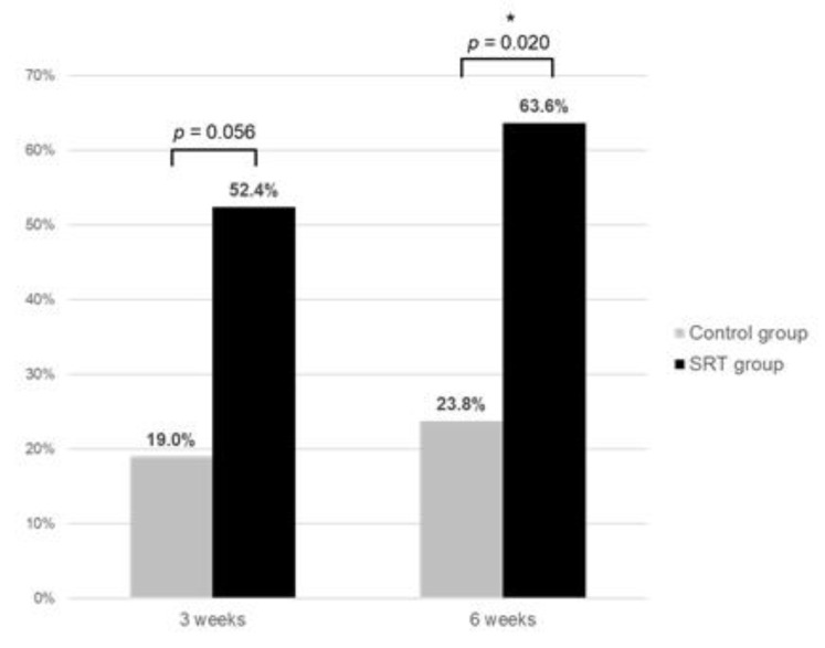
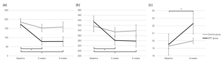
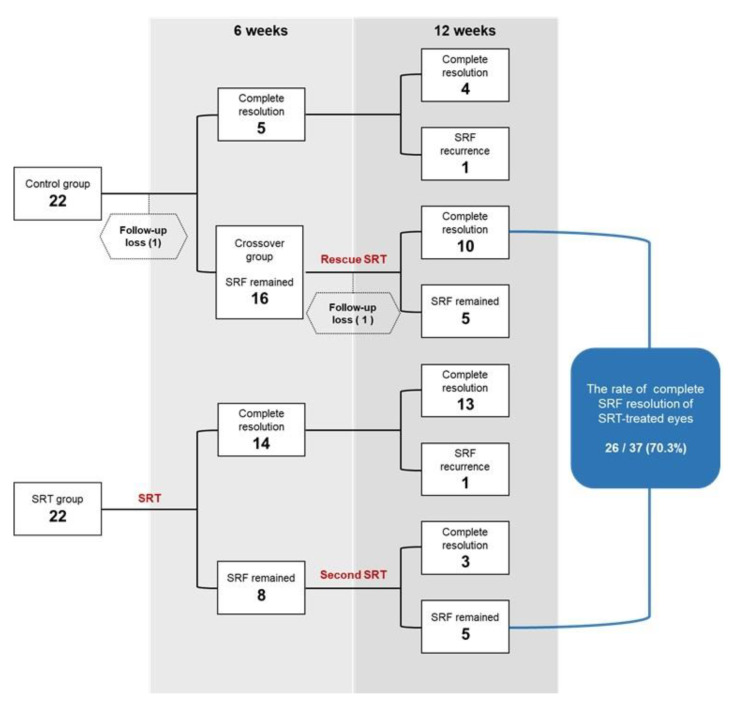
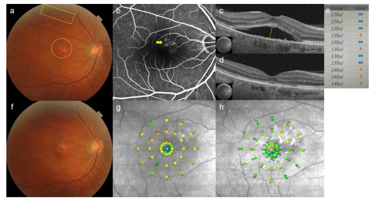
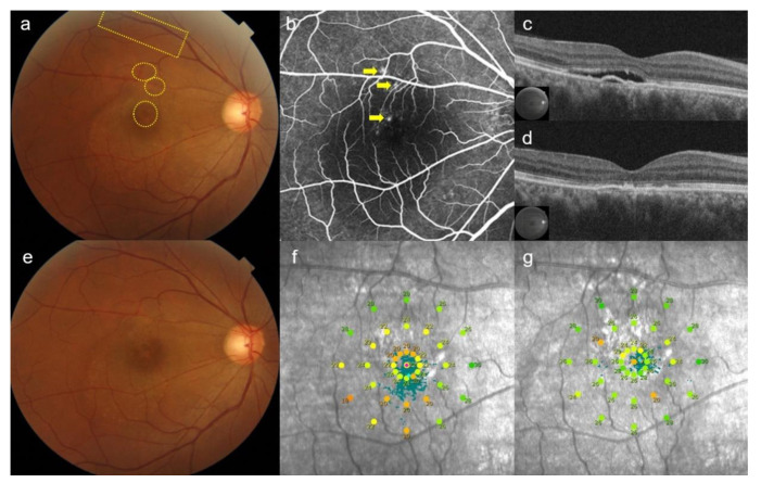
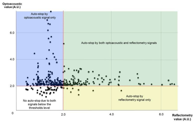
This prospective randomized controlled trial evaluated the safety and efficacy of real-time feedback-controlled dosimetry (RFD)-guided selective retina therapy (SRT) in chronic central serous chorioretinopathy (CSC). Forty-four participants with chronic CSC were included and randomly assigned to the control group or SRT group. The SRT laser system with RFD-guidance was applied to cover the entire leakage area. If SRF remained at the 6-week follow-up visit, re-treatment and rescue SRT was performed for the SRT group and crossover group, respectively. The rate of complete resolution of subretinal fluid (SRF), mean SRF height, and mean retinal sensitivity were compared between the two groups at 6-weeks post-treatment. The complete SRF resolution rate in all SRT-treated eyes was evaluated at 12-weeks post-treatment. The rate of complete SRF resolution was significantly higher in the SRT group (63.6%) than in the control group (23.8%) at 6-weeks post-treatment (p = 0.020). The mean SRF height at 6 weeks after SRT was significantly lower in the SRT group (p = 0.041). Overall, SRT-treated eyes showed complete SRF resolution in 70.3% of eyes at 12-weeks post-treatment. RFD-guided SRT was safe and effective to remove SRF in chronic CSC patients during the 3-month follow-up period.

Keywords: central serous chorioretinopathy; real-time feedback-controlled dosimetry; retinal sensitivity; selective retina therapy.


Conflict of interest statement

All authors have completed and submitted the ICMJE Form for Disclosure of Potential Conflicts of Interest and have disclosed the following: Young-Jung Roh have a patent related to real-time feedback dosimetry in South Korea. (Patent no.: 10-1966906). The SRT laser system (R:Gen, Lutronic) was approved for patients with central serous chorioretinopathy in South Korea. Lutronic provided the SRT laser system and technical support but had no role in the design of the study; in the collection, analyses, or interpretation of data; in the writing of the manuscript, or in the decision to publish the results.

Figures

Figure 1
Figure 1
Flow-diagram showing patient allocation to the SRT and control group.
Figure 2
Figure 2
Comparison of the rate of complete resolution of subretinal fluid between the SRT group and control group at 3-weeks and 6-weeks posttreatment. * p < 0.05.
Figure 3
Figure 3
Comparison of change in (a) subretinal fluid height, (b) central macular thickness, and (c) mean retinal sensitivity between the SRT group and control group during the 6-week follow-up period. * p < 0.05.
Figure 4
Figure 4
Flow-chart representing the rate of complete resolution of subretinal fluid (SRF) in the SRT group and control group during the 12-week follow-up period.
Figure 5
Figure 5
Case 1: A 51-year-old man presented with a 9-month history of blurred vision in the left eye. Subretinal fluid at baseline was observed at the macula, while 11 test spots (yellow rectangular) and 17 treatment spots (yellow circle) were invisible on color fundus photography (a). Diffuse leakages (yellow arrow) were shown on fundus fluorescent angiography (b). Subretinal fluid height (yellow line) was measured by using optical coherence tomography (OCT) (c). At 6-weeks post-selective retina therapy (SRT), subretinal fluid was completely resolved on OCT (d). Screen image of real-time feedback-controlled dosimetry (RFD) after test spot irradiation. Based on the algorithm of RFD, the sideways-pointing arrow indicates appropriate pulse energy. The downward-pointing arrow indicates an alarm for overtreatment. The pulse energy of 130 µJ yields different signals (sideways-pointing and downward-pointing arrows), because auto-stops occurred at different places of micropulses by RFD for each spot. The initial pulse energy for a treatment spot was 120 µJ, as the pulse energy constantly showed appropriateness (sideways-pointing arrow). (e). No SRT spots were visible on color fundus photography (f). Mean retinal sensitivity (17 dB) on microperimetry at baseline (g) was improved to (25 dB) on microperimetry (h) at 6 weeks after treatment.
Figure 6
Figure 6
Case 2: A 45-year-old man presented with an 8-month history of blurred vision in the right eye. After the 6-week observation period, subretinal fluid was still observed at the macula on color fundus photography. The area of 13 test spots (yellow rectangular) and 15 rescue treatment spots (yellow circles) were invisible (a). Three focal leakages (yellow arrows) were shown on fundus fluorescent angiography (b). Subretinal fluid and pigment epithelial detachment was observed on optical coherence tomography (OCT) before rescue with selective retina therapy (SRT) (c). At 6-weeks post-SRT, subretinal fluid was completely resolved on OCT (d). No SRT spots were visible on fundus photography (e). Mean retinal sensitivity (20 dB) on microperimetry at baseline (f) was increased to (22 dB) on microperimetry (g) at 6-weeks post-treatment.
Figure 7
Figure 7
Scatter plot of optoacoustic and reflectometry values in selective retina therapy treatment spots.

References

    1. Nicholson B., Noble J., Forooghian F., Meyerle C. Central serous chorioretinopathy: Update on pathophysiology and treatment. Surv. Ophthalmol. 2013;58:103–126. doi: 10.1016/j.survophthal.2012.07.004. - DOI - PMC - PubMed
    1. Wang M., Munch I.C., Hasler P.W., Prünte C., Larsen M. Central serous chorioretinopathy. Acta Ophthalmol. 2008;86:126–145. doi: 10.1111/j.1600-0420.2007.00889.x. - DOI - PubMed
    1. Daruich A., Matet A., Dirani A., Bousquet E., Zhao M., Farman N., Jaisser F., Behar-Cohen F. Central serous chorioretinopathy: Recent findings and new physiopathology hypothesis. Prog. Retin. Eye Res. 2015;48:82–118. doi: 10.1016/j.preteyeres.2015.05.003. - DOI - PubMed
    1. Singh S.R., Matet A., van Dijk E.H.C., Daruich A., Fauser S., Yzer S., Peiretti E., Sivaprasad S., Lotery A.J., Boon C.J.F., et al. Discrepancy in current central serous chorioretinopathy classification. Br. J. Ophthalmol. 2019;103:737–742. doi: 10.1136/bjophthalmol-2018-312435. - DOI - PubMed
    1. Piccolino F.C., de la Longrais R.R., Ravera G., Eandi C.M., Ventre L., Abdollahi A., Manea M. The foveal photoreceptor layer and visual acuity loss in central serous chorioretinopathy. Am. J. Ophthalmol. 2005;139:87–99. doi: 10.1016/j.ajo.2004.08.037. - DOI - PubMed

LinkOut - more resources