. 2021 Sep 26;26(19):5829.
doi: 10.3390/molecules26195829.

Identification of the Protein Glycation Sites in Human Myoglobin as Rapidly Induced by d-Ribose

Affiliations

Identification of the Protein Glycation Sites in Human Myoglobin as Rapidly Induced by d-Ribose

Jing-Jing Liu et al. Molecules. .

Abstract

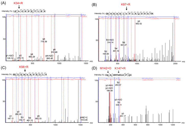
Protein glycation is an important protein post-translational modification and is one of the main pathogenesis of diabetic angiopathy. Other than glycated hemoglobin, the protein glycation of other globins such as myoglobin (Mb) is less studied. The protein glycation of human Mb with ribose has not been reported, and the glycation sites in the Mb remain unknown. This article reports that d-ribose undergoes rapid protein glycation of human myoglobin (HMb) at lysine residues (K34, K87, K56, and K147) on the protein surface, as identified by ultra-high performance liquid chromatography-mass spectrometry (UHPLC-MS) and electrospray ionization tandem mass spectrometry (ESI-MS/MS). Moreover, glycation by d-ribose at these sites slightly decreased the rate of the met heme (FeIII) in reaction with H2O2 to form a ferryl heme (FeIV=O). This study provides valuable insight into the protein glycation by d-ribose and provides a foundation for studying the structure and function of glycated heme proteins.

Keywords: d-ribose; diabetes; glycosylation sites; human myoglobin; protein glycation.


Conflict of interest statement

The authors declare no conflict of interest.

Figures

Figure 1
Figure 1
The X-ray crystal structure of the K45R/C110A HMb mutant (left, PDB code 3RGK) [30]. The heme active site and the location of the Lys residues are shown for clarification. Note that the conformation of the side chain of K56 was too flexible to be determined. The protein glycation of the HMb by d-ribose is indicated by the arrow (right).
Figure 2
Figure 2
ESI-MS spectra of the HMb after reacting with d-ribose for 1 h (A), 2 h (B), and 4 h (C), respectively. The spectrum of HMb after reacting with d-glucose for 24 h (D) was shown for comparison.
Figure 3
Figure 3
The amino acid sequence of HMb and the expected peptide fragments (T1–T22) by trypsin digestion. The molecular weight of each peptide fragment and the four identified Lys residues are labeled.
Figure 4
Figure 4
ESI-MS spectra (A,C) and extracted ion chromatograms (EICs) (B,D) of the trypsin digestion products of HMb induced by d-ribose at different times (0, 2, and 4 h).
Figure 5
Figure 5
ESI-MS/MS spectra of peptide fragments: The triply protonated tryptic glycosylated HMb peptide LFK(+132.04)GHPETLEK at m/z 477.58 (A); the triply protonated tryptic glycosylated HMb peptide GHHEAEIK(+132.04)PLAQSHATK at m/z 662.67 (B); the doubly protonated tryptic glycosylated HMb peptide SEDEMK(+132.04)ASEDLK at m/z 757.33 (C); and the triply protonated tryptic glycosylated HMb peptide DM(+15.99)ASNYK(+132.04)ELGFQG at m/z 804.35 (D), respectively. The glycation sites of the Lys and the oxidation of the Met142 are indicated by arrows.
Figure 6
Figure 6
(A) The stopped-flow spectra of the ribosylated HMb (R-HMb) in reaction with H2O2 (1 mM) at pH 7.0 for 30 s. Inset: The decay of the Soret band at 409 nm. (B) The plots of the observed rate constants versus the H2O2 concentrations, with the results of the HMb shown for comparison.

Similar articles

Cited by

References

    1. Guariguata L., Whiting D.R., Hambleton I., Beagley J., Linnenkamp U., Shaw J.E. Global estimates of diabetes prevalence for 2013 and projections for 2035. Diabetes Res. Clin. Pract. 2014;103:137–149. doi: 10.1016/j.diabres.2013.11.002. - DOI - PubMed
    1. Giardino I., Edelstein D., Brownlee M. Nonenzymatic glycosylation in vitro and in bovine endothelial cells alters basic fibroblast growth factor activity. A model for intracellular glycosylation in diabetes. J. Clin. Investig. 1994;94:110–117. doi: 10.1172/JCI117296. - DOI - PMC - PubMed
    1. Schalkwijk C.G., Ligtvoet N., Twaalfhoven H., Jager A., Blaauwgeers H.G., Schlingemann R.O., Tarnow L., Parving H.H., Stehouwer C.D., Van Hinsbergh V.W. Amadori albumin in type 1 diabetic patients: Correlation with markers of endothelial function, association with diabetic nephropathy, and localization in retinal capillaries. Diabetes. 1999;48:2446–2453. doi: 10.2337/diabetes.48.12.2446. - DOI - PubMed
    1. Lin Y.-W. Structure and function of heme proteins regulated by diverse post-translational modifications. Arch. Biochem. Biophys. 2018;641:1–30. doi: 10.1016/j.abb.2018.01.009. - DOI - PubMed
    1. Adrover M., Mariño L., Sanchis P., Pauwels K., Kraan Y., Lebrun P., Vilanova B., Muñoz F., Broersen K., Donoso J. Mechanistic Insights in Glycation-Induced Protein Aggregation. Biomacromolecules. 2014;15:3449–3462. doi: 10.1021/bm501077j. - DOI - PubMed

LinkOut - more resources