

The Short-Term Exposure to SDHI Fungicides Boscalid and Bixafen Induces a Mitochondrial Dysfunction in Selective Human Cell Lines
- PMID: 34641386
- PMCID: PMC8510389
- DOI: 10.3390/molecules26195842
The Short-Term Exposure to SDHI Fungicides Boscalid and Bixafen Induces a Mitochondrial Dysfunction in Selective Human Cell Lines
Abstract
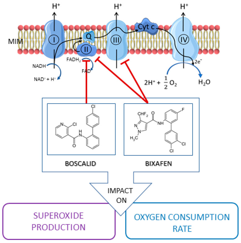
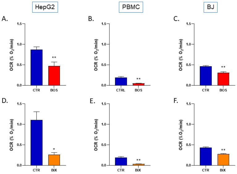
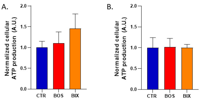
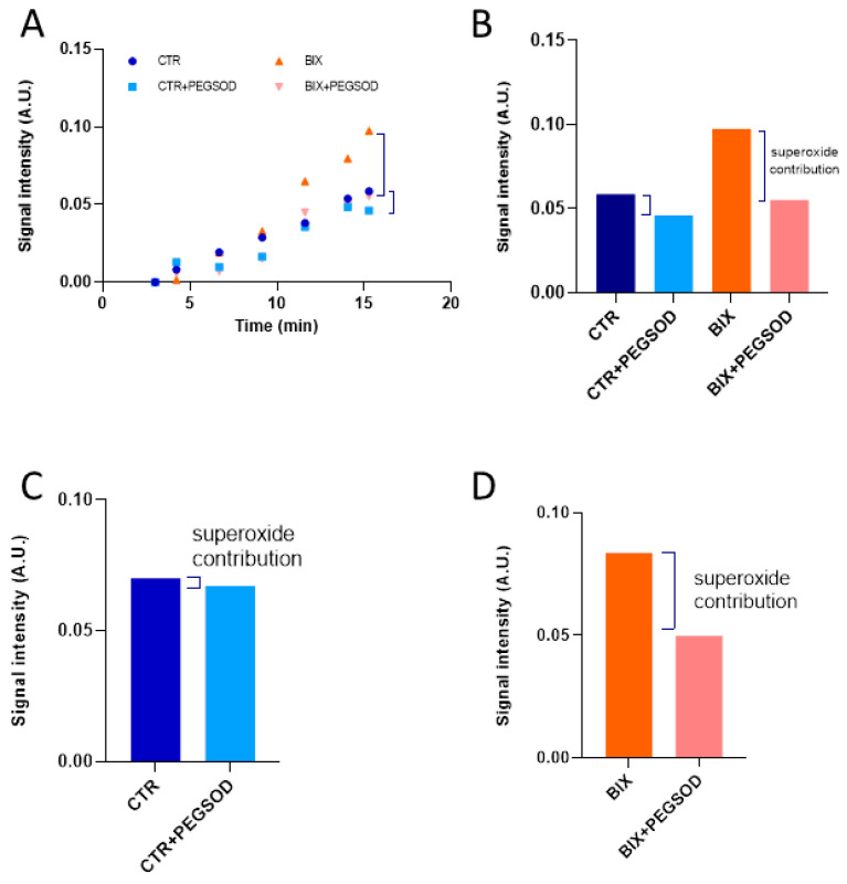
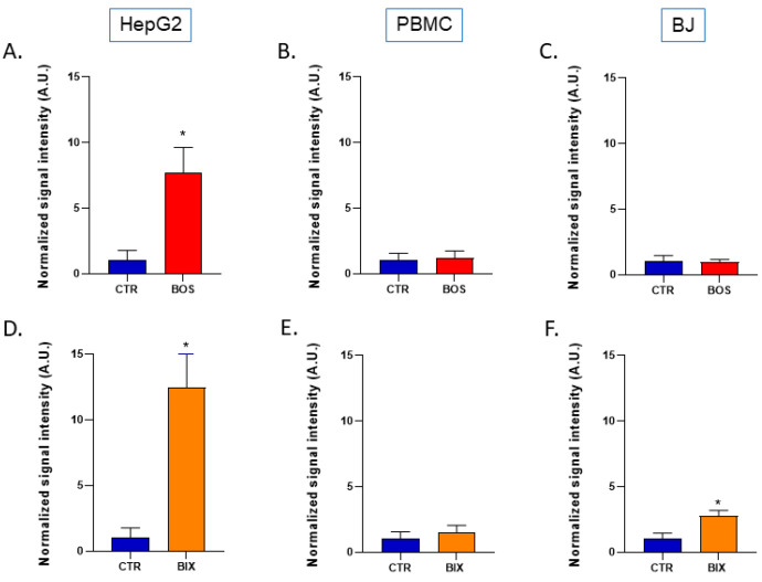
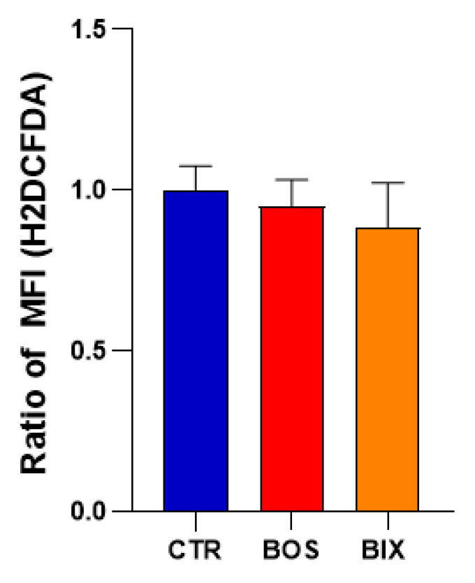
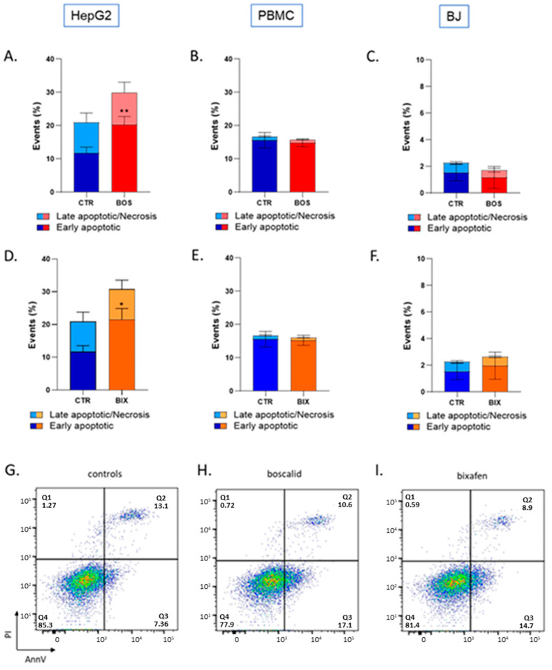
Fungicides are used to suppress the growth of fungi for crop protection. The most widely used fungicides are succinate dehydrogenase inhibitors (SDHIs) that act by blocking succinate dehydrogenase, the complex II of the mitochondrial electron transport chain. As recent reports suggested that SDHI-fungicides could not be selective for their fungi targets, we tested the mitochondrial function of human cells (Peripheral Blood Mononuclear Cells or PBMCs, HepG2 liver cells, and BJ-fibroblasts) after exposure for a short time to Boscalid and Bixafen, the two most used SDHIs. Electron Paramagnetic Resonance (EPR) spectroscopy was used to assess the oxygen consumption rate (OCR) and the level of mitochondrial superoxide radical. The OCR was significantly decreased in the three cell lines after exposure to both SDHIs. The level of mitochondrial superoxide increased in HepG2 after Boscalid and Bixafen exposure. In BJ-fibroblasts, mitochondrial superoxide was increased after Bixafen exposure, but not after Boscalid. No significant increase in mitochondrial superoxide was observed in PBMCs. Flow cytometry revealed an increase in the number of early apoptotic cells in HepG2 exposed to both SDHIs, but not in PBMCs and BJ-fibroblasts, results consistent with the high level of mitochondrial superoxide found in HepG2 cells after exposure. In conclusion, short-term exposure to Boscalid and Bixafen induces a mitochondrial dysfunction in human cells.
Keywords: Bixafen; Boscalid; EPR; SDHI; mitochondria; oxygen consumption rate (OCR); superoxide.
Conflict of interest statement
The authors declare no conflict of interest.
Figures
Similar articles
-
Impact of Exposure to Pyraclostrobin and to a Pyraclostrobin/Boscalid Mixture on the Mitochondrial Function of Human Hepatocytes.Molecules. 2023 Oct 10;28(20):7013. doi: 10.3390/molecules28207013. Molecules. 2023. PMID: 37894492 Free PMC article.
-
Mitochondrial dysfunction induced in human hepatic HepG2 cells exposed to the fungicide kresoxim-methyl and to a mixture kresoxim-methyl/boscalid.Redox Rep. 2024 Dec;29(1):2424677. doi: 10.1080/13510002.2024.2424677. Epub 2024 Nov 14. Redox Rep. 2024. PMID: 39541499 Free PMC article.
-
Lack of cross-resistance to a novel succinate dehydrogenase inhibitor, fluopyram, in highly boscalid-resistant isolates of Corynespora cassiicola and Podosphaera xanthii.Pest Manag Sci. 2011 Apr;67(4):474-82. doi: 10.1002/ps.2092. Epub 2011 Jan 11. Pest Manag Sci. 2011. PMID: 21394880
-
SDHI Fungicide Toxicity and Associated Adverse Outcome Pathways: What Can Zebrafish Tell Us?Int J Mol Sci. 2021 Nov 16;22(22):12362. doi: 10.3390/ijms222212362. Int J Mol Sci. 2021. PMID: 34830252 Free PMC article. Review.
-
A review of current knowledge of resistance aspects for the next-generation succinate dehydrogenase inhibitor fungicides.Phytopathology. 2013 Sep;103(9):880-7. doi: 10.1094/PHYTO-01-13-0009-RVW. Phytopathology. 2013. PMID: 23593940 Review.
Cited by
-
Impact of Exposure to Pyraclostrobin and to a Pyraclostrobin/Boscalid Mixture on the Mitochondrial Function of Human Hepatocytes.Molecules. 2023 Oct 10;28(20):7013. doi: 10.3390/molecules28207013. Molecules. 2023. PMID: 37894492 Free PMC article.
-
Environmental pollution effect on honey bees and their derived products: a comprehensive analysis.Environ Sci Pollut Res Int. 2025 Apr;32(16):10370-10391. doi: 10.1007/s11356-024-33754-4. Epub 2024 Jun 7. Environ Sci Pollut Res Int. 2025. PMID: 38847955 Free PMC article. Review.
-
Bixafen, Prothioconazole, and Trifloxystrobin Alone or in Combination Have a Greater Effect on Health Related Gene Expression in Honey Bees from Nutritionally Deprived than from Protein Supplemented Colonies.Insects. 2024 Jul 11;15(7):523. doi: 10.3390/insects15070523. Insects. 2024. PMID: 39057256 Free PMC article.
-
Analysis of Pesticide Residues on Fruit Using Swab Spray Ionization Mass Spectrometry.Molecules. 2023 Sep 14;28(18):6611. doi: 10.3390/molecules28186611. Molecules. 2023. PMID: 37764387 Free PMC article.
-
Selected Fungicides as Potential EDC Estrogenic Micropollutants in the Environment.Molecules. 2023 Nov 5;28(21):7437. doi: 10.3390/molecules28217437. Molecules. 2023. PMID: 37959855 Free PMC article.
References
-
- ANSES Report 2018-SA-0113. [(accessed on 26 July 2021)]. Available online: https://www.anses.fr/fr/system/files/PHYTO2018SA0113Ra.pdf.
-
- Kamp H., Wahrheit J., Stinchcombe S., Walk T., Stauber F., Ravenzwaay B. Succinate dehydrogenase inhibitors: In silico flux analysis and in vivo metabolomics investigations show no severe metabolic consequences for rats and humans. Food Chem. Toxicol. 2021;150:112085. doi: 10.1016/j.fct.2021.112085. - DOI - PubMed
-
- Bénit P., Kahn A., Chretien D., Bortoli S., Huc L., Schiff M., Gimenez-Roqueplo A.-P., Favier J., Gressens P., Rak M., et al. Evolutionarily conserved susceptibility of the mitochondrial respiratory chain to SDHI pesticides and its consequence on the impact of SDHIs on human cultured cells. PLoS ONE. 2019;14:e0224132. doi: 10.1371/journal.pone.0224132. - DOI - PMC - PubMed
MeSH terms
Substances
Grants and funding
LinkOut - more resources
Full Text Sources
Research Materials
