Review
. 2021 Sep 27;26(19):5847.
doi: 10.3390/molecules26195847.

An Insight into FDA Approved Antibody-Drug Conjugates for Cancer Therapy

Affiliations
Review

An Insight into FDA Approved Antibody-Drug Conjugates for Cancer Therapy

Juliana T W Tong et al. Molecules. .

Abstract

The large number of emerging antibody-drug conjugates (ADCs) for cancer therapy has resulted in a significant market 'boom', garnering worldwide attention. Despite ADCs presenting huge challenges to researchers, particularly regarding the identification of a suitable combination of antibody, linker, and payload, as of September 2021, 11 ADCs have been granted FDA approval, with eight of these approved since 2017 alone. Optimism for this therapeutic approach is clear, despite the COVID-19 pandemic, 2020 was a landmark year for deals and partnerships in the ADC arena, suggesting that there remains significant interest from Big Pharma. Herein we review the enthusiasm for ADCs by focusing on the features of those approved by the FDA, and offer some thoughts as to where the field is headed.

Keywords: ADCs; FDA approved; antibody-drug conjugates; cancer; targeted therapy.


Conflict of interest statement

The authors declare no conflict of interest.

Figures

Figure 1
Figure 1
The general structure of an antibody-drug conjugate (ADC) and key points about the different components. (Created with BioRender.com, accessed 27 September 2021).
Figure 2
Figure 2
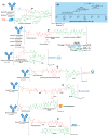
(A) Structures of FDA approved antibody-drug conjugates (ADCs). The antibody is shown in blue, and chemical structures for linker and payload are in red and green, respectively. Scissors indicate the cleavage site (if applicable). Pharmaceutical makers and drug-to-antibody ratio for each ADC is indicated. (B) Comparison of approximate payload potency ranges (Created with BioRender.com, accessed September 2021).
Figure 3
Figure 3
The general mechanism of action of an antibody-drug conjugate (ADC). (Adapted from “Antibody-Drug Conjugate Release”, by BioRender.com (accessed 27 September 2021). Retrieved from https://app.biorender-templates, accessed 27 September 2021).
Figure 4
Figure 4
Structure for Mylotarg® (gemtuzumab ozogamicin). The antibody is shown in blue, and chemical structures for linker and payload are in red and green, respectively. The cleavage site is indicated by scissors.
Figure 5
Figure 5
Mechanism for double-strand (ds) DNA cleavage by N-acetyl-γ-calicheamicin. The enediyne warhead is shown in red.
Figure 6
Figure 6
Structure of Adcetris® (brentuximab vedotin). The antibody is shown in blue, and chemical structures for linker and payload are in red and green, respectively. Spontaneous 1,6-elimination mechanism for the PABC-substituted MMAE, leading to release of MMAE, p-iminoquinone methide, and carbon dioxide.
Figure 7
Figure 7
Schematic showing partial reduction of IgG1 antibody interchain disulfide bonds to generate two nucleophilic free thiol groups that can be reacted with an electrophilic linker-payload construct, such as maleimide (DAR = 2). Maleimide conjugation to cysteine is shown in this example.
Figure 8
Figure 8
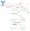
(A) Structure of Kadcyla® (ado-trastuzumab emtansine). The antibody is shown in blue, and chemical structures for linker and payload are in red and green, respectively. (B) The chemical structure for maytansine and DM1. The thiopropanoyl group of DM1, which allows for conjugation to a maleimidomethyl cyclohexane-1-carboxylate (MCC) group is shown in the red box.
Figure 9
Figure 9
Structure of Besponsa® (inotuzumab ozogamicin). The antibody is shown in blue, and chemical structures for linker and payload are in red and green, respectively.
Figure 10
Figure 10
Structure of Polivy® (polatuzumab vedotin-piiq) and Padcev® (enfortumab vedotin-ejfv). The antibody is shown in blue, and chemical structures for linker and payload are in red and green, respectively.
Figure 11
Figure 11
Structure of Enhertu® (fam-trastuzumab deruxtecan-nxki). The antibody is shown in blue, and chemical structures for linker and payload are in red and green, respectively.
Figure 12
Figure 12
Structure of Trodelvy® (sacituzumab govitecan-hziy). The antibody is shown in blue, and chemical structures for linker and payload are in red and green, respectively. PEG, polyethyleneglycol.
Figure 13
Figure 13
Structure of Blenrep® (belantamab mafodotin-blmf). The antibody is shown in blue, and chemical structures for linker and payload are in red and green, respectively.
Figure 14
Figure 14
Structure of Zynlonta® (loncastuximab tesirine-lpyl). The antibody is shown in blue, and chemical structures for linker and payload are in red and green, respectively. PEG, polyethyleneglycol. Spontaneous 1,6-elimination mechanism for the PABC-substituted SG3199, leading to release of SG3199, p-iminoquinone methide, and carbon dioxide.
Figure 15
Figure 15
Schematic showing binding of the PBD dimer, SG3199, to the minor groove of DNA. The N2 of guanine binds the electrophilic C11 position on the PBD dimer.
Figure 16
Figure 16
Structure of Tivdak® (tisotumab vedotin-tftv). The antibody is shown in blue, and chemical structures for linker and payload are in red and green, respectively.

References

    1. Strebhardt K., Ullrich A. Paul Ehrlich’s magic bullet concept: 100 years of progress. Nat. Rev. Cancer. 2008;8:473–480. doi: 10.1038/nrc2394. - DOI - PubMed
    1. DeVita V.T., Chu E. A history of cancer chemotherapy. Cancer Res. 2008;68:8643–8653. doi: 10.1158/0008-5472.CAN-07-6611. - DOI - PubMed
    1. Köhler G., Milstein C. Continuous cultures of fused cells secreting antibody of predefined specificity. Nature. 1975;256:495–497. doi: 10.1038/256495a0. - DOI - PubMed
    1. Ford C.H., Newman E.C., Johnson J.R., Woodhouse C.S., Reeder A.T., Rowland G.F., Simmonds R.G. Localisation and toxicity study of a vindesine-anti-CEA conjugate in patients with advanced cancer. Br. J. Cancer. 1983;47:35–42. doi: 10.1038/bjc.1983.4. - DOI - PMC - PubMed
    1. Tolcher A.W. The evolution of antibody-drug conjugates: A positive inflexion point. Am. Soc. Clin. Oncol. Educ. Book. 2020:127–134. doi: 10.1200/EDBK_281103. - DOI - PubMed