. 2021 Sep 28;26(19):5886.
doi: 10.3390/molecules26195886.

A Systematic Review of the Biological Effects of Cordycepin

Affiliations

A Systematic Review of the Biological Effects of Cordycepin

Masar Radhi et al. Molecules. .

Abstract

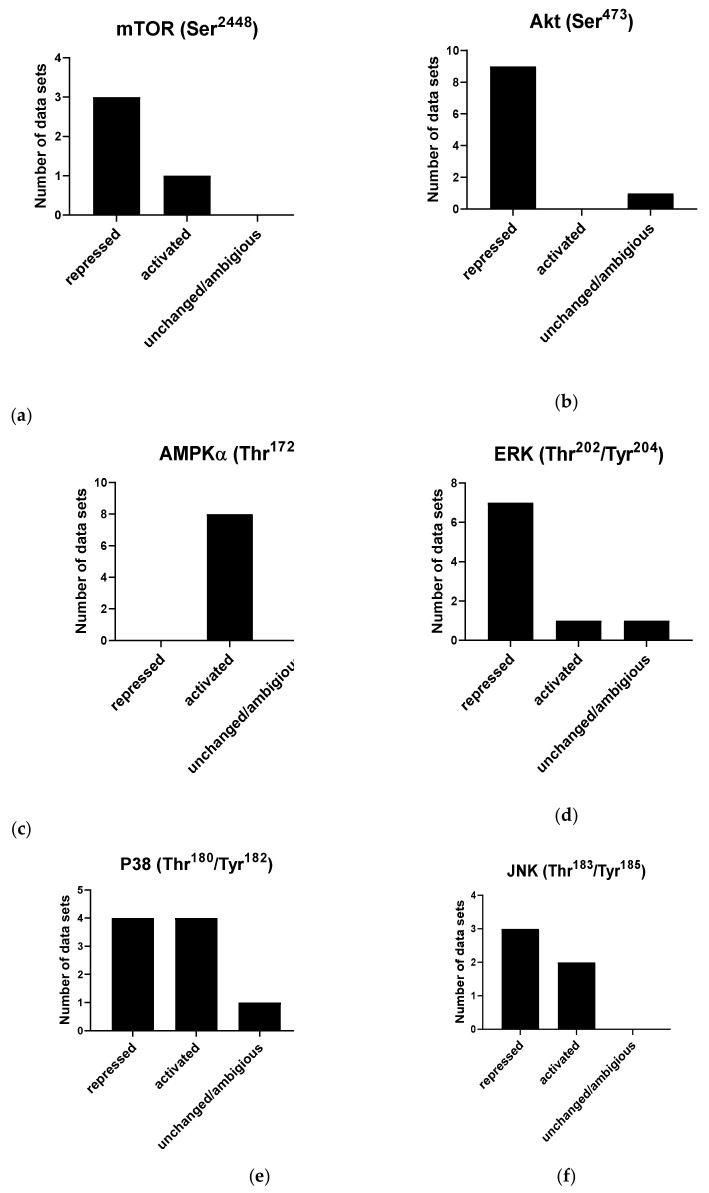
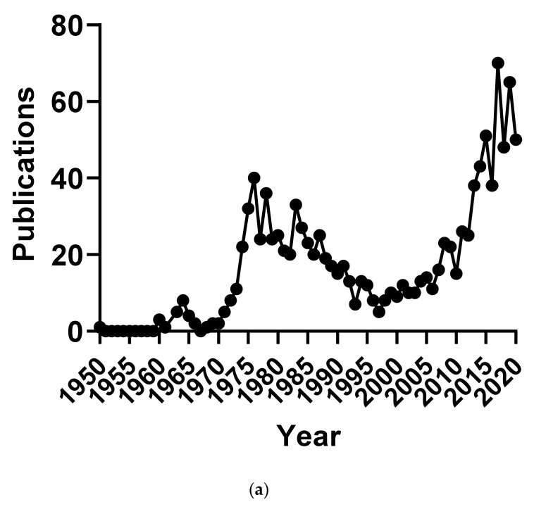
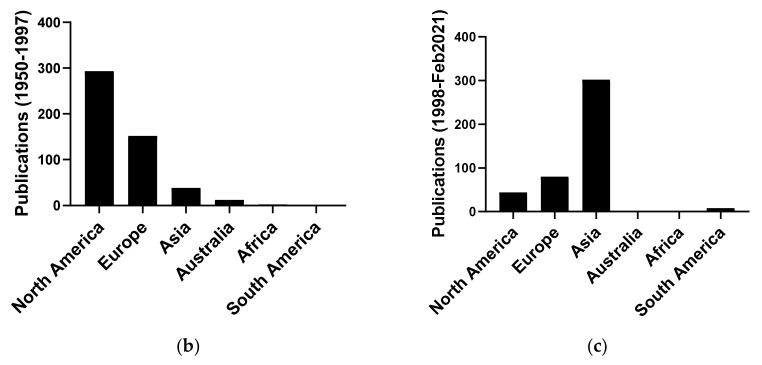
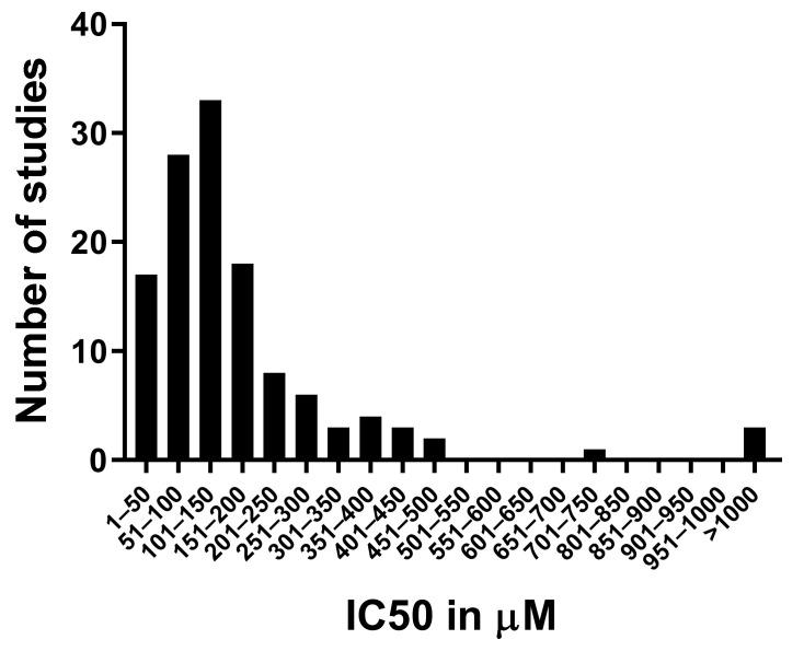
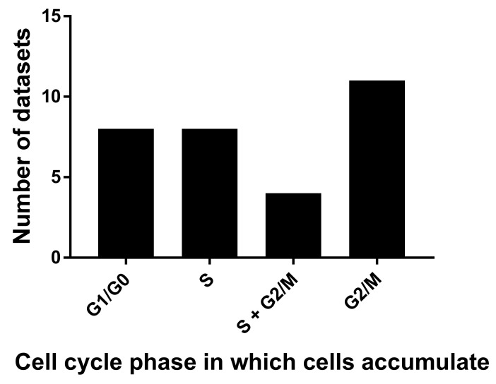
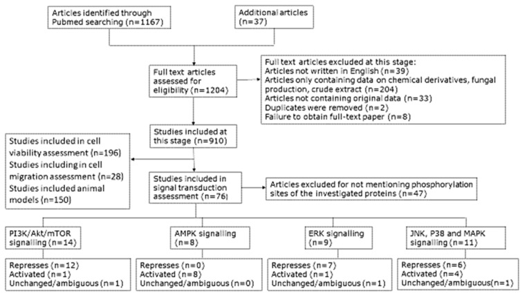
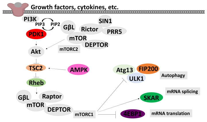
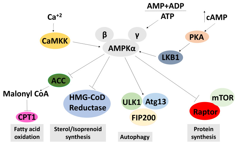
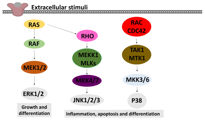
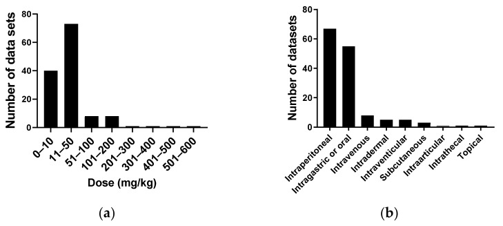
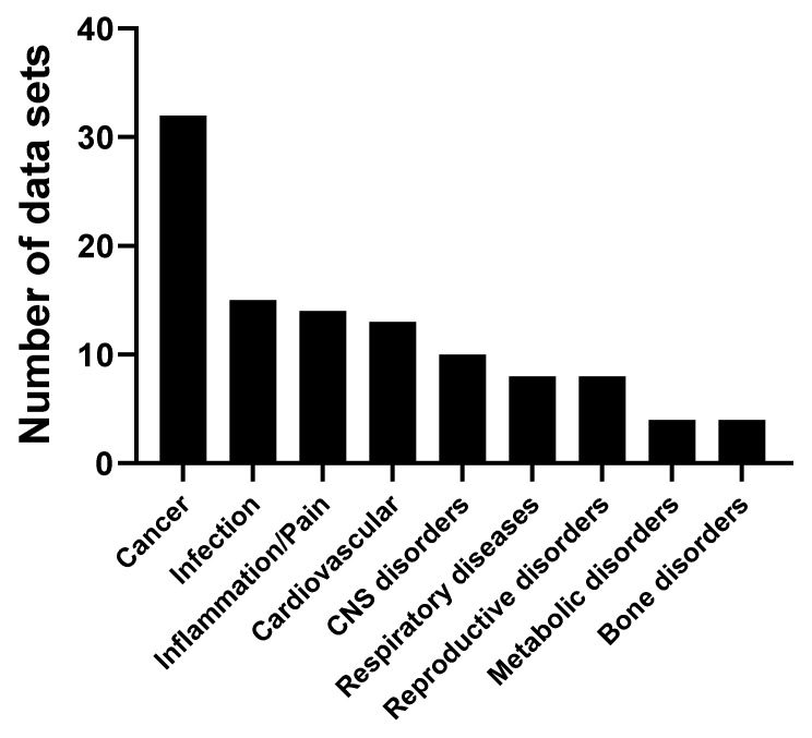
We conducted a systematic review of the literature on the effects of cordycepin on cell survival and proliferation, inflammation, signal transduction and animal models. A total of 1204 publications on cordycepin were found by the cut-off date of 1 February 2021. After application of the exclusion criteria, 791 papers remained. These were read and data on the chosen subjects were extracted. We found 192 papers on the effects of cordycepin on cell survival and proliferation and calculated a median inhibitory concentration (IC50) of 135 µM. Cordycepin consistently repressed cell migration (26 papers) and cellular inflammation (53 papers). Evaluation of 76 papers on signal transduction indicated consistently reduced PI3K/mTOR/AKT and ERK signalling and activation of AMPK. In contrast, the effects of cordycepin on the p38 and Jun kinases were variable, as were the effects on cell cycle arrest (53 papers), suggesting these are cell-specific responses. The examination of 150 animal studies indicated that purified cordycepin has many potential therapeutic effects, including the reduction of tumour growth (37 papers), repression of pain and inflammation (9 papers), protecting brain function (11 papers), improvement of respiratory and cardiac conditions (8 and 19 papers) and amelioration of metabolic disorders (8 papers). Nearly all these data are consistent with cordycepin mediating its therapeutic effects through activating AMPK, inhibiting PI3K/mTOR/AKT and repressing the inflammatory response. We conclude that cordycepin has excellent potential as a lead for drug development, especially for age-related diseases. In addition, we discuss the remaining issues around the mechanism of action, toxicity and biodistribution of cordycepin.

Keywords: AKT; AMPK; ERK; cell viability; cordycepin; inflammation; mTOR; natural product; review; signal transduction.


Conflict of interest statement

The authors declare no conflict of interest.

Figures

Figure 8
Figure 8
Effects of cordycepin on widely studied signal transduction pathways. Papers were selected as described in the Materials and Methods section and classified according to the effect observed on the indicated phosphorylation sites. (a) Effects of cordycepin on the Ser2448 phosphorylation site on mTOR; (b) Effects of cordycepin on the Ser4738 phosphorylation site on AKT; (c) Effects of cordycepin on the Thr172 phosphorylation site on AMPKα; (d) Effects of cordycepin on the Thr202/Tyr204 phosphorylation sites on ERK; (e) Effects of cordycepin on the Thr180/Tyr182 phosphorylation site on the p38 kinase; (f) Effects of cordycepin on the Thr183/Tyr185 phosphorylation site on the Jun kinase. The papers from which these data were extracted can be found in Table 1 in the Methods.
Figure 1
Figure 1
Number of publications on cordycepin by year and geographical origin. (a) Number of publications on cordycepin per year of publication 1950–2018; (b,c) Geographical distribution of the affiliation of the corresponding authors of publications on cordycepin in two periods: (b) 1950–1997 and (c) 1998–February 2021.
Figure 1
Figure 1
Number of publications on cordycepin by year and geographical origin. (a) Number of publications on cordycepin per year of publication 1950–2018; (b,c) Geographical distribution of the affiliation of the corresponding authors of publications on cordycepin in two periods: (b) 1950–1997 and (c) 1998–February 2021.
Figure 2
Figure 2
Distribution of the IC50 data for cordycepin from the literature. The 50% inhibitory concentration in µM was retrieved from 128 datasets described in 57 papers (listed in the Methods section). The number of datasets with an IC50 in each concentration bracket indicated on the Y axis was counted and graphed.
Figure 3
Figure 3
Cordycepin arrests cells in different cell cycle stages. 31 flow cytometry datasets from 18 papers were examined for the cell cycle stage in which cell numbers are significantly increased after cordycepin treatment.
Figure 4
Figure 4
Diagram presenting the selection process for inclusion of publications in this review; n = number of articles.
Figure 5
Figure 5
A schematic model of the PI3K/Akt/mTOR signalling pathways. This model integrates PI3K/mTOR/Akt and AMPK signal transduction pathways. Arrows indicate activation and T ends indicate inhibition. Triggered PI3K activates Akt/mTOR cascade, through activation of PDK1 and phosphorylation of AKT in the activation T-loop. mTOR complex 2 (mTORC2), is activated by an as-yet unknown pathway and contributes to the activation of AKT by phosphorylation in the C-terminal. AKT inhibits TSC2, which leads to activation of the small GTPase Rheb and activation of the mTOR complex 2 (mTORC2). The pathway is negatively regulated by AMPK, through activation of TSC2. The PI3K/Akt/mTOR signalling pathway increases protein synthesis through the phosphorylation of the cap-dependent translation inhibitor protein 4EBP1 and inhibits autophagy, leading to the promotion of growth and the anabolic state.
Figure 6
Figure 6
A schematic model of the AMPK signalling pathways. This model shows the activation of AMPK in response to low adenosine triphosphate (ATP) levels, and an increased adenosine diphosphate (ADP) and adenosine monophosphate (AMP). As a result, it activates pathways that produce ATP, thus increasing ATP levels. Conversely, pathways that deplete ATP are repressed by AMPK. AMPK is activated by an increased AMP + ADP to ATP ratio and phosphorylation by CAMKK or LKB1. Activated AMPK inhibits acetyl-CoA carboxylase (ACC), HMG-CoD reductase and mTORC1, leading to an increase of fatty acid oxidation and a reduction in sterol and protein synthesis. Active AMPK inhibits autophagy. An arrow indicates an upregulation of the process and a T end represents a downregulation of the process.
Figure 7
Figure 7
A schematic model of the MAPK signalling pathways. This model shows the three major pathways of MAPK: extracellular signal-regulated kinase (ERK) 1 and 2, c-Jun N-terminal kinases (JNK) 1–3 and p38 MAPK. In all these pathways, the activation of a small GTPase (RAS, RHO or RAC/CDC42) leads to the activation of a MAP kinase kinase kinase (MAPKKK: RAF, MEKK1/MLK, TAK/MTK1), which activates a MAP kinase kinase (MAPKK) by phosphorylation. These MAPKKs finally phosphorylate and activate the MAPKs (ERK1/2, JNK1/2/3 and p38).
Figure 9
Figure 9
Dose of cordycepin administered to animal models in mg/kg. (a) A total of 133 studies were classified according to the range of cordycepin dose administered to the animal model; (b) The route of administration of cordycepin in 146 studies.In this systematic review, 167 articles studying the effects of purified cordycepin in a variety of animal models were found (see Table 3). We noted the details of the experimental set-up (species, model of human disease, dose in mg/kg basis and route of administration). The disease models were classified and counted (Figure 10). The majority of studies were of animal models of cancer, closely followed by cardiovascular diseases, infections and central nervous system disorders.
Figure 10
Figure 10
Classification of animal models. A total of 139 articles were classified according to the type of human disease modelled. CNS: central nervous system. Categories which had only one entry were excluded, as described in the Methods section. The exact allocation of papers to disease classes can be found in the Methods section (Table 3).

References

    1. Cunningham K., Manson W., Spring F., Hutchinson S. Cordycepin, a metabolic product isolated from cultures of Cordyceps militaris (Linn.) Link. Nature. 1950;166:949. doi: 10.1038/166949a0. - DOI - PubMed
    1. Winkler D. Cordyceps sinensis—a precious parasitic fungus infecting Tibet. Field Mycol. 2010;11:60. doi: 10.1016/j.fldmyc.2010.04.009. - DOI
    1. Penman S., Rosbash M., Penman M. Messenger and heterogeneous nuclear RNA in HeLa cells: Differential inhibition by cordycepin. Proc. Natl. Acad. Sci. USA. 1970;67:1878–1885. doi: 10.1073/pnas.67.4.1878. - DOI - PMC - PubMed
    1. Philipson L., Wall R., Glickman G., Darnell J. Addition of polyadenylate sequences to virus-specific RNA during adenovirus replication. Proc. Natl. Acad. Sci. USA. 1971;68:2806–2809. doi: 10.1073/pnas.68.11.2806. - DOI - PMC - PubMed
    1. Wu A., Ting R., Paran M., Gallo R. Cordycepin inhibits induction of murine leukovirus production by 5-iodo-2′-deoxyuridine. Proc. Natl. Acad. Sci. USA. 1972;69:3820–3824. doi: 10.1073/pnas.69.12.3820. - DOI - PMC - PubMed

Publication types