Review
. 2021 Oct 7;26(19):6057.
doi: 10.3390/molecules26196057.

Drugs That Changed Society: History and Current Status of the Early Antibiotics: Salvarsan, Sulfonamides, and β-Lactams

Affiliations
Review

Drugs That Changed Society: History and Current Status of the Early Antibiotics: Salvarsan, Sulfonamides, and β-Lactams

Søren Brøgger Christensen. Molecules. .

Abstract

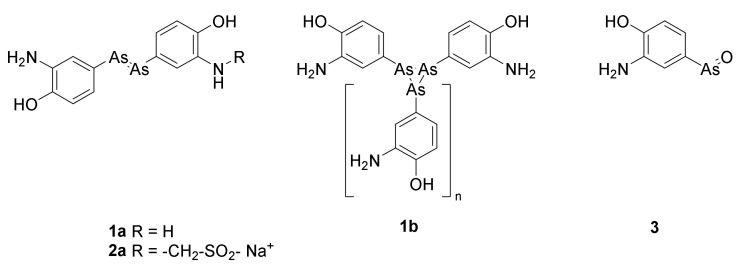
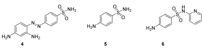
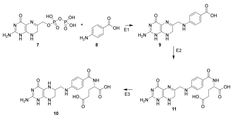
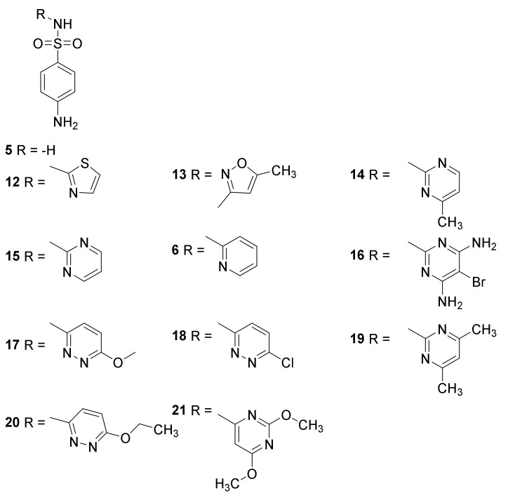
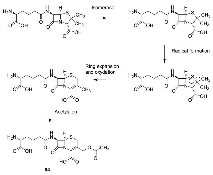
The appearance of antibiotic drugs revolutionized the possibilities for treatment of diseases with high mortality such as pneumonia, sepsis, plaque, diphtheria, tetanus, typhoid fever, and tuberculosis. Today fewer than 1% of mortalities in high income countries are caused by diseases caused by bacteria. However, it should be recalled that the antibiotics were introduced in parallel with sanitation including sewerage, piped drinking water, high standard of living and improved understanding of the connection between food and health. Development of salvarsan, sulfonamides, and β-lactams into efficient drugs is described. The effects on life expectancy and life quality of these new drugs are indicated.

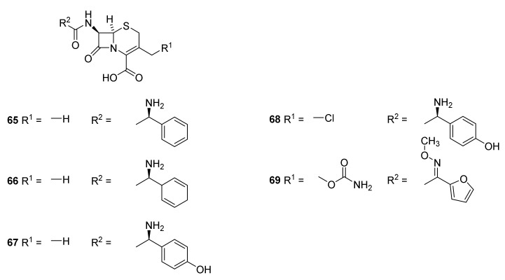
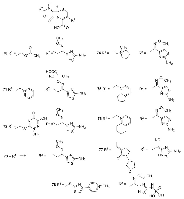
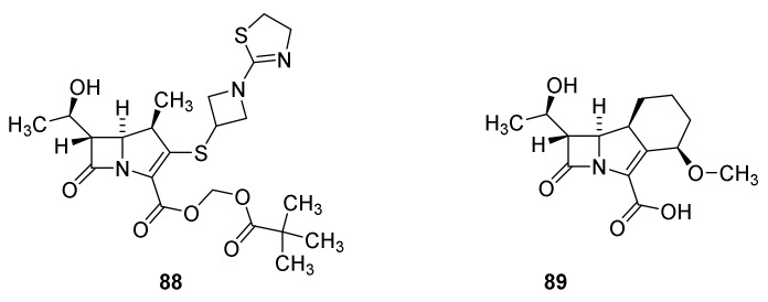
Keywords: bacterial infectious diseases; carbapenems; cephalosporins; monobactams; penicillins; salvarsan; sulfonamides; thiopenems; β-lactamases.


Conflict of interest statement

The authors declare no conflict of interest.

Figures

Figure 1
Figure 1
The commonly shown structure for arsphenamine (salvarsan) is 1a. Arsphenamine is now known to be a mixture of compounds with the formula 1b, in which n varies from 1 to 7. The predominant values are n = 1 and n = 3. Neosalvarsan (2a) was designed to increase the solubility [18]. Oxphenarsine 3 is the expected active metabolite of arspehamine [24].
Figure 2
Figure 2
Prontosil (4), sulfonamide (4-aminobenzenesulfonamiode, 5) and 4-sulfapyridine (6).
Scheme 1
Scheme 1
Biosynthesis of tetrahydrofolate (10) from 7,8-dihydropteridinepyrophosphate (7) and 4-aminobenzoic acid (8) via 7,8-dihydropteroate (9) and dihydrofolate (11) [36]. Dihydropteroate synthase (E1), dihydrofolate synthase (E2) and dihydrofoate reductase (E3) [37].
Figure 3
Figure 3
Clinically used sulfonamides for treatment of infectious diseases: sulfonilamide (5), sulfapyridine (6), sulfathiazole (12), sulfamehoxazole (13), sulfamerazine (14), sulfadiazine (15), sulfabromomethazine (16), sulfamethoxypyridazine (17), sulfachloropyridazine (18), sulfametazine (19), sulfaethoxypyridazine (20) and sulfadimethoxine (21) [36].
Figure 4
Figure 4
Naturally occurring β-lactams backbones [37].
Figure 5
Figure 5
Structures of penicillins with British and American names. Penicillin-I/F-penicillin (22), dihydropenicillin-I (23), penicillin-II/G-penicillin (24), penicillin-III/X-penicillin (25) and K-penicillin (26). The side chain is dependent on the Pencillium species and broth from which the penicillin is isolated.
Scheme 2
Scheme 2
Structure elucidation of penicillin. Hydrolysis with mineral acid affords carbon dioxide, penicillamine (28) and penaldic acid (27), which is labile and spontaneously converted into penillo-aldehyde (29). Treatment with benzylamine yields aminolysis of the β-lactam to give the benzylammonium salt of the benzylamide 30. Methanolysis affords the monomethyl ester of methyl penicilloate (31), which may be converted into methyl penaldoate and (32) and penicillamine (28). The suggested structures have been confirmed by syntheses [47,48].
Scheme 3
Scheme 3
In aqueous solution pencillin is hydrolyzed (black arrows) to penicilloic acid (33), which rearranges into penillic acid (34). Catalyzed by mercury ions 34 is converted into penillamine (35), or catalyzed by barium sulfate (red arrows) isomerize into isopenicillic acid (36) by a retro Hetero-Michael addition [47,48].
Scheme 4
Scheme 4
Resonance of penicillin and reaction with penicillin-binding peptide to acylate the penicillin-binding peptide (37) followed by hydrolysis [45,47,48].
Scheme 5
Scheme 5
Biosynthesis of the non-ribosomal peptide penicillin G [37]. EnzFe: isopenicillin N-synthase-FeII, transferase: isopenicillin N N-acyltransferase [54].
Figure 6
Figure 6
6-Aminopenicillanic acid (38).
Figure 7
Figure 7
Cartoon illustrating the cross binding of peptidoglycan strands in the cell walls of Staphyllococcus aureus [52,58]. Pe indicates the attachment of an additional cross binding peptide by a peptide bond to N-acetymuramic acid. G indicates prolongation with other units of the dimer N-acetylglucosamine-N-acetylmuramic acid.
Figure 8
Figure 8
Mechanism for cross binding [44,58]. Pe indicates the attachment of an additional cross binding peptide by a peptide bond to N-acetymuramic acid. G indicates prolongation with other units of the dimer N-acetylglucosamine-N-acetylmuramic acid.
Figure 9
Figure 9
Semisynthetic penicillins. Phenoxyacyl penicillins: phenoxymethyl penicillin/penicillin V (Vepicombin, 39), phenoxyethyl penicillin (phenithicillin, 40), Phenoxypropylpenicillin (41) [63,64]. Aminoacylpenicillins ampicillin (42), epicillin (43), amoxicillin (44) and cyclacillin (45).
Figure 10
Figure 10
Semisynthetic penicillins with activity toward Pseudomonas aeruginosa Carbenicillin (46), sulbenicillin (47), ticarcillin (48) [44], piperacillin (49) and mezlocillin (50).
Figure 11
Figure 11
Examples of β-lactamase resistant penicillins, methicillin (51), nafcillin (52) and oxacillin (53).
Scheme 6
Scheme 6
Conversion of penicillin G into the 6-methoxy derivative 54 [82].
Figure 12
Figure 12
Temocillin (55) and formadicillin (56) [44]. Temocillin is approved by European Medicine Agency.
Figure 13
Figure 13
Ureidopenicillins: Azlocillin (51), mezlocillin (52) and piperazillin (53) [38].
Figure 14
Figure 14
Prodrugs of penicillin: talampicillin (60), bacampicillin (61), pivampicillin (62) and lenampenicillin (63) [42,44].
Figure 15
Figure 15
Cephalosporin C (64).
Scheme 7
Scheme 7
Pencillin cephalosporin rearrangement [95].
Scheme 8
Scheme 8
Cleavage of the 7-amide group of cephalosporins [44,97].
Scheme 9
Scheme 9
Biosynthesis of cephalosporin C (64). Isomerase: isopenicillin N isomerase, ring expansion, oxidation and acetylation: deacetylcephalosporin C/deacetylcephalosporin C synthase-FeII, acetylation deacetylcephalosprin C acetyltransferase [37,54].
Figure 16
Figure 16
Examples of first generation cephalosporins. Cephalexin (65), cefadrine (66) and cefadroxil (67). Second generation cephalosporins cefalclor (68) and cefuroxime (69).
Figure 17
Figure 17
Third generation cephalosporins. Cefotaxime (70), ceftazidime (71), ceftriaxone (72) and ceftizoxime (73). Fourth generation cephalosporins. Cefepime (74), cefpirome (75) and cefquinome (76). Fifth generation cephalosporins: ceftobiprole (77) and ceftaroline fosamil (78) [98,99].
Figure 18
Figure 18
Carbapenems and 1-methylcarbapenems. Olivanic acid (79), thienamycin (80), imipenem (81), panipenem (82), biapenem (83), meropenem (84), ertapenem (85) and doripenem (86) [101].
Figure 19
Figure 19
Tebipenem pivoxil (88) and sanfetrinem (89).
Figure 20
Figure 20
Faropenem (90), faropenem medoximil (91), sulopenem (92) and sulopenem etzadrozil (93).
Scheme 10
Scheme 10
Formation of the thipenem skeleton [113].
Scheme 11
Scheme 11
Hydrolysis of sulfazecin (95).
Figure 21
Figure 21
Aztreonam (96).
Figure 22
Figure 22
β-Lactamase inhibitors containing a β-lactam residue in the skeleton: clavulanic acid (97), sulbactam (98), tazobactam (99) and enmetazobactam (100) [119].
Scheme 12
Scheme 12
Irreversibel reaction between sulbactam (98) and β-lactamase [123].
Scheme 13
Scheme 13
Reaction of lactivicin (101) and phenoxylactivicin (102) with pencillin binding proteins [58].
Figure 23
Figure 23
Avibactam (103) and NXL-105 (104).
Scheme 14
Scheme 14
Reaction of avibactam (103) with PBP. The carbamoyl-lactamase complex is more stable than the analogous complexes between lactamase inhibitors of the β-lactam type [58].
Figure 24
Figure 24
Boronic acid β-lactamase inhibitors. Vaborbactam (105) and taniborbactam (106).
Figure 25
Figure 25
Serine-β-lactamase-varbobactam complex [129] and metallo-β-lactamase-taniborbactam complex [119].

References

    1. Koefoed M. Befolkningsforholdene i Danmark. Statens Stistiske Buerau; Copenhagen, Denmark: 1905.
    1. Christensen G. Døden Skifter Årsag. Dansk Sygeplejehistoriske Museum; Copenhagen, Denmark: 2017. p. 15.
    1. Singer C., Underwood E.A. A Short History of Medicine. 2nd ed. Clarendon Press; Oxford, UK: 1962.
    1. Slavkin H.C. Biofilms, microbial ecology and Antoni van Leeuwenhoek. J. Am. Dent. Assoc. 1997;128:492–495. doi: 10.14219/jada.archive.1997.0238. - DOI - PubMed
    1. Müller O.F. In: Animalcula Infusoria Fluviatilia er Marina. Mölleri T.N., editor. Hauniæ; Copenhagen, Denmark: 1786.