. 2021 Oct 8;26(19):6079.
doi: 10.3390/molecules26196079.

In Vitro Analyses of Spinach-Derived Opioid Peptides, Rubiscolins: Receptor Selectivity and Intracellular Activities through G Protein- and β-Arrestin-Mediated Pathways

Affiliations

In Vitro Analyses of Spinach-Derived Opioid Peptides, Rubiscolins: Receptor Selectivity and Intracellular Activities through G Protein- and β-Arrestin-Mediated Pathways

Yusuke Karasawa et al. Molecules. .

Abstract

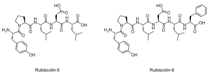
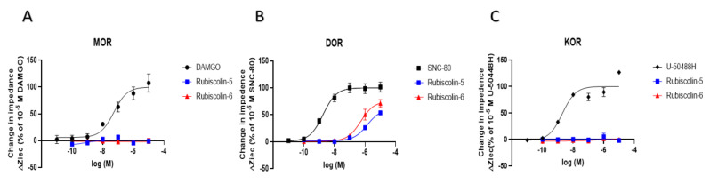
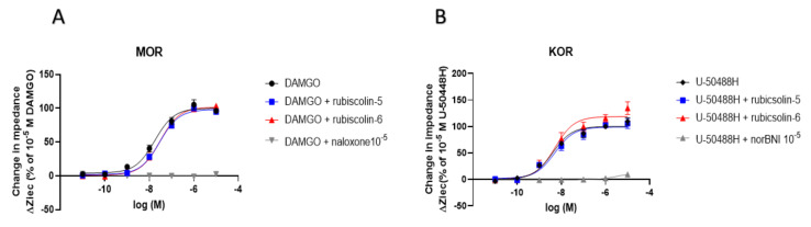
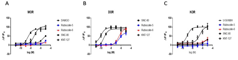
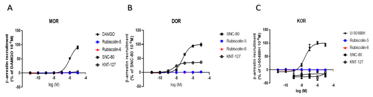
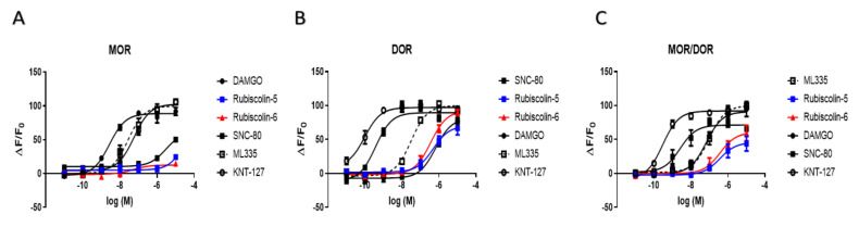
Activated opioid receptors transmit internal signals through two major pathways: the G-protein-mediated pathway, which exerts analgesia, and the β-arrestin-mediated pathway, which leads to unfavorable side effects. Hence, G-protein-biased opioid agonists are preferable as opioid analgesics. Rubiscolins, the spinach-derived naturally occurring opioid peptides, are selective δ opioid receptor agonists, and their p.o. administration exhibits antinociceptive effects. Although the potency and effect of rubiscolins as G-protein-biased molecules are partially confirmed, their in vitro profiles remain unclear. We, therefore, evaluated the properties of rubiscolins, in detail, through several analyses, including the CellKeyTM assay, cADDis® cAMP assay, and PathHunter® β-arrestin recruitment assay, using cells stably expressing µ, δ, κ, or µ/δ heteromer opioid receptors. In the CellKeyTM assay, rubiscolins showed selective agonistic effects for δ opioid receptor and little agonistic or antagonistic effects for µ and κ opioid receptors. Furthermore, rubiscolins were found to be G-protein-biased δ opioid receptor agonists based on the results obtained in cADDis® cAMP and PathHunter® β-arrestin recruitment assays. Finally, we found, for the first time, that they are also partially agonistic for the µ/δ dimers. In conclusion, rubiscolins could serve as attractive seeds, as δ opioid receptor-specific agonists, for the development of novel opioid analgesics with reduced side effects.

Keywords: G-protein-biased agonist; analgesic; opioid peptide; rubiscolins; δ opioid receptor.


Conflict of interest statement

The principal author and one coauthor (M.Y.) are employees of a pharmaceutical company (Viatris Pharmaceuticals Japan Inc. and Pfizer Japan Inc., respectively). However, the present study has no financial or other relationships with these companies, as it was entirely sponsored by and performed at the Jikei University School of Medicine, National Cancer Center Research Institute, and Juntendo University Graduate School of Medicine.

Figures

Figure 1
Figure 1
Molecular structures of rubiscolins.
Figure 2
Figure 2
Effect of rubiscolins on MOR, DOR, and KOR, observed using the CellKeyTM system. The cells expressing MOR (A), DOR (B), and KOR (C) were treated with each compound (10−11–10−5 M), and changes in impedance (ΔZiec) were measured using the CellKeyTM system. Concentration–response curves were prepared by calculating ΔZiec relative to the data obtained for each positive control: 10−5 M DAMGO for MOR (A), 10−5 M SNC-80 for DOR (B), and 10−5 M U-50488H for KOR (C). All data points are presented as means ± S.E.M. for three independent experiments (n = 3–5).
Figure 3
Figure 3
Evaluation of antagonistic effects induced by rubiscolins combined with positive control for MOR or KOR, observed using the CellKeyTM system. The cells expressing MOR (A) and KOR (B) were treated with each positive control alone or in combination with rubiscolin-5, rubiscolin-6, or 10−5 concentration of each negative control (10−11–10−5 M), and changes in impedance (ΔZiec) were measured using the CellKeyTM system. Concentration–response curves were prepared by calculating ΔZiec relative to the data obtained for each positive control: 10−5 M DAMGO for MOR (A) and 10−5 M U-50488H for KOR (B). All data points are presented as means ± S.E.M. for three independent experiments (n = 3–4).
Figure 4
Figure 4
Changes in intracellular cAMP levels induced by rubiscolin-5, rubiscolin-6, and opioid compounds. Cells expressing MOR (A), DOR (B), or KOR (C) were treated with the listed compounds (10−11–10−5 M), and intracellular cAMP levels were measured with the cADDis® cAMP assay. Concentration–response curves were prepared by calculating cAMP levels relative to the data obtained with 10−5 M DAMGO for MOR (A), 10−5 M SNC-80 for DOR (B), and 10−5 M U-50488H for KOR (C). Data are presented as means ± S.E.M. for three independent experiments (n = 3–5).
Figure 5
Figure 5
Levels of β-arrestin recruitment through OR induced by rubiscolin-5, rubiscolin-6, and opioid compounds. PathHunter® β-arrestin assay was performed in cells expressing MOR (A), DOR (B), and KOR (C) by treating with each compound (10−11–10−5 M). Concentration–response curves were prepared by calculating intracellular β-arrestin levels relative to the data obtained for each positive control: 10−5 M DAMGO for MOR (A), 10−5 M SNC-80 for DOR (B), and 10−6 M of U-50488H for KOR (C). All data points are presented as means ± S.E.M. for three independent experiments (n = 3–6).
Figure 6
Figure 6
Changes in intracellular cAMP levels induced by rubiscolin-5, rubiscolin-6, and opioid compounds. Cells expressing MOR (A), DOR (B), or MOR/DOR (C) were treated with the listed compounds (10−11–10−5 M), and the intracellular cAMP levels were measured with the cADDis® cAMP assay. Concentration–response curves were prepared by calculating cAMP levels relative to the data obtained with 10−5 M DAMGO for MOR (A), 10−5 M SNC-80 for DOR (B), and 10−5 M ML335 for MOR/DOR (C). Data are presented as means ± S.E.M. for three independent experiments (n = 6–8).

References

    1. Benyamin R., Trescot A.M., Datta S., Buenaventura R., Adlaka R., Sehgal N., Glaser S.E., Vallejo R. Opioid complications and side effects. Pain Physician. 2008;11((Suppl. 2)):S105–S120. doi: 10.36076/ppj.2008/11/S105. - DOI - PubMed
    1. Khademi H., Kamangar F., Brennan P., Malekzadeh R. Opioid Therapy and its Side Effects: A Review. Arch. Iran. Med. 2016;19:870–876. - PubMed
    1. Machelska H., Celik M. Advances in Achieving Opioid Analgesia Without Side Effects. Front. Pharmacol. 2018;9:1388. doi: 10.3389/fphar.2018.01388. - DOI - PMC - PubMed
    1. Trescot A.M., Datta S., Lee M., Hansen H. Opioid pharmacology. Pain Physician. 2008;11((Suppl. 2)):S133–S153. doi: 10.36076/ppj.2008/11/S133. - DOI - PubMed
    1. Raehal K.M., Schmid C.L., Groer C.E., Bohn L.M. Functional selectivity at the μ-opioid receptor: Implications for understanding opioid analgesia and tolerance. Pharmacol. Rev. 2011;63:1001–1019. doi: 10.1124/pr.111.004598. - DOI - PMC - PubMed

MeSH terms

LinkOut - more resources