. 2021 Oct 12;25(1):356.
doi: 10.1186/s13054-021-03775-3.

Exosomal miR-30d-5p of neutrophils induces M1 macrophage polarization and primes macrophage pyroptosis in sepsis-related acute lung injury

Affiliations

Exosomal miR-30d-5p of neutrophils induces M1 macrophage polarization and primes macrophage pyroptosis in sepsis-related acute lung injury

Yang Jiao et al. Crit Care. .

Abstract

Background: Polymorphonuclear neutrophils (PMNs) play an important role in sepsis-related acute lung injury (ALI). Accumulating evidence suggests PMN-derived exosomes as a new subcellular entity acting as a fundamental link between PMN-driven inflammation and tissue damage. However, the role of PMN-derived exosomes in sepsis-related ALI and the underlying mechanisms remains unclear.

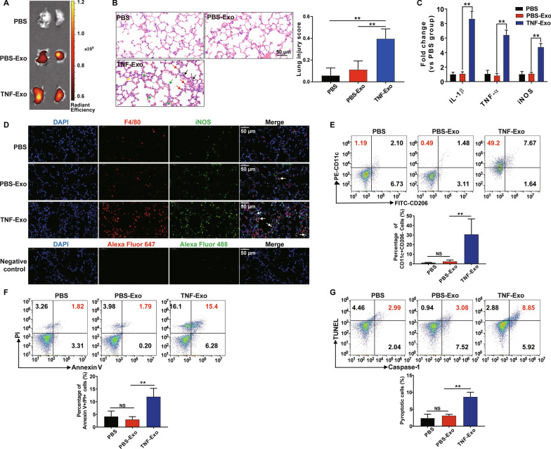
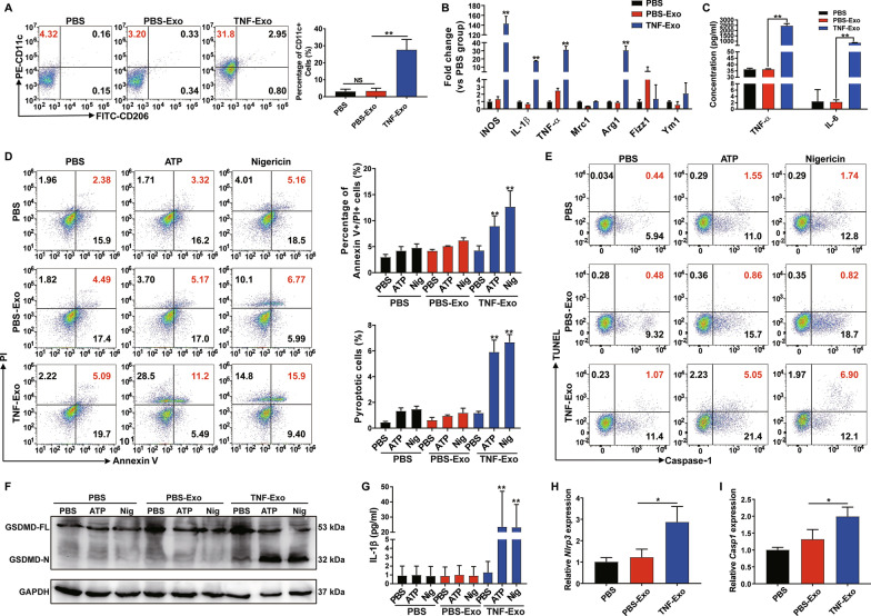
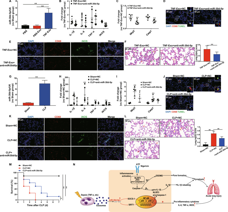
Methods: Tumor necrosis factor-α (TNF-α), a key regulator of innate immunity in sepsis-related ALI, was used to stimulate PMNs from healthy C57BL/6J mice in vitro. Exosomes isolated from the supernatant were injected to C57BL/6J wild-type mice intraperitoneally (i.p.) and then examined for lung inflammation, macrophage (Mϕ) polarization and pyroptosis. In vitro co-culture system was applied where the mouse Raw264.7 macrophages or bone marrow-derived macrophages (BMDMs) were co-cultured with PMN-derived exosomes to further confirm the results of in vivo animal study and explore the potential mechanisms involved.

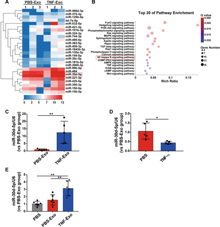
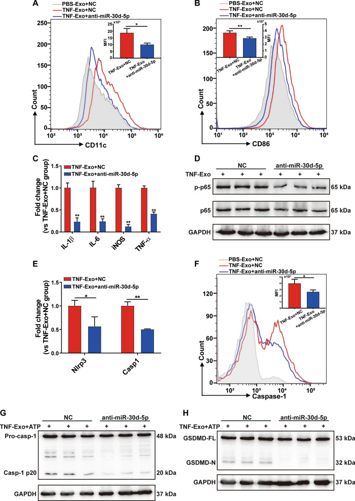
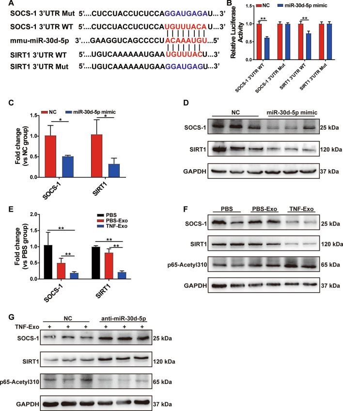
Results: Exosomes released by TNF-α-stimulated PMNs (TNF-Exo) promoted M1 macrophage activation after in vivo i.p. injection or in vitro co-culture. In addition, TNF-Exo primed macrophage for pyroptosis by upregulating NOD-like receptor 3 (NLRP3) inflammasome expression through nuclear factor κB (NF-κB) signaling pathway. Mechanistic studies demonstrated that miR-30d-5p mediated the function of TNF-Exo by targeting suppressor of cytokine signaling (SOCS-1) and sirtuin 1 (SIRT1) in macrophages. Furthermore, intravenous administration of miR-30d-5p inhibitors significantly decreased TNF-Exo or cecal ligation and puncture (CLP)-induced M1 macrophage activation and macrophage death in the lung, as well as the histological lesions.

Conclusions: The present study demonstrated that exosomal miR-30d-5p from PMNs contributed to sepsis-related ALI by inducing M1 macrophage polarization and priming macrophage pyroptosis through activating NF-κB signaling. These findings suggest a novel mechanism of PMN-Mϕ interaction in sepsis-related ALI, which may provide new therapeutic strategies in sepsis patients.

Keywords: Exosomes; Macrophage; Neutrophil; Pyroptosis; Sepsis-related acute lung injury; miR-30d-5p.


Conflict of interest statement

The authors declare that they have no competing interests.

Figures

Fig. 1
Fig. 1
TNF-Exo induces lung injury by affecting M1 macrophage activation and pyroptosis in vivo. WT C57BL/6 mice were administered with PBS-Exo/TNF-Exo (300 μg/mouse) intraperitoneally for 24 h. An equal volume of PBS was used as negative control. a Ex vivo fluorescence signals in the lungs of mice injected i.p. with Dil-labeled exosomes. b Evaluation of lung histology by H&E staining (magnification × 400). Green arrows indicate neutrophils in the alveolar and interstitial space, red arrows indicate alveolar macrophages, yellow arrows indicate hyaline membranes, and black arrows indicate thickening of the alveolar walls. Scale bar, 50 μm. c Detection of inflammatory cytokine mRNA (IL-1β, TNF-α) and iNOS mRNA expression in the lung tissues by RT-qPCR. d Representative images of direct immunofluorescence staining of DNA (blue), F4/80 (red) and iNOS (green) in the lung sections, and white arrows indicate iNOS positive macrophages. Negative control represents immunofluorescence images stained with species-specific secondary antibodies coupled with Alexa Fluor Dyes alone. Scale bar, 50 μm. e Flow cytometry detection of CD11c and CD206 expression on peritoneal macrophage (PMϕ) after being gated with macrophage marker F4/80. f Representative flow cytometry plots of Annexin V/PI staining of PMϕ, and analysis of Annexin V/PI double-stained cells by dying. g Representative flow cytometry plots and quantitation of PMϕ pyroptosis (Caspase-1/TUNEL double-positive cells). One-way analysis of variance with Tukey's multiple comparisons test was used for the analysis. Graphs represent means ± SEM, n ≥ 3; *P < 0.05, **P < 0.01 compared within two groups
Fig. 2
Fig. 2
TNF-Exo promotes M1 macrophage activation and primes macrophage pyroptosis through NF-κB pathway in an in vitro co-culture model. ac Treatment of Raw264.7 macrophages with PBS-Exo/TNF-Exo for 24 h. An equal volume of PBS was used as negative control. a Flow cytometry detection of CD11c and CD206 expression on Raw264.7 macrophages. b Detection of expression levels of iNOS, IL-1β, TNF-α, Mrc1, Arg1, Fizz1 and Ym1 mRNA by RT-qPCR. c Detection of the concentration of inflammatory cytokines (IL-6, TNF-α) in the supernatant of Raw264.7 macrophages by ELISA. d–g Stimulation of Raw264.7 macrophages with PBS/PBS-Exo/TNF-Exo for 24 h and treatment with PBS, 5 mM ATP or 20 mM nigericin for 2 h. d Flow cytometry evaluation of macrophage death by Annexin-V and PI double-staining. e Flow cytometry evaluation of macrophage pyroptosis by Caspase-1 and TUNEL double-staining. f Cleavage of GSDMD by western blot. GSDMD-FL: full-length GSDMD, GSDMD-N: N-terminal cleavage products of GSDMD. g Analysis of culture supernatants for IL-1β secretion by ELISA. h–i Treatment of Raw264.7 macrophages with PBS/PBS-Exo/TNF-Exo for 24 h, and detection of NLRP3 and caspase-1 mRNA expression by RT-qPCR. One-way analysis of variance with Tukey's multiple comparisons test was used for the analysis. Graphs represent means ± SEM, n ≥ 3; *P < 0.05, **P < 0.01 compared within two groups
Fig. 3
Fig. 3
miRNA analysis of PMN-derived exosomes. a Heat map of exosomal miRNA-seq (n = 3). The fluorescence intensity of 26 differentially expressed miRNAs (≥ twofold) is illustrated from high (red) to low (blue). b Kyoto Encyclopedia of Genes and Genomes (KEGG) analysis on differentially expressed exosomal miRNAs; the 20 most enriched pathways related to signaling transduction are shown. The rich ratio indicates the number of genes in the miRNA target list over the total genes in the respective canonical pathway. c–e Expression of miR-30d-5p in PMN-derived exosomes, in PMNs stimulated with PBS/TNF-α and in recipient Raw264.7 macrophages treated with PBS/PBS-Exo/TNF-Exo by RT-qPCR. Student’s t test (c, d) or one-way analysis of variance with Tukey's multiple comparisons test (E) was used for the analysis. Graphs represent means ± SEM, n ≥ 3; *P < 0.05, **P < 0.01 compared within two groups
Fig. 4
Fig. 4
TNF-Exo activates NF-κB signaling pathway in macrophage via miR-30d-5p. a–e Prior to co-culturing with TNF-Exo for 24 h, Raw264.7 macrophages were transfected with negative control (NC) or miR-30d-5p inhibitors (anti-miR-30d-5p) for 24 h. a, b Flow cytometry detection of CD11c and CD86 expression. c Detection of inflammatory cytokine mRNA (IL-6, IL-1β, TNF-α) and iNOS mRNA expression by RT-qPCR. d Western blot of NF-κB p-p65 and p65 in Raw264.7 macrophages. e Detection of NLRP3 and caspase-1 mRNA expression by RT-qPCR. f–h Transfection of Raw264.7 macrophages with negative control (NC) or miR-30d-5p inhibitors for 24 h, followed by stimulation with TNF-Exo plus ATP. f Flow cytometry detection of Alexa Fluor 488-labeled caspase-1 FLICA expression. g Western blots of pro-caspase-1 (Pro-casp-1) and activated/cleaved caspase-1 (Casp-1 p20) in whole-cell lysates of Raw264.7. h Cleavage of GSDMD by immunoblotting. Student’s t test was used for analysis. Graphs represent means ± SEM, n ≥ 3; *P < 0.05, **P < 0.01 compared within two groups
Fig. 5
Fig. 5
Exosomal miR-30d-5p activates NF-κB in macrophage via targeting SOCS-1 and SIRT1. a Sequence alignment between miR-30d-5p and its putative binding sites (in red letters) in the SOCS-1/SIRT1 3′-UTR. Mutation of the miR-30d-5p target sites (in blue letters) is also shown. b Detection of the relative luciferase activities of WT and Mut SOCS-1/SIRT1 reporters by luciferase reporter assay, using Renilla luciferase vector as the internal control. RT-qPCR analysis of relative SOCS-1/SIRT1 mRNA level (c) and Western blot (d) of SOCS-1 and SIRT1 in Raw264.7 macrophages transfected with miR-30d-5p mimics as indicated. e, f Treatment of Raw264.7 macrophages with PBS/PBS-Exo/TNF-Exo for 24 h. e Detection of mRNA levels of SOCS-1 and SIRT1 by RT-qPCR. f Western blot analysis of SOCS-1, SIRT1 and p65-Acetyl 310. g Prior to co-culturing with TNF-Exo for 24 h, Raw264.7 macrophages were transfected with control or miR-30d-5p inhibitors for 24 h. The expression levels of SOCS-1, SIRT1 and p65-Acetyl 310 in Raw264.7 cells were measured by Western blot. Student’s t test (b, c) or one-way analysis of variance with Tukey's multiple comparisons test (e) was used for the analysis. Graphs represent means ± SEM, n ≥ 3; *P < 0.05, **P < 0.01 compared within two groups
Fig. 6
Fig. 6
miR-30d-5p inhibition alleviates TNF-Exo or CLP-induced lung injury. Mice were injected with TNF-Exo (300 μg/mouse) intraperitoneally (a–f) or subjected to sham or CLP (g–l) for 24 h. The miR-30d-5p inhibitor or negative control (NC) was transferred into each mouse 1 day before TNF-Exo injection or CLP surgery. Relative expression levels of miR-30d-5p (a, g), inflammatory cytokine mRNA (IL-6, IL-1β, TNF-α) and iNOS mRNA (b, h), NLRP3 and caspase-1 mRNA expression (c, i) in the lung tissues were measured by RT-qPCR. d, j Representative images of direct immunofluorescence staining of DNA (blue), CD68 (red) and TUNEL (green) in the lung sections. Scale bar, 50 μm. e, k Representative images of direct immunofluorescence staining of DNA (blue), CD68 (red) and iNOS (green) in the lung sections, and white arrows indicate iNOS positive macrophages. Scale bar, 50 μm. f, l Evaluation of lung histology by H&E staining (magnification × 400). Green arrows indicate neutrophils in the alveolar and interstitial space, red arrows indicate alveolar macrophages, yellow arrows indicate hyaline membranes, blue arrows indicate proteinaceous debris filling the airspaces, and black arrows indicate thickening of the alveolar walls. Lung injury scores were assessed. Scale bar, 50 μm. Student’s t test or one-way analysis of variance with Tukey's multiple comparisons test was used for the analysis. m Survival rate of CLP mice with or without miR-30d-5p inhibition (n = 8) and log-rank test was used for the analysis. n Schematic representation of the mechanism by which neutrophil-derived exosomes induce M1 macrophage polarization and prime macrophage pyroptosis in sepsis-related ALI. Graphs represent means ± SEM, n ≥ 3; *P < 0.05, **P < 0.01 compared within two groups (h, i *,**P compared with Sham + NC group, #,##P compared with CLP + NC group)

References

    1. Fleischmann C, Scherag A, Adhikari N, Hartog C et al (2016) Assessment of global incidence and mortality of hospital-treated sepsis. current estimates and limitations. Am J Respir Crit Care Med 193(3): 259-272 - PubMed
    1. Singer M, Deutschman C, Seymour C, Shankar-Hari M, et al. The third international consensus definitions for sepsis and septic shock (sepsis-3) JAMA. 2016;315(8):801–810. doi: 10.1001/jama.2016.0287. - DOI - PMC - PubMed
    1. Moreira J. Severe sepsis and septic shock. N Engl J Med. 2013;369(21):2063. - PubMed
    1. Liu L, Sun B. Neutrophil pyroptosis: new perspectives on sepsis. Cell Mol Life Sci CMLS. 2019;76(11):2031–2042. doi: 10.1007/s00018-019-03060-1. - DOI - PMC - PubMed
    1. Thebaud B, Stewart DJ. Exosomes: cell garbage can, therapeutic carrier, or Trojan horse? Circulation. 2012;126(22):2553. doi: 10.1161/CIRCULATIONAHA.112.146738. - DOI - PubMed

Publication types