. 2021 Dec 1;41(48):9988-10003.
doi: 10.1523/JNEUROSCI.0881-21.2021. Epub 2021 Oct 12.

Contralateral Projection of Anterior Cingulate Cortex Contributes to Mirror-Image Pain

Affiliations

Contralateral Projection of Anterior Cingulate Cortex Contributes to Mirror-Image Pain

Su-Wan Hu et al. J Neurosci. .

Abstract

Long-term limb nerve injury often leads to mirror-image pain (MIP), an abnormal pain sensation in the limb contralateral to the injury. Although it is clear that MIP is mediated in part by central nociception processing, the underlying mechanisms remain poorly understood. The anterior cingulate cortex (ACC) is a key brain region that receives relayed peripheral nociceptive information from the contralateral limb. In this study, we induced MIP in male mice, in which a unilateral chronic constrictive injury of the sciatic nerve (CCI) induced a decreased nociceptive threshold in both hind limbs and an increased number of c-Fos-expressing neurons in the ACC both contralateral and ipsilateral to the injured limb. Using viral-mediated projection mapping, we observed that a portion of ACC neurons formed monosynaptic connections with contralateral ACC neurons. Furthermore, the number of cross-callosal projection ACC neurons that exhibited c-Fos signal was increased in MIP-expressing mice, suggesting enhanced transmission between ACC neurons of the two hemispheres. Moreover, selective inhibition of the cross-callosal projection ACC neurons contralateral to the injured limb normalized the nociceptive sensation of the uninjured limb without affecting the increased nociceptive sensation of the injured limb in CCI mice. In contrast, inhibition of the non-cross-callosal projection ACC neurons contralateral to the injury normalized the nociceptive sensation of the injured limb without affecting the MIP exhibited in the uninjured limb. These results reveal a circuit mechanism, namely, the cross-callosal projection of ACC between two hemispheres, that contributes to MIP and possibly other forms of contralateral migration of pain sensation.SIGNIFICANCE STATEMENT Mirror-image pain (MIP) refers to the increased pain sensitivity of the contralateral body part in patients with chronic pain. This pathology requires central processing, yet the mechanisms are less known. Here, we demonstrate that the cross-callosal projection neurons in the anterior cingulate cortex (ACC) contralateral to the injury contribute to MIP exhibited in the uninjured limb, but do not affect nociceptive sensation of the injured limb. In contrast, the non-cross-callosal projection neurons in the ACC contralateral to the injury contribute to nociceptive sensation of the injured limb, but do not affect MIP exhibited in the uninjured limb. Our study depicts a novel cross-callosal projection of ACC that contributes to MIP, providing a central mechanism for MIP in chronic pain state.

Keywords: anterior cingulate cortex; chronic constrictive injury of the sciatic nerve; excitatory pyramidal neuron; mirror-image pain; projection.


Figures

Figure 1.
Figure 1.
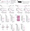
CCI-induced chronic neuropathic pain produces MIP in the contralateral paw. A, Experimental timeline. PWTs and PWLs of the two hind paws in mice were assessed at day 1 before, and at days 3, 7, 14, and 21 after the sham or CCI surgery. TFT and FMT tests, and SPT, OFT, EPM, and TST were conducted in different batches of mice from day 14 after the CCI surgery. B, Schematic for the unilateral sham or CCI surgery and bilateral pain threshold measurements. C, Statistics showing that, compared with the sham mice, CCI mice exhibited decreased PWTs and PWLs in the injured paw at days 3, 7, 14, and 21 following the CCI surgery. nsham = 14; nCCI = 18. PWT: group, F(1,30) = 113.2, p < 0.0001; sham versus CCI, 3 d p < 0.0001, 7 d p = 0.0017, 14 d p = 0.0002, 21 d p = 0.0002. PWL: group, F(1,30) = 263.1, p < 0.0001; 3 d p < 0.0001, 7 d p < 0.0001, 14 d p < 0.0001, 21 d p < 0.0001. D, Statistics showing that, compared with sham mice, CCI mice showed decreased PWTs at days 14 and 21, and decreased PWLs at days 7, 14, and 21 in the contralateral paw, suggesting an occurrence of MIP. nsham = 14; nCCI = 18. PWT: group, F(1,30) = 14.46, p = 0.0007; sham versus CCI, 14 d p = 0.0006, 21 d p < 0.0001. PWL: group, F(1,30) = 36.87, p < 0.0001; sham versus CCI, 7 d p < 0.0001, 14 d p < 0.0001, 21 d p = 0.0004. E, F, Summary data showing that sham and CCI mice did not differ in (E) tail flick latency or (F) FMT. TFT: mean ± SD, sham, 3.982 ± 0.4416; CCI, 4.011 ± 0.7325; t(11) = 0.08,524, p = 0.9336. FMT: sham, 0.4050 ± 0.2393; CCI, 0.4743 ± 0.2934; t(11) = 0.461, p = 0.6538. G, Statistics showing that, in SPT, the percent of sucrose preference did not differ between sham (70.14 ± 4.162) and CCI mice (68.95 ± 8.367). t(11) = 0.3163, p = 0.7577. H, Typical traveling trace of sham and CCI mice in open field. I, J, Summary data showing that, compared with sham mice, CCI mice exhibited similar levels of (I) number of entries into the center and (J) time spent in the center zone in the OFT. OFT: time spent in the center, sham, 33.67 ± 15.22; CCI, 32.07 ± 7.523; t(11) = 0.2463, p = 0.8100; number of entries, sham, 26 ± 9.92; CCI, 21.14 ± 4.1; t(11) = 1.189, p = 0.2594. K, Typical traveling traces of sham and CCI mice in EPM test. L, M, Statistics showing that sham and CCI mice exhibited similar levels of (L) number of entries into the open arm and (M) time spent in the open arm in EPM test. EPM: entries into the open arms: sham, 9 ± 7.772; CCI, 8.143 ± 6.176; t(11) = 0.2218, p = 0.8286; time spent in the open arms, sham, 15.12 ± 9.636; CCI, 17.44 ± 13.08; t(11) = 0.3591, p = 0.7263. M, Summary results showing that the immobility time in TST did not differ between sham (112.2 ± 12.38) and CCI mice (108.2 ± 11.37). t(11) = 0.6028, p = 0.5588. **p < 0.01. ***p < 0.001. ****p < 0.0001. Error bars indicate SEM. Data analyzed by (C,D) two-way repeated-measures ANOVA with Bonferroni post-tests, or (E-G,I,J,L-N) unpaired t test. Ipsi, Ipsilateral; contra, contralateral; BL, baseline.
Figure 2.
Figure 2.
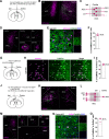
c-Fos signals are increased bilaterally in ACC on chronic neuropathic pain induced by CCI. A, Representative confocal images of c-Fos staining in the two sides of ACC from sham and 14 d CCI mice. Scale bars: top column, 200 µm; bottom column, 100 µm. B, Quantitative summary of c-Fos-positive neurons in two sides of ACC, indicating that both sides of the ACC are hyperactivated on CCI-induced chronic neuropathic pain. n = 20 from 6 mice/group. Contra: sham, 50.65 ± 19.18; CCI, 128.9 ± 63.7; Ipsi: sham, 52.5 ± 21.84; CCI, 136.6 ± 34.29. Group, F(1,76) = 96.67, p < 0.0001, two-way ANOVA; sham versus CCI, Contra p < 0.0001, Ipsi p < 0.0001; Bonferroni post-tests. C, Representative confocal images for cell type identification of c-Fos-positive neurons in ACC from 14 d CCI mice. White arrows indicate colabeled neurons. Scale bar, 50 µm. D, Quantitative results showing that the majority of the c-Fos-positive neurons in the two sides of ACC from CCI mice were colabeled by CaMKII. n = 9 from 4 mice/group. Sham, 92.7 ± 3.428; CCI, 92.26 ± 4.658. t(8) = 0.1973, p = 0.8485; paired t test. E, Quantitative results showing that few c-Fos-positive neurons were labeled by GABA. GABA neurons were labeled by the local injection of AAV-vGAT1-mCherry. n = 8 from 4 mice/group. Sham, 2.912 ± 1.331; CCI, 2.783 ± 0.7378. t(7) = 0.3075, p = 0.7674; paired t test. ****p < 0.0001. Error bars indicate SEM. Ipsi, Ipsilateral; contra, contralateral.
Figure 3.
Figure 3.
Chemogenetic inhibition of the two unilateral ACC exerts different effects on the original pain and MIP induced by CCI. A, G, Experimental timeline and figure legends for behavioral summary. Mice were injected with AAV vectors into the ACC contralateral or ipsilateral to the CCI at day 14 before the CCI surgery. PWTs and PWLS were assessed in 2 d from day 14 after the CCI surgery. B, H, Schematic of virus injection and typical confocal image for mCherry expression in the ACC contralateral or ipsilateral to the CCI. Scale bar, 100 µm. C, D, Statistics demonstrating that chemogenetic inhibition of the ACC excitatory pyramidal neurons contralateral to the CCI relieved (C) mechanical allodynia and (D) thermal hyperalgesia in the ipsilateral paw induced by CCI. PWT: group, F(3,24) = 11.61, p < 0.0001; CCI+mCherry versus CCI+Gi-mCherry, CNO p = 0.0047. PWL: group, F(3,24) = 110, p < 0.0001; CCI+mCherry versus CCI+Gi-mCherry, CNO p = 0.0009. E, F, Statistics showing that chemogenetic inhibition of the ACC excitatory pyramidal neurons contralateral to the CCI did not change the (E) PWTs and (F) PWLs in the contralateral paw of CCI mice. PWT: group, F(3,24) = 10.92, p = 0.0001; CCI+mCherry versus CCI+Gi-mCherry, CNO p > 0.9999. PWL: group, F(3,24) = 33.96, p < 0.0001; CCI+mCherry versus CCI+Gi-mCherry, CNO p > 0.9999. I, J, Summary showing that chemogenetic inhibition of the ACC excitatory pyramidal neurons ipsilateral to the CCI did not change the (I) PWTs and (J) PWLs in the ipsilateral paw of CCI mice. PWT: group, F(3,24) = 25.67, p < 0.0001; CCI+mCherry versus CCI+Gi-mCherry, CNO p > 0.9999. PWL: group, F(3,24) = 69.76, p < 0.0001; CCI+mCherry versus CCI+Gi-mCherry, CNO p > 0.9999. K, L, Summary showing that chemogenetic inhibition of the ACC excitatory pyramidal neurons ipsilateral to the CCI relieved MIP in the contralateral paw induced by CCI. PWT: group, F(3,24) = 7.472, p = 0.0011; CCI+mCherry versus CCI+Gi-mCherry, CNO p = 0.0486. PWL: group, F(3,24) = 17.72, p < 0.0001; CCI+mCherry versus CCI+Gi-mCherry, CNO p = 0.0024. *p < 0.05. **p < 0.01. ***p < 0.001. ****p < 0.0001. C-F, I-L, Data analyzed by two-way repeated-measures ANOVA with Bonferroni post-tests. Error bars indicate SEM. Ipsi, Ipsilateral; contra, contralateral; BL, baseline.
Figure 4.
Figure 4.
Genetic ablation or long-term inhibition of the excitatory pyramidal neurons in the ACC contralateral to the CCI relieves MIP induced by CCI. A, Timeline for the microinjections, CCI surgery, and PWT and PWL tests. B, Schematic of the virus injection for genetic ablation of the excitatory pyramidal neurons in the ACC contralateral to the CCI and typical confocal image indicating the efficacy of caspase 3. Scale bar, 200 µm. Summary showing that, compared with that in the ACC ipsilateral to the CCI, the number of NeuN-labeled cells was significantly decreased in the ACC contralateral to the CCI. n = 8 from 4 mice. Ipsi, 329.9 ± 34.98; Contra, 45.13 ± 9.553. t(7) = 24.67, p < 0.0001. C, Summary results showing that, compared with their control counterparts, the CCI mice with caspase 3 expression exhibited increased PWTs and PWLs at days 14 and 21 following the CCI surgery. PWT: group, F(3,23) = 18.21, p < 0.0001; CCI-ctrl versus CCI-cas3, 14 d p = 0.0247, 21 d p = 0.0309. PWL: group, F(3,23) = 321.3, p < 0.0001; CCI-ctrl versus CCI-cas3, 14 d p = 0.0001, 21 d p < 0.0001. D, Summary showing that depleting the ACC excitatory pyramidal neurons contralateral to the CCI increased the PWTs at days 14 and 21, and the PWLs at days 7, 14, and 21 following the CCI surgery. PWT: group, F(3,23) = 5.111, p = 0.0074; CCI-ctrl versus CCI-cas3, 14 d p = 0.0235, 21 d p = 0.0398. PWL: group, F(3,23) = 25.33, p < 0.0001; CCI-ctrl versus CCI-cas3, 7 d p = 0.0002, 14 d p < 0.0001, 21 d p = 0.0064. E, Timeline for the behavioral tests in mice with repeated CNO administrations. Five day CNO injections (1 mg/kg, i.p., twice per day, 8:00 A.M. and 8:00 P.M.) began at day 10 following the CCI surgery. PWTs and PWLs were evaluated at days 1, 3, and 5 after the CNO injection. F, Schematic of the virus injection and typical confocal image showing the mCherry expression in the ACC contralateral to the CCI. Scale bar, 100 µm. G, Summary showing that repeated inhibition of the excitatory pyramidal neurons in the ACC contralateral to the CCI increased the PWTs and PWLs in the ipsilateral paw in CCI mice. Ipsilateral PWT: group, F(3,25) = 11.79, p < 0.0001; CCI+mCherry versus CCI+Gi-mCherry, 1 d p = 0.0261. Ipsilateral PWL: group, F(3,25) = 108.6, p < 0.0001; CCI+mCherry versus CCI+Gi-mCherry, 1 d p < 0.0001, 3 d p = 0.0025. H, Summary showing that the repeated inhibition relieved MIP in the contralateral paw in CCI mice. Contralateral PWT: group, F(3,25) = 6.458, p = 0.0022; CCI+mCherry versus CCI+Gi-mCherry, 1 d p = 0.0257. Contralateral PWL: group, F(3,25) = 15.19, p < 0.0001; CCI+mCherry versus CCI+Gi-mCherry, 1 d p = 0.0033. *p < 0.05. **p < 0.01. ***p < 0.001. ****p < 0.0001. Data analyzed by (B) paired t test and (C,D,G,H) two-way repeated-measures ANOVA with Bonferroni post-tests. Error bars indicate SEM. Ipsi, Ipsilateral; contra, contralateral; BL, baseline; cas3, caspase 3.
Figure 5.
Figure 5.
ACC contralateral to the CCI sends direct projection to the ACC ipsilateral to the CCI. A, Schematic of virus injections into the ACC contralateral to the CCI. The virus injections consisted of two stages: (1) mix of AAV-CaMKII-cre and AAV-DIO-TK-EGFP; and (2) 21 d later, HSV-△TK-tdTomato. B, Representative confocal images showing the tdTomato expression in the two sides of ACC and EGFP expression in the ACC contralateral to the CCI. The neurons labeled by both tdTomato and EGFP in the ACC contralateral to the CCI were defined as starter cells. Scale bar, 200 µm. C, Schematic of the main pain-associated brain regions that receive monosynaptic inputs from the starter cells. D, Example confocal images of the tdTomato-positive neurons from the PFC, secondary motor cortex (M2), lateral occipital cortex (LO), anteromedial thalamus (AM), and mediodorsal thalamus (MD). Scale bar, 200 µm. E, Example confocal images of the CaMKIIα-stained section of the ACC ipsilateral to the CCI. Scale bar, 50 µm. F, Quantified summary of the percent of CaMKIIα-positive neurons to tdTomato-positive cells in the ACC ipsilateral to the CCI: 87.9 ± 4.71; n = 12 from 4 mice. G, Schematic of virus injections for labeling the ACC neurons ipsilateral to the CCI that receive contralateral inputs. H, Example images showing the mCherry-labeled ACC sections stained with CaMKIIα. White arrows indicate colabeled neurons. Scale bar, 100 µm. I, Summary showing the percent of CaMKIIα-positive neurons to mCherry-positive cells: 81.2 ± 2.96; n = 12 from 4 mice. J, Schematic of virus injections into the ACC ipsilateral to the CCI. The virus injections consisted of two stages: (1) mix of AAV-CaMKII-cre, AAV-DIO-TVA-EGFP, and AAV-DIO-RG; and (2) 21 d later, RV-EnVA-△G-DsRed. K, Example confocal images showing the tdTomato expression in the two sides of ACC and EGFP expression in the ACC ipsilateral to the CCI. The neurons labeled by both DsRed and EGFP in the ACC ipsilateral to the CCI were defined as starter cells. Scale bar, 200 µm. L, Schematic of the main pain-associated brain regions that send monosynaptic projections onto the starter cells. M, Representative confocal images of the DsRed-positive neurons from the M2, PFC, claustrum (Cl), BLA, AM, MD, S1, and retrosplenial granular cortex (RSG). Scale bar, 200 µm. N, Example confocal images of the CaMKIIα-stained section of ACC contralateral to the CCI. Scale bar, 50 µm. O, Quantified summary of the percent of CaMKIIα-positive neurons to DsRed-positive cells in the ACC contralateral to the CCI: 95.1 ± 10.5; n = 15 from 5 mice. Error bars indicate SEM. Ipsi, Ipsilateral; contra, contralateral.
Figure 6.
Figure 6.
c-Fos signals of the cross-callosal projection excitatory pyramidal neurons in the ACC contralateral to the CCI are increased on chronic neuropathic pain induced by CCI. A, Schematic of virus injections for labeling the cross-callosal projection excitatory pyramidal neurons in the ACC contralateral to the CCI. B, Example confocal images showing the ACC sections stained with c-Fos from sham and 14 d CCI mice. White arrows indicate colabeled neurons. Scale bar, 50 µm. C, Statistics showing that the percent of c-Fos-positive neurons to mCherry-labeled population were higher in 14 d CCI mice than that in sham mice: sham, 9.455 ± 3.514, n = 10 from 4 mice; CCI, 46.69 ± 8.616, n = 15 from 5 mice (t(19.93) = 14.97, p < 0.0001, unpaired t test with Welch correction). ****p < 0.0001. Error bars indicate SEM. Ipsi, Ipsilateral; contra, contralateral.
Figure 7.
Figure 7.
Chemogenetic inhibition of the cross-callosal projection excitatory pyramidal neurons in the ACC contralateral to the CCI with a single CNO injection does not affect nociceptive sensation of CCI mice. A, Timeline of microinjections, CCI surgery, and behavioral tests, and figure legends for behavioral summary. PWTs and PWLs, respectively, were assessed in 2 d from day 14 after the CCI surgery. B, Schematic of virus injections to express Gi-mCherry or mCherry (as control) in the cross-callosal projection excitatory pyramidal neurons in the ACC contralateral to the CCI, and typical image showing the mCherry expression in the ACC contralateral to the CCI. Scale bar, 100 µm. C-F, Behavioral summary showing that inhibition of these projecting neurons by a single CNO injection (1 mg/kg, i.p.) did not change the pain thresholds of the two sides of paws in CCI mice. Ipsilateral PWT: group, F(3,24) = 18.67, p < 0.0001; CCI+mCherry versus CCI+Gi-mCherry, CNO p = 0.9611. Ipsilateral PWL: group, F(3,23) = 117.8, p < 0.0001; CCI+mCherry versus CCI+Gi-mCherry, CNO p = 0.9510. Contralateral PWT: group, F(3,24) = 9.347, p = 0.0003; CCI+mCherry versus CCI+Gi-mCherry, CNO p = 0.9894. Contralateral PWL: group, F(3,23) = 18.58, p < 0.0001; CCI+mCherry versus CCI+Gi-mCherry, CNO p = 0.8634. C-F, Data analyzed by two-way repeated-measures ANOVA with Bonferroni post-tests. Error bars indicate SEM. Ipsi, Ipsilateral; contra, contralateral; BL, baseline.
Figure 8.
Figure 8.
Long-term inhibition or genetic ablation of the cross-callosal projection excitatory pyramidal neurons in the ACC contralateral to the CCI relieves MIP without changing the original pain in CCI mice. A, Timeline of microinjections, CCI surgery, CNO injection, and behavioral tests. CNO was given twice every day for 5 d (1 mg/kg, i.p.). B, Schematic of the virus injection and typical confocal image showing the mCherry expression in putative cross-callosal projection excitatory pyramidal neurons in the ACC contralateral to the CCI. Scale bar, 100 µm. C, Schematic of the inhibited neurons in the ACC contralateral to the CCI. D, Summary showing that the repeated inhibition had no effect on PWTs and PWLs in the ipsilateral paw in CCI mice. Ipsilateral PWT: group, F(3,23) = 11.79, p < 0.0001; Ipsilateral PWL: group, F(3,23) = 190.9, p < 0.0001. E, Summary showing that the repeated inhibition of these projecting neurons relieved MIP in the contralateral paw in CCI mice. Contralateral PWT: group, F(3,23) = 4.722, p = 0.0012; CCI+mCherry versus CCI+Gi-mCherry, 1 d p = 0.0458. Contralateral PWL: group, F(3,23) = 18.25, p < 0.0001; CCI+mCherry versus CCI+Gi-mCherry, 1 d p = 0.0162, 3 d p = 0.0452. F, Timeline for the microinjections, CCI surgery, and PWT and PWL tests. G, Schematic of the virus injection for genetic ablation of the cross-callosal projection excitatory pyramidal neurons in the ACC contralateral to the CCI, and example confocal image indicating the efficacy of caspase 3. Scale bar, 200 µm. H, Schematic of the ablated neurons in the ACC contralateral to the CCI. I, Summary results showing that this genetic ablation had no effect on pain thresholds in the ipsilateral paw of CCI. PWT: group, F(3,22) = 55.46, p < 0.0001. PWL: group, F(3,22) = 195.8, p < 0.0001. J, Summary showing that depleting the cross-callosal projection excitatory pyramidal neurons in the ACC contralateral to the CCI increased the PWTs at day 21, and the PWLs at days 14 and 21 following the CCI surgery. PWT: group, F(3,22) = 13.1, p < 0.0001; CCI-ctrl versus CCI-cas3, 21 d p = 0.0063. PWL: group, F(3,22) = 14.85, p < 0.0001; CCI-ctrl versus CCI-cas3, 14 d p = 0.0013, 21 d p = 0.0002. *p < 0.05. **p < 0.01. ***p < 0.001. D, E, I, J, Data analyzed by two-way repeated-measures ANOVA with Bonferroni post-tests. Error bars indicate SEM. Ipsi, Ipsilateral; contra, contralateral; BL, baseline; cas3, caspase 3.
Figure 9.
Figure 9.
Chemogenetic inhibition of the non–cross-callosal projection excitatory pyramidal neurons in the ACC contralateral to the CCI reduces nociception in the ipsilateral paw but has no effect on contralateral MIP in CCI mice. A, Timeline of microinjections, CCI surgery, and behavioral tests. PWTs and PWLS were assessed in 2 d from day 14 after the CCI surgery. B, Schematic of virus injections and typical confocal image showing the mCherry expression. Scale bar, 100 µm. C, Schematic of the inhibited non–cross-callosal projection excitatory pyramidal neurons in the ACC contralateral to the CCI. D, Statistics showing that chemogenetic inhibition by the CNO injection (1 mg/kg, i.p.) reduced the pain thresholds in the ipsilateral paw of CCI mice. PWT: group, F(3,24) = 22, p < 0.0001; CCI+DO-mCherry versus CCI+DO-Gi-mCherry, CNO p = 0.0026. PWL: group, F(3,24) = 112, p < 0.0001; CCI+DO-mCherry versus CCI+DO-Gi-mCherry, CNO p = 0.0008. E, Summary results suggesting that MIP was not affected by inhibiting the non–cross-callosal projection excitatory pyramidal neurons in the ACC contralateral to the CCI. PWT: group, F(3,24) = 10.62, p = 0.0001; CCI+DO-mCherry versus CCI+DO-Gi-mCherry, CNO p = 0.9328. PWL: group, F(3,24) = 36.49, p < 0.0001; CCI+DO-mCherry versus CCI+DO-Gi-mCherry, CNO p = 0.6897. F, Schematic of virus injections for labeling the cross-callosal projection neurons with EGFP and non–cross-callosal projection neurons with mCherry in the ACC contralateral to the CCI. G, Typical confocal image showing the mCherry and EGFP expression in the ACC contralateral to the CCI. Scale bar, 100 µm. H, Timeline of microinjections, CCI surgery, CNO injections, and behavioral tests. CNO was injected twice per day for 5 d (1 mg/kg, i.p.). I, Schematic of virus injections and typical confocal image showing the mCherry expression. Scale bar, 100 µm. J, Summary showing that the repeated inhibition increased PWTs and PWLs in the ipsilateral paw in CCI mice at day 1. Ipsilateral PWT: group, F(3,24) = 25.47, p < 0.0001; CCI+DO-mCherry versus CCI+DO-Gi-mCherry, 1 d p = 0.0008. Ipsilateral PWL: group, F(3,24) = 130, p < 0.0001; CCI+DO-mCherry versus CCI+DO-Gi-mCherry, 1 d p = 0.0011. K, Summary showing that the repeated inhibition had no effect on MIP in the contralateral paw in CCI mice. Contralateral PWT: group, F(3,24) = 13.66, p < 0.0001. Contralateral PWL: group, F(3,24) = 28.51, p < 0.0001. *p < 0.05. **p < 0.01. ***p < 0.001. D, E, J, K, Data analyzed by two-way repeated-measures ANOVA with Bonferroni post-tests. Error bars indicate SEM. Ipsi, Ipsilateral; contra, contralateral; BL, baseline; cas3, caspase 3.
Figure 10.
Figure 10.
Schematic of the theoretical pathways through which the ACC contralateral to the CCI mediates the original pain and MIP induced by CCI. The ACC excitatory pyramidal neurons contralateral to the CCI receive nociceptive information via the ascending pathway, and exert pro-nociceptive effect on the original pain processing through the non–cross-callosal projection excitatory pyramidal neurons via the descending pro-nociceptive pathway. After receiving the nociceptive information, the ACC contralateral to the CCI conveys this information to the other side via the cross-callosal projection, and the ACC ipsilateral to the CCI sequentially transmits the pain signals, thus producing MIP at the contralateral side via the descending MIP-promoting pathway. Ipsi, Ipsilateral; contra, contralateral.

Similar articles

Cited by

References

    1. An K, Zhao H, Miao Y, Xu Q, Li YF, Ma YQ, Shi YM, Shen JW, Meng JJ, Yao YG, Zhang Z, Chen JT, Bao J, Zhang M, Xue T (2020) A circadian rhythm-gated subcortical pathway for nighttime-light-induced depressive-like behaviors in mice. Nat Neurosci 23:869–880. 10.1038/s41593-020-0640-8 - DOI - PubMed
    1. Arguis MJ, Perez J, Martinez G, Ubre M, Gomar C (2008) Contralateral neuropathic pain following a surgical model of unilateral nerve injury in rats. Reg Anesth Pain Med 33:211–216. 10.1016/j.rapm.2007.12.003 - DOI - PubMed
    1. Bliss TV, Collingridge GL, Kaang BK, Zhuo M (2016) Synaptic plasticity in the anterior cingulate cortex in acute and chronic pain. Nat Rev Neurosci 17:485–496. 10.1038/nrn.2016.68 - DOI - PubMed
    1. Bonin RP, Bories C, De Koninck Y (2014) A simplified up-down method (SUDO) for measuring mechanical nociception in rodents using von Frey filaments. Mol Pain 10:26. 10.1186/1744-8069-10-26 - DOI - PMC - PubMed
    1. Callaway EM, Luo L (2015) Monosynaptic circuit tracing with glycoprotein-deleted rabies viruses. J Neurosci 35:8979–8985. 10.1523/JNEUROSCI.0409-15.2015 - DOI - PMC - PubMed

Publication types