. 2021 Oct 12;12(1):5948.
doi: 10.1038/s41467-021-26095-0.

Endurance exercise training-responsive miR-19b-3p improves skeletal muscle glucose metabolism

Affiliations

Endurance exercise training-responsive miR-19b-3p improves skeletal muscle glucose metabolism

Julie Massart et al. Nat Commun. .

Abstract

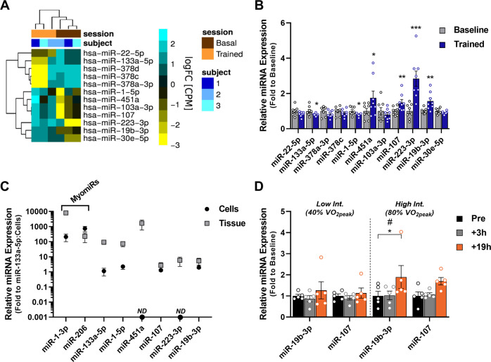
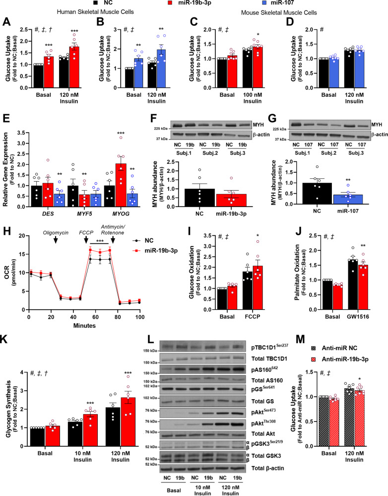
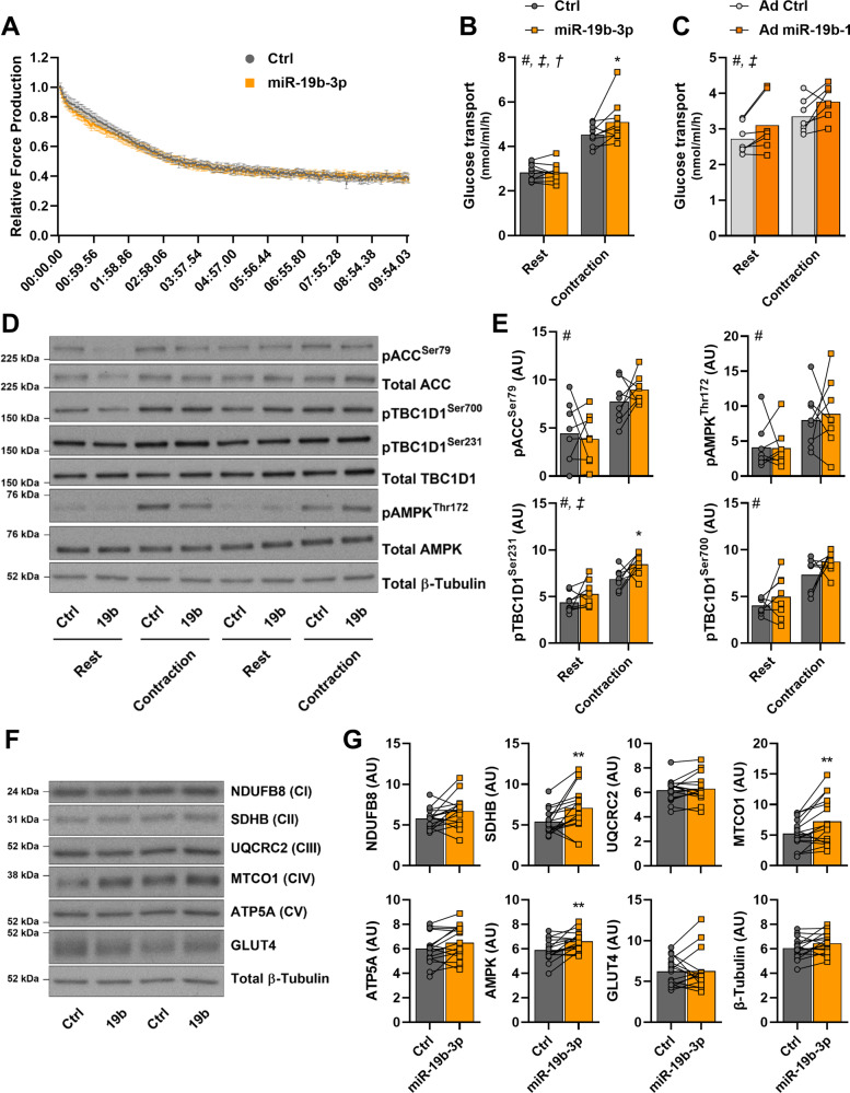
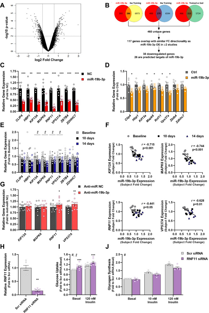
Skeletal muscle is a highly adaptable tissue and remodels in response to exercise training. Using short RNA sequencing, we determine the miRNA profile of skeletal muscle from healthy male volunteers before and after a 14-day aerobic exercise training regime. Among the exercise training-responsive miRNAs identified, miR-19b-3p was selected for further validation. Overexpression of miR-19b-3p in human skeletal muscle cells increases insulin signaling, glucose uptake, and maximal oxygen consumption, recapitulating the adaptive response to aerobic exercise training. Overexpression of miR-19b-3p in mouse flexor digitorum brevis muscle enhances contraction-induced glucose uptake, indicating that miR-19b-3p exerts control on exercise training-induced adaptations in skeletal muscle. Potential targets of miR-19b-3p that are reduced after aerobic exercise training include KIF13A, MAPK6, RNF11, and VPS37A. Amongst these, RNF11 silencing potentiates glucose uptake in human skeletal muscle cells. Collectively, we identify miR-19b-3p as an aerobic exercise training-induced miRNA that regulates skeletal muscle glucose metabolism.


Conflict of interest statement

The authors declare no competing interests.

Figures

Fig. 1
Fig. 1. Aerobic exercise training regulates multiple miRNAs in human skeletal muscle.
Skeletal muscle biopsies were obtained from healthy volunteers before and after an aerobic exercise training program. A Altered miRNAs as determined by short RNA sequencing (n = 3 individuals; FDR < 0.05). B RT-qPCR determination of miRNA expression before (Baseline) and after 14-day aerobic exercise training (Trained) of miRNAs identified by short RNA sequencing shown in (A) (n = 8 individuals) (p = 0.024 for miR-133a-5p; p = 0.0236 for miR-1-5p; p = 0.0224 for miR-451a; p = 0.0029 for miR-107; p = 0.0008 for miR-223.3p; p = 0.0013 for miR-19b-3p). C Relative miRNA expression in human skeletal muscle biopsies versus myotubes differentiated in vitro (data are mean ± SD for n = 3 cell culture independent samples and n = 8 tissue independent samples). D miR-19b-3p and miR-107 expression determined by RT-qPCR in human skeletal muscle biopsies obtained at baseline, 3 h, and 19 h after either low intensity (40% VO2peak) or high intensity (80% VO2peak) aerobic exercise (p = 0.0228 for miR-19b-3p 19 h following high-intensity exercise; n = 5 individuals). miRNA RT-qPCR data were normalized to miR-186-5p. Data are mean ± SEM. # Exercise effect (Friedman’s test); *p < 0.05, **p < 0.01, ***p < 0.001 using two-tailed paired Student’s t-test or Friedman’s test followed by Dunn’s multiple comparison test.
Fig. 2
Fig. 2. miR-19b-3p improves glucose metabolism and insulin sensitivity in skeletal muscle.
Myotubes were transfected with miR-19b-3p or miR-107 precursors or a negative control (NC) miRNA precursor. A, B Human skeletal muscle myotubes were incubated in the absence (Basal) or presence of insulin (120 nM) to determine the effect of A) miR-19b-3p (p = 0.0003 for basal and p = 0.0001 for insulin) and B miR-107 overexpression (p = 0.0019 for basal and p = 0.0014 for insulin) on glucose uptake (n = 6 independent cell experiments). C, D Mouse skeletal muscle myotubes were incubated in the absence (Basal) or presence of insulin (100 nM) to determine effects of C miR-19b-3p (p = 0.0307 for insulin) and D miR-107 overexpression on glucose uptake (n = 7 independent cell experiments). E Expression of DES, MYF5, and MYOG was determined by RT-qPCR in human myotubes overexpressing either miR-19b-3p or miR-107 (n = 6 independent cell experiments). Data for human skeletal muscle cells were normalized to the geometrical mean of GUSB and TBP expression (miR-19b-3p: p = 0.0035 for MYF5; p = 0.0008 for MYOG; miR-107: p = 0.001 for DES; p = 0.0057 for MYOG). F, G Western blot analysis and quantification of Myosin Heavy Chain 1 and 2 (MYH) abundance in human myotubes overexpressing F miR-19b-3p and G miR-107 (miR-107: p = 0.0067) (n = 6 independent cell experiments). H Oxygen Consumption Rate (OCR) in human myotubes overexpressing miR-19b-3p (n = 7 independent cell experiments). Addition of chemical substrates are indicated by arrows. p = 0.0001 following addition of FCCP. I Human myotubes were incubated in DMSO alone (Basal) or in the presence of 1 µM of the mitochondrial uncoupler FCCP to determine the effects of miR-19b-3p overexpression on glucose oxidation (n = 6 independent cell experiments) (p = 0.0241 for FCCP). J Human myotubes were incubated in DMSO alone (Basal) or in the presence of 10 nM GW1516 (PPARδ agonist) for 96 h prior to determination of miR-19b-3p overexpression on palmitate oxidation (n = 6 independent cell experiments) (p = 0.0026 for basal and p = 0.0013 for GW1516). K Human myotubes were incubated in the absence (Basal) or presence of insulin (10 or 120 nM) to determine the effect of miR-19b-3p overexpression on glucose incorporation into glycogen (n = 6 independent cell experiments) (p = 0.0009 for 10 nM insulin and p = 0.0001 for 120 nM insulin). L Representative immunoblots of human myotubes incubated for 10 min in the absence (Basal) or presence of insulin (10 or 120 nM) to determine the effects of miR-19b-3p overexpression on protein phosphorylation and protein abundance. Data of immunoblot quantification are found in Table S2. M Human myotubes were transfected with a miR-19b-3p-specific inhibitor (Anti-miR-19b-3p) or a negative control inhibitor (Anti-miR NC) and incubated in the absence (Basal) or presence of insulin (120 nM) to determine the effects of miR-19b-3p inhibition on glucose uptake (n = 7). Data are mean ± SEM. # Treatment effect, ‡ miRNA effect, † Interaction; and *p < 0.05, **p < 0.01, ***p < 0.001 by paired Student’s t-test or repeated measures 2-way ANOVA with Sidak’s post hoc testing when appropriate.
Fig. 3
Fig. 3. miR-19b-3p improves contraction-stimulated glucose transport in mouse skeletal muscle.
Mouse flexor digitorum brevis (FDB) skeletal muscle was electroporated with a control (Ctrl) or a pri-miR-19b-3p-encoding plasmid (miR-19b-3p) and 1 week later, was subjected to electrical pulse-stimulated contraction in vitro. A Relative force produced during each contraction measured over 10 min (n = 9 independent mouse muscles per construct). B In vitro glucose transport in FDB skeletal muscle electroporated with either control or pri-miR-19b-3p-encoding plasmid at rest or following electrical pulse-stimulated contraction (p = 0.0163) (n = 10 for rest and n = 9 for contraction independent mouse muscles per construct). C In vitro glucose transport in FDB skeletal muscle transduced with a control (Ad Ctrl) or mmu-miR-19b-1-expressing adenovirus (Ad miR-19b-1) at rest or following electrical pulse-stimulated contraction (n = 7 independent mouse muscles). D Representative immunoblots of FDB skeletal muscle to determine the effects of miR-19b-3p overexpression on basal and contraction-induced protein phosphorylation or abundance. E Quantification of (D) for phosphorylation of ACC (Ser79), AMPK (Thr172), and TBC1D1 (Ser231 and Ser700) (pTBC1D1 Ser231: p = 0.0479 following contraction) (n = 8 independent mouse muscles). F Representative immunoblots of FDB skeletal muscle to determine the effects of miR-19b-3p overexpression on protein abundance. G Quantification of (F) for total abundance of NDUFB8, SDHB (p = 0.0085), UQCRC2, MTCO1 (p = 0.005), ATP5A, AMPK (α-subunits; p = 0.0025), and GLUT4 (n = 16 independent mouse muscles). Data are mean. # Contraction effect; ‡ miRNA effect; † Interaction; and *p < 0.05 and **p < 0.01 using two-tailed paired Student’s t-test or repeated measures two-way ANOVA with Sidak’s post hoc testing when appropriate.
Fig. 4
Fig. 4. miR-19b-3p modulates expression of genes altered by endurance exercise training in human skeletal muscle.
A Volcano plot showing changes in gene expression determined by microarray analysis of myotubes overexpressing miR-19b-3p. Dashed lines indicate value cut-off at p < 0.01 and fold change of at least ±50% (n = 4 independent cell culture experiments). B Overview of workflow to identify genes with altered expression profiles following miR-19b-3p overexpression in human skeletal muscle cells and following aerobic exercise training (analysis of publicly available data,,). C Expression of miR-19b-3p predicted target genes CLIP4 (p = 0.0001), HBP1 (p = 0.0032), KIF13A (p = 0.0001), MAPK6 (p = 0.0007), RNF11 (p = 0.0001), VPS37A (p = 0.0004), ZBTB4 (p = 0.0001), and ZDHHC7 (p = 0.0001) as determined by RT-qPCR following negative control (NC) or miR-19b-3p overexpression in human skeletal muscle cells (n = 6 independent cell culture experiments). D Effect of miR-19b-3p overexpression in mouse tibialis anterior skeletal muscle on expression of Clip4 (p = 0.0275), Hbp1, Kif13a (p = 0.0388), Mapk6 (p = 0.0116), Rnf11 (p = 0.034), Vps37a (p = 0.0206), Zbtb4, and Zdhhc7 as determined by RT-qPCR (n = 6 independent mouse samples). E Gene expression of CLIP4, HBP1, KIF13A (p = 0.0497 at day 10), MAPK6 (p = 0.0143 at day 10 and p = 0.0031 at day 14), RNF11 (p = 0.038 at day 10 and p = 0.017 at day 14), VPS37A (p = 0.0051 at day 10 and p = 0.0222 at day 14), ZBTB4, and ZDHHC7 as determined by RT-qPCR in human skeletal muscle biopsies collected before (Baseline) and after 10 or 14 days of endurance exercise training (n = 8 individuals). F Pearson correlation of fold changes in miR-19b-3p expression and fold change of KIF13A (p = 0.0001), MAPK6 (p = 0.0001), RNF11 (p = 0.0312), or VPS37A (p = 0.001) expression in human skeletal muscle biopsies collected prior to (Baseline) and after 10 or 14 days of endurance exercise training (n = 24 biopsies). G Human myotubes were transfected with a miR-19b-3p-specific inhibitor (Anti-miR-19b-3p) or a negative control inhibitor (Anti-miR NC) and gene expression of KIF13A, MAPK6, RNF11 (p = 0.024), and VPS37A (p = 0.0034) was determined by RT-qPCR (n = 10 independent cell culture experiments). H Human skeletal muscle myotubes were transfected with a scramble siRNA sequence (Scr siRNA) or a RNF11 siRNA and gene expression of RNF11 was determined by RT-qPCR (p = 0018, n = 6 independent cell culture experiments). I Human skeletal muscle myotubes were incubated in the absence (Basal, p = 0.0004) or presence of insulin (120 nM, p = 0.0008) to determine the effect of RNF11 silencing on glucose uptake (n = 8 independent cell culture experiments). J Glycogen synthesis rate was determined in human skeletal muscle cells in the absence (Basal) or presence of insulin (10 or 120 nM) following RNF11 silencing (n = 5 independent cell culture experiments). Data are mean ± SEM. RT-qPCR data in human skeletal muscle biopsies and cells were normalized to the geometrical mean of GUSB and TBP expression. For mouse TA muscle, gene expression was normalized to Tbp expression. # Insulin effect; ‡ Endurance exercise training effect; and *p < 0.05, **p < 0.01, ***p < 0.001 by two-tailed paired Student’s t-test, repeated measures one-way ANOVA with Dunnett’s post hoc testing, or repeated measures two-way ANOVA with Sidak’s post hoc testing when appropriate.

References

    1. Egan B, Zierath JR. Exercise metabolism and the molecular regulation of skeletal muscle adaptation. Cell Metab. 2013;17:162–184. doi: 10.1016/j.cmet.2012.12.012. - DOI - PubMed
    1. Rogers MA, King DS, Hagberg JM, Ehsani AA, Holloszy JO. Effect of 10 days of physical inactivity on glucose tolerance in master athletes. J. Appl. Physiol. 1990;68:1833–1837. doi: 10.1152/jappl.1990.68.5.1833. - DOI - PubMed
    1. Morrison DJ, Kowalski GM, Grespan E, Mari A, Bruce CR, Wadley GD. Measurement of postprandial glucose fluxes in response to acute and chronic endurance exercise in healthy humans. Am. J. Physiol. Endocrinol. Metab. 2018;314:E503–E511. doi: 10.1152/ajpendo.00316.2017. - DOI - PubMed
    1. Devlin JT, Hirshman M, Horton ED, Horton ES. Enhanced peripheral and splanchnic insulin sensitivity in NIDDM men after single bout of exercise. Diabetes. 1987;36:434–439. doi: 10.2337/diab.36.4.434. - DOI - PubMed
    1. Booth FW, Roberts CK, Laye MJ. Lack of exercise is a major cause of chronic diseases. Compr. Physiol. 2012;2:1143–1211. doi: 10.1002/cphy.c110025. - DOI - PMC - PubMed

Publication types

MeSH terms